Translate this page into:
Oral compound nevus: an unusual presentation
Correspondence Address:
Srilekha Pulivarthi
Department of Oral Medicine and Radiology, SVS Institute of Dental Sciences, Appanapally, Mahaboobnagar, Telangana
India
| How to cite this article: Pulivarthi S, Ramlal G, Divya Shree P. Oral compound nevus: an unusual presentation. Indian J Dermatol Venereol Leprol 2020;86:284-285 |
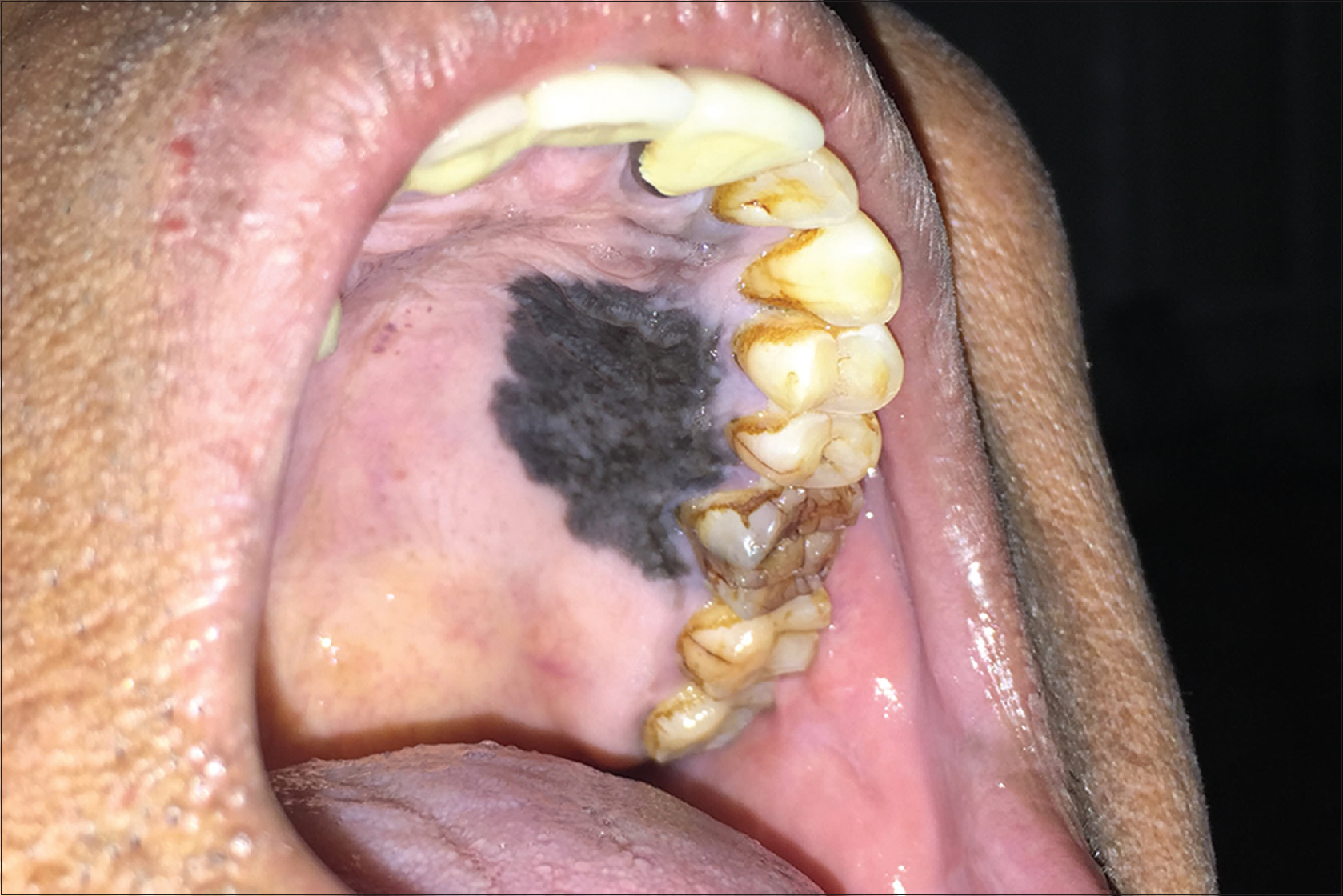
A 65-year-old man presented with a bluish-black coloured interconnected lesion in his oral cavity involving the labial gingiva and vestibule and hard palate. [Figure - 1] Slight lesional elevation was noted on the palatal and labial gingiva, blending with adjacent mucosa in labial vestibule. It was nontender, could not be scraped off and the surface was rough on palpation. Histopathological report revealed a compound nevus with epidermal nevus cells and underlying dermal connective tissue.
 |
| Figure 1: Bluish-black diffuse pigmented patch in the hard palate region |
Intraoral lesions of compound nevi are rare and occur as small macules (usually <2–3 mm). The widespread presentation of this lesion is a notable characteristic.
Declaration of patient consent
The authors certify that they have obtained all appropriate patient consent forms. In the form, the patient has given his consent for his images and other clinical information to be reported in the journal. The patient understands that name and initials will not be published and due efforts will be made to conceal identity, but anonymity cannot be guaranteed.
Financial support and sponsorship
Nil.
Conflicts of interest
There are no conflicts of interest.
Fulltext Views
3,653
PDF downloads
2,603


