Translate this page into:
Pagetoid dyskeratosis of hands: Report of two cases and the usefulness of dermoscopy
2 Department of Dermatology, General Hospital la Mancha Centro, Alcázar de San Juan, Ciudad Real, Granada, Spain
3 Department of Dermatology,University Hospital Virgen de las Nieves; Faculty of Medicine, Institute of Biosanitary Investigation Ibs, University of Granada, Granada, Spain
4 Department of Dermatology, University Hospital San Cecilio, Granada, Spain
Correspondence Address:
María Librada Porrino-Bustamante
University Hospital La Zarzuela, Calle de Pleyades, 25, 28023, Madrid
Spain
| How to cite this article: Porrino-Bustamante ML, Sánchez-López J, Arias-Santiago S, Fernández-Pugnaire MA. Pagetoid dyskeratosis of hands: Report of two cases and the usefulness of dermoscopy. Indian J Dermatol Venereol Leprol 2020;86:424-427 |
Sir,
Pagetoid dyskeratosis is an incidental pathologic finding which can be noted in skin biopsies from different lesions and body locations.[1] Friction has been proposed as an inducing factor.[1] Actually, the real nature of these cells and their causes are not well known. Pagetoid dyskeratosis has been described in different benign skin lesions, such as naevi, soft fibromas, acrochordons, lentigos, fibrous papules, milia and seborrheic keratoses.[1]
Only two papers have been published describing the dermoscopic findings of lesions with pagetoid dyskeratosis which consisted of a parallel ridge pattern.[2],[3] Herein, we report two new cases of pagetoid dyskeratosis in patients with brown pigmentation of hands and parallel ridge pattern on dermoscopy.
Case 1
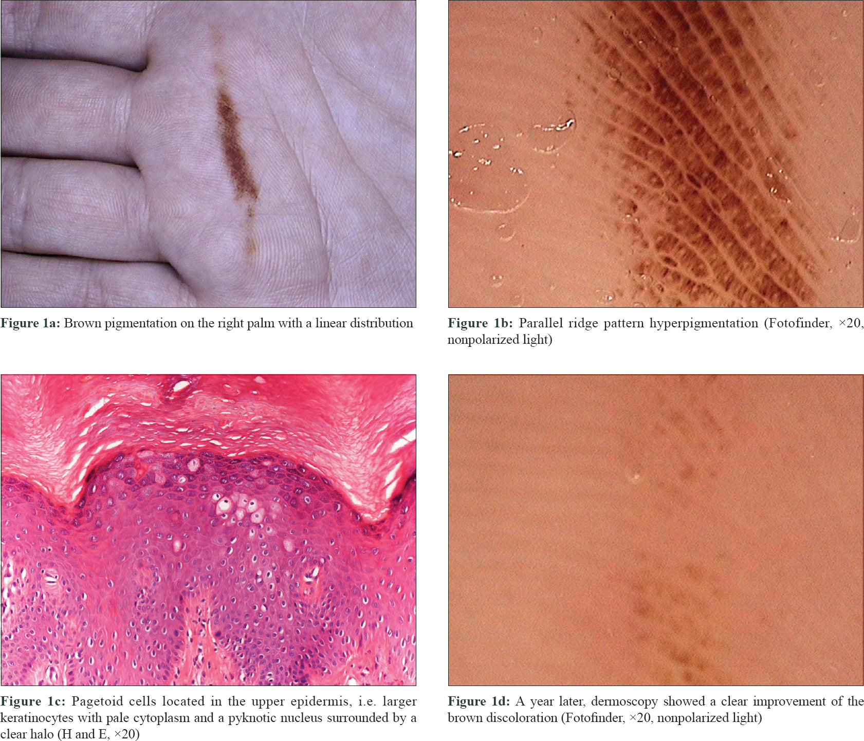
A 26-year-old woman presented with brown patches on her right palm lasting for 1 year [Figure - 1]a. She referred that she had started working as a housecleaner one year ago. Dermoscopy showed a brown pigmentation with parallel ridge pattern sparing the furrows [Figure - 1]b. A skin biopsy demonstrated some large keratinocytes in the upper layers of the epidermis, with pale cytoplasm and pyknotic nucleus, surrounded by a clear halo [Figure - 1]c. A year later, she started working as a salesperson and the pigmentation almost completely disappeared [Figure - 1]d.
 |
| Figure 1: |
Case 2
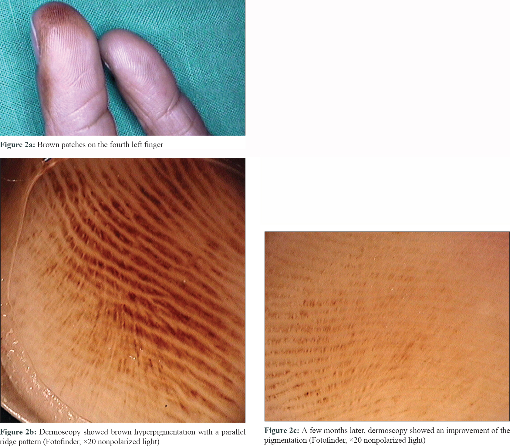
A 31-year-old woman presented with a 3-year history of brown spots on her fourth left finger [Figure - 2]a, which progressively increased in size. She did not recall any suspected trigger. Dermoscopy showed a parallel ridge pattern [Figure - 2]b. A skin biopsy showed pagetoid cells with no other findings, matching the description of pagetoid dyskeratosis. A few months later, the discoloration had almost disappeared spontaneously [Figure - 2]c.
 |
| Figure 2: |
Pagetoid dyskeratosis is thought to be a reactive keratinocyte response to mechanical trauma which leads to premature keratinization of a small part of the normal keratinocyte population.[1],[4] This is probably the reason why it is more frequent in areas more commonly exposed to physical injury, such as intertriginous areas, trunk, buttocks, face and limbs.[1]
Keratinocytes in pagetoid dyskeratosis have a size larger than normal, usually a roundish shape, a pale eosinophilic cytoplasm and a pyknotic nucleus surrounded by a clear halo. These cells usually predominate in the upper layers of the epidermis. They are similar to the cells present in Paget's disease, although the latter show large atypical nuclei.[4]
Clinically, pagetoid dyskeratosis tends to appear as hyperpigmented lesions, probably because of its relation to friction.[3],[4] Both our patients had brown discoloration of fingers or palms. In our first patient, the repetitive friction because of the use of a mop at workplace may had caused the hyperpigmentation; this is supported by the resolution of the discoloration when she changed her job. No triggering factor was identified in the second woman, but the improvement of the pigmentation suggested that there was probably a trigger which later was removed.
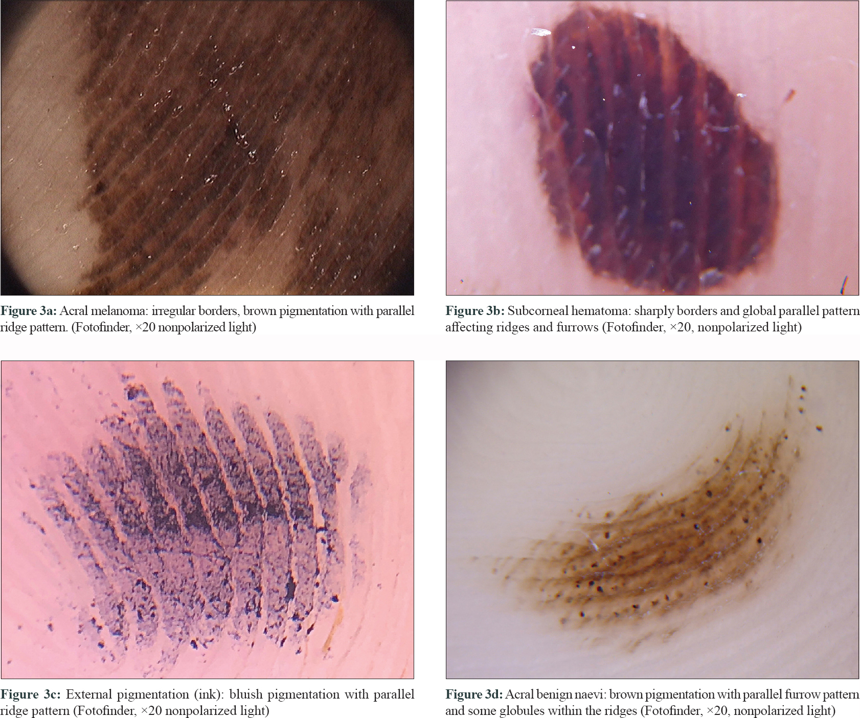
In dermoscopy, the parallel ridge pattern is characterized by the presence of pigment on the dermatoglyph's ridges, which appear larger than the furrows and with the sweat-gland openings located in the centre. This pattern is one of the two acral malignant ones described, along with irregular diffuse pigmentation.[5] The parallel ridge pattern is highly specific for acral melanoma [Figure - 3]a. However, there are exceptions wherein benign lesions may show such a pattern, such as lentiginoses, congenital naevi, acral angioma, subcorneal hemorrhages [Figure - 3]b, exogenous pigmentation [Figure - 3]c, pigmented warts, blue naevi, chemotherapy-induced hyperpigmentation, postraumatic purpura due to repetitive microtrauma and rare cases of benign acral melanocytic nevus [the parallel furrow pattern is more frequent in acral naevi–[Figure - 3]d.[5] Two cases of brownish pigmentation of finger[2] and palm,[3] respectively, have been reported, both showing a parallel ridge pattern on dermoscopy and pagetoid dyskeratosis on the skin biopsy. The brownish discoloration is presumed to result from the distribution of pale cells along the crista cutis which triggers scattered reflection.[2]
 |
| Figure 3: |
In conclusion, we herein report two cases of brownish hyperpigmentation of hands showing a parallel ridge pattern on dermoscopy and pagetoid dyskeratosis on the skin biopsy. A proper clinical history may help to identify possible causes of the discoloration and dermoscopy may help providing further support to the diagnosis of frictional dermatosis.
Declaration of patient consent
The authors certify that they have obtained all appropriate patient consent forms. In the form, the patients have given their consent for their images and other clinical information to be reported in the journal. The patients understand that their names and initials will not be published and due efforts will be made to conceal their identity, but anonymity cannot be guaranteed.
Acknowledgements
To Dr. Aneiros-Fernández for providing the histological image.
Financial support and sponsorship
Nil.
Conflicts of interest
There are no conflicts of interest.
| 1. |
Santos-Briz A, Cañueto J, del Carmen S, Mir-Bonafe JM, Fernandez E. Pagetoid dyskeratosis in dermatopathology. Am J Dermatopathol 2015;37:261-5.
[Google Scholar]
|
| 2. |
Toyonaga E, Inokuma D, Abe Y, Abe R, Shimizu H. Pagetoid dyskeratosis with parallel ridge pattern under dermoscopy. JAMA Dermatol 2013;149:109-11.
[Google Scholar]
|
| 3. |
Loidi L, Mitxelena J, Córdoba A, Yanguas I. Dermoscopic features of pagetoid dyskeratosis of the palm. Actas Dermosifiliogr 2014;105:804-5.
[Google Scholar]
|
| 4. |
Salphale P, Thomas M. Pagetoid dyskeratosis of the forehead. Indian Dermatol Online J 2014;5:215-6.
[Google Scholar]
|
| 5. |
Thomas L, Phan A, Pralong P, Poulalhon N, Debarbieux S, Dalle S. Special locations dermoscopy: Facial, acral, and nail. Dermatol Clin 2013;31:615-24, ix.
[Google Scholar]
|
Fulltext Views
3,891
PDF downloads
2,058


