Translate this page into:
Peri-ocular, papulo-nodular plaques over bilateral eyelids
Corresponding author: Dr. Jaspriya Sandhu, Departments of Dermatology, Venereology & Leprology, Dayanand Medical College & Hospital, Ludhiana, Punjab, India. sandhu.jaspriya@gmail.com
-
Received: ,
Accepted: ,
How to cite this article: Sandhu J, Singh A, Gupta SK, Singh A, Garg B. Peri-ocular, papulo-nodular plaques over bilateral eyelids. Indian J Dermatol Venereol Leprol. 2024;90:385-7. doi: 10.25259/IJDVL_377_2023
A 33-year-old woman presented with an insidious onset, gradually progressive, mildly pruritic, multiple, raised lesions over her bilateral eyelids for one year. She did not have any systemic disease and her primary concern was aesthetic. Cutaneous examination revealed multiple discrete to coalescent, yellow to reddish-brown, papulo-nodular plaques present symmetrically and circumferentially over bilateral periocular area [Figure 1]. There was no involvement of other sites such as the flexures and mucosae. Haematological examination including lipid profile was within normal limits. A biopsy from the lesions showed dense, spindle cell proliferation arranged in the form of short fascicles and focal storiform pattern in the dermis [Figure 2]. On high-power magnification, mononuclear histiocytic cell proliferation was seen in the dermis [Figure 3]. Immunohistochemistry (IHC) showed positive staining for vimentin and CD-68 and negative staining for CD-34, smooth muscle actin, S-100, desmin, myogenin and beta-catenin [Figures 4a–4d]. A bone marrow biopsy was done, which revealed a hypercellular marrow with myeloid prominence with leukocytosis and eosinophilia. JAK-2 Exon 14 mutation assay revealed no abnormality. A haematology consult was taken to rule out systemic involvement.
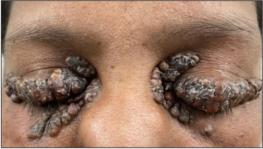
- Multiple yellow to red-brown papulo-nodular plaques over peri ocular area.
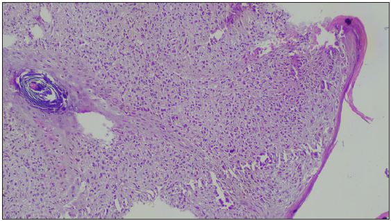
- Diffuse infiltration by histiocytes exhibiting focal storiform pattern (Haematoxylin and Eosin, 100x).
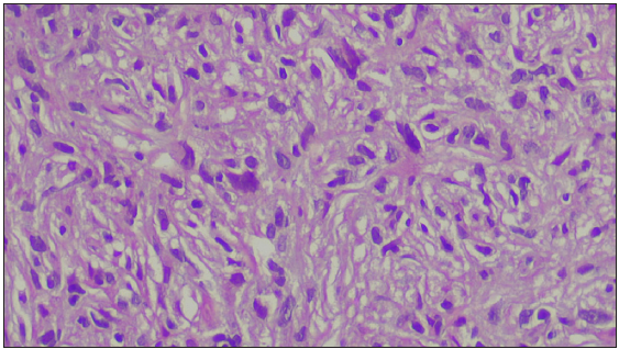
- Mononuclear histiocytic cell proliferation seen in the dermis (Haematoxylin and Eosin, 400x).
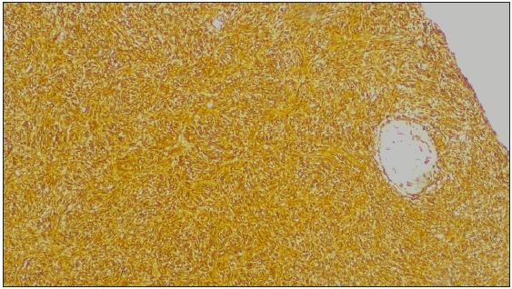
- Immuno-histochemistry (100x): Positivity of vimentin.
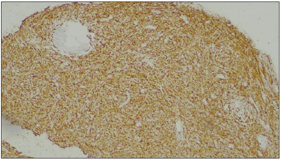
- Immuno-histochemistry (100x): Positivity for CD-68.
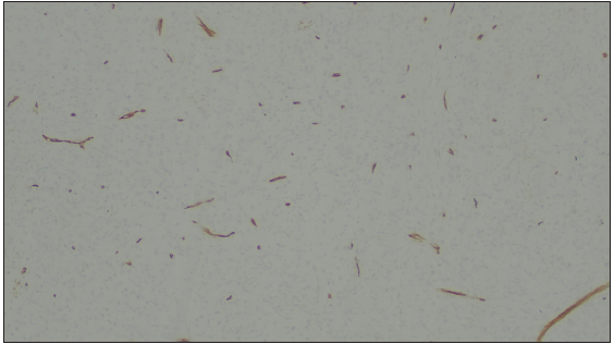
- Immuno-histochemistry (IHC) (100x): Negative IHC for CD-34.
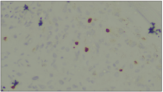
- Immuno-histochemistry (IHC) (100x): Negative IHC for Ki-67.
Question
What is your diagnosis?
Answer
Diagnosis: Progressive nodular histiocytosis
Discussion
Progressive nodular histiocytosis (PNH) is a rare variant of non-langerhans cell histiocytosis affecting the skin and mucous membranes. It was first described by Taunton et al. in 1978.1 PNH presents as persistent, progressive, disseminated papulo-nodules involving head, neck and trunk.2 Severe facial involvement in some cases may lead to leonine facies, and ophthalmological involvement may occur.2 Present case was an unusual case of progressive nodular histiocytosis without dissemination and localised to the eyelids. Skin biopsy was performed with the differential diagnosis of necrobiotic xanthogranuloma (NXG) and lipoid proteinosis. However, on the basis of clinical, histopathological examination and IHC, we arrived at a diagnosis of progressive nodular histiocytosis.
To the best of our knowledge, less than 25 cases of progressive nodular histiocytosis (PNH) have been reported so far. It is usually characterised by its progressive course and multiple disseminated, typical pedunculated nodules. Our case is unusual because the patient did not have disseminated lesions. The histopathology and IHC, however, were consistent with the diagnosis of PNH. A possible association with chronic myeloid leukaemia has been reported with long-standing disease.3 Therefore, JAK-2 Exon-14 mutation assay was performed to rule out myeloproliferative association.However, more data is needed to substantiate this association.
PNH should be differentiated from other types of histiocytosis by its distribution, morphology and systemic involvement. It has a prominent facial distribution with a progressive but benign course as compared to juvenile xanthogranuloma and benign cephalic histiocytosis which usually shows spontaneous regression.3 Xanthoma disseminatum is commonly associated with diabetes insipidus and respiratory tract involvement, which is not seen in PNH. Generalised eruptive histiocytosis has a generalised and symmetrical distribution, which is usually benign and may show spontaneous resolution.2 There are reports with lesions predominantly involving head and neck but disseminated lesions were also present in these patients.2,4–7
This case is of particular interest due to its unusual clinical presentation as well as lack of dissemination. PNH is a rare and has esoteric cutaneous pathology, which may pose a diagnostic challenge to the dermatologists.
Declaration of patient consent
The authors certify that they have obtained all appropriate patient consent.
Financial support and sponsorship
Nil.
Conflicts of interest
There are no conflicts of interest.
Use of artificial intelligence (AI)–assisted technology for manuscript preparation
The authors confirm that there was no use of artificial intelligence (AI)-assisted technology for assisting in the writing or editing of the manuscript and no images were manipulated using AI.
References
- Progressive nodular histiocytosis: A case report and literature review. Indian J Dermatol. 2011;50:1546-51.
- [Google Scholar]
- Progressive nodular histiocytosis accompanied by systemic disorders. Br J Dermatol. 2000;143:628-31.
- [CrossRef] [PubMed] [Google Scholar]
- Progressive nodular histiocytosis: Report of a case and review of the literature. Case Rep Pathol. 2021;2021:5531820.
- [CrossRef] [PubMed] [PubMed Central] [Google Scholar]
- Progressive nodular histiocytosis associated with Eale’s disease. Indian J Dermatol. 2015;60:388-90.
- [CrossRef] [PubMed] [PubMed Central] [Google Scholar]
- Progressive nodular histiocytosis: An unusual disorder. Dermatol Online J. 2021;27:13030/qt4t37r77d.
- [Google Scholar]
- Progressive nodular histiocytosis: A rare type of xanthogranuloma. Dermatol Sin. 2011;29:98-100.
- [Google Scholar]


