Generic selectors
Exact matches only
Search in title
Search in content
Post Type Selectors
Search in posts
Search in pages
Filter by Categories
15th National Conference of the IAOMFP, Chennai, 2006
Abstract
Abstracts from current literature
Acne in India: Guidelines for management - IAA Consensus Document
Addendum
Announcement
Art & Psychiatry
Article
Articles
Association Activities
Association Notes
Award Article
Book Review
Brief Report
Case Analysis
Case Letter
Case Letters
Case Notes
Case Report
Case Reports
Clinical and Laboratory Investigations
Clinical Article
Clinical Studies
Clinical Study
Commentary
Conference Oration
Conference Summary
Continuing Medical Education
Correspondence
Corrigendum
Cosmetic Dermatology
Cosmetology
Current Best Evidence
Current Issue
Current View
Derma Quest
Dermato Surgery
Dermatopathology
Dermatosurgery Specials
Dispensing Pearl
Do you know?
Drug Dialogues
e-IJDVL
Editor Speaks
Editorial
Editorial Remarks
Editorial Report
Editorial Report - 2007
Editorial report for 2004-2005
Errata
Erratum
Focus
Fourth All India Conference Programme
From Our Book Shelf
From the Desk of Chief Editor
General
Get Set for Net
Get set for the net
Guest Article
Guest Editorial
History
How I Manage?
IADVL Announcement
IADVL Announcements
IJDVL Awards
IJDVL AWARDS 2015
IJDVL Awards 2018
IJDVL Awards 2019
IJDVL Awards 2020
IJDVL International Awards 2018
Images in Clinical Practice
Images in Dermatology
In Memorium
Inaugural Address
Index
Knowledge From World Contemporaries
Leprosy Section
Letter in Response to Previous Publication
Letter to Editor
Letter to the Editor
Letter to the Editor - Case Letter
Letter to the Editor - Letter in Response to Published Article
LETTER TO THE EDITOR - LETTERS IN RESPONSE TO PUBLISHED ARTICLES
Letter to the Editor - Observation Letter
Letter to the Editor - Study Letter
Letter to the Editor - Therapy Letter
Letter to the Editor: Articles in Response to Previously Published Articles
Letters in Response to Previous Publication
Letters to the Editor
Letters to the Editor - Letter in Response to Previously Published Articles
Letters to the Editor: Case Letters
Letters to the Editor: Letters in Response to Previously Published Articles
Media and news
Medicolegal Window
Messages
Miscellaneous Letter
Musings
Net Case
Net case report
Net Image
Net Images
Net Letter
Net Quiz
Net Study
New Preparations
News
News & Views
Obituary
Observation Letter
Observation Letters
Oration
Original Article
ORIGINAL CONTRIBUTION
Original Contributions
Pattern of Skin Diseases
Pearls
Pediatric Dermatology
Pediatric Rounds
Perspective
Presedential Address
Presidential Address
Presidents Remarks
Quiz
Recommendations
Regret
Report
Report of chief editor
Report of Hon : Treasurer IADVL
Report of Hon. General Secretary IADVL
Research Methdology
Research Methodology
Resident page
Resident's Page
Resident’s Page
Residents' Corner
Residents' Corner
Residents' Page
Retraction
Review
Review Article
Review Articles
Reviewers 2022
Revision Corner
Self Assessment Programme
SEMINAR
Seminar: Chronic Arsenicosis in India
Seminar: HIV Infection
Short Communication
Short Communications
Short Report
Snippets
Special Article
Specialty Interface
Studies
Study Letter
Study Letters
Supplement-Photoprotection
Supplement-Psoriasis
Symposium - Contact Dermatitis
Symposium - Lasers
Symposium - Pediatric Dermatoses
Symposium - Psoriasis
Symposium - Vesicobullous Disorders
SYMPOSIUM - VITILIGO
Symposium Aesthetic Surgery
Symposium Dermatopathology
Symposium-Hair Disorders
Symposium-Nails Part I
Symposium-Nails-Part II
Systematic Review and Meta-Analysis
Systematic Reviews and Meta-analyses
Systematic Reviews and Meta-analysis
Tables
Technology
Therapeutic Guideline-IADVL
Therapeutic Guidelines
Therapeutic Guidelines - IADVL
Therapeutics
Therapy
Therapy Letter
Therapy Letters
View Point
Viewpoint
What’s new in Dermatology
View/Download PDF

Translate this page into:

Net Quiz
ARTICLE IN PRESS
doi:
10.25259/IJDVL_1464_2024

Pigmented nodular lesion below eyelid in a retro positive patient

Department of Pathology, All India Institute of Medical Sciences Mangalagiri, Guntur, Andhra Pradesh, India
Department of Plastic Surgery, All India Institute of Medical Sciences Mangalagiri, Guntur, Andhra Pradesh, India
Department of Dermatology, All India Institute of Medical Sciences Mangalagiri, Guntur, Andhra Pradesh, India.

Corresponding author: Dr. Likhitha Reddy C, Department of Plastic Surgery, AIIMS, Mangalagiri, Guntur, India. drlikhitha.pls@aiimsmangalagiri.edu.in

Licence
This is an open-access article distributed under the terms of the Creative Commons Attribution-Non Commercial-Share Alike 4.0 License, which allows others to remix, transform, and build upon the work non-commercially, as long as the author is credited and the new creations are licensed under the identical terms.

How to cite this article: Rajagopal P, C LR, Gopinath H, Shankaralingappa A. Pigmented nodular lesion below eyelid in a retro positive patient. Indian J Dermatol Venereol Leprol. doi: 10.25259/IJDVL_1464_2024

A 39-year-old lady presented to the outpatient department with complaints of an itchy black nodule below her right eyes. She reported that the lesion had slowly grown to its present size over the last one year. She was HIV positive and was on highly active antiretroviral therapy. She did not report weight loss, fever, or any significant past or family history. On clinical examination, a solitary pigmented dome-shaped exophytic papule (1.2×1.0×0.8 cm in size) with a keratotic center was seen below the right eyelid [Figure 1]. The lesion was excised and sent for histopathological evaluation. Histopathology revealed an exo- and endophytic cup-shaped lesion lined by squamous epithelium with marked hyperkeratosis, parakeratosis, filiform papillomatosis, and lobular configuration at the epithelial stromal interface [Figure 2a]. The lobules had peripheral basaloid and central squamous cells with the presence of a few squamous eddies and microvesicle formation [Figure 2b]. Melanin deposition and increased dendritic melanocytes were also seen [Figure 2c]. No ragged edges of squamous epithelium, overt nuclear atypia, or glassy eosinophilic cytoplasm were seen.
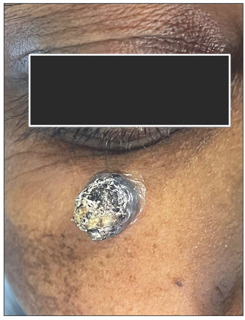
Hyperpigmented dome-shaped lesion with central keratotic crust near the right lower eyelid.
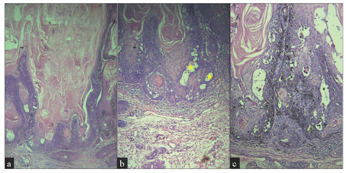
Figure 1:
Hyperpigmented dome-shaped lesion with central keratotic crust near the right lower eyelid.
(a) Exo and endophytic proliferative squamous lesion with a central cup-shaped area filled with hyperkeratotic, parakeratinised material with blunt edges at epithelial-stromal interface (Haematoxylin and eosin, 40x), (b) microvesicles (stars) and squamous eddies (400x) and (c) increased dendritic melanocytes (100x).
Figure 2:
(a) Exo and endophytic proliferative squamous lesion with a central cup-shaped area filled with hyperkeratotic, parakeratinised material with blunt edges at epithelial-stromal interface (Haematoxylin and eosin, 40x), (b) microvesicles (stars) and squamous eddies (400x) and (c) increased dendritic melanocytes (100x).

Question

What is your diagnosis?

Diagnosis

Pigmented inverted follicular keratosis

Treatment

The lesion was already excised in toto with margins considering a possibility of malignancy clinically.

Discussion

Inverted follicular keratosis is a benign tumour of follicular infundibulum. It usually presents as a solitary skin-coloured papule or nodule, usually on the face and eyelid.1 Clinically, a nodular dome-shaped lesion on the face raises the differential diagnosis of basal cell carcinoma, keratoacanthoma, squamous cell carcinoma, and at times malignant melanoma, seborrheic keratosis, and verruca vulgaris.24 Knowledge of the differential diagnostic possibilities could enable the clinician to make an appropriate decision; however, at times it can be difficult to differentiate a benign from a malignant lesion.

Histopathological examination is mandatory in arriving at a correct diagnosis. Benign and malignant neoplasms can create diagnostic confusion with inverted follicular keratosis not only clinically but also histopathologically.13 The differential diagnosis1-4 of inverted follicular keratosis is given in Table 1. Even though, in this case, histologically the cup-shaped nature of the lesion created a diagnostic confusion with keratoacanthoma, the endophytic extension, blunt edge interface with stroma, squamous eddies, and microvesicles in the absence of glassy eosinophilic cytoplasm and ragged infiltrative edges guided us to the diagnosis of pigmented inverted follicular keratosis.

Table 1: Histopathological differential diagnosis of inverted follicular keratosis
S. No Histopathology features Inverted follicular keratosis Keratoacanthoma Seborrheic keratosis Verruca vulgaris Squamous cell carcinoma Basal cell carcinoma
1 Nature of lesion Exo- and endophytic Usually endophytic Exophytic Exophytic Ulcero-proliferative Nodular
2 Koliocytes Can be seen Absent Absent Present Usually absent Absent
3 Chunky hypergranulosis Can be seen Absent Absent Present Usually absent Absent
4 Epithelial stromal interface Pushing with blunt edges Can be infiltrative Pushing Pushing Infiltrative Pushing
5 Glassy eosinophilic squamous cells Absent Present Absent Absent Absent Absent
6 Mitosis Not uncommon in basal layers Common throughout the epithelium Absent or limited to basal layer Absent or limited to basal layer Common throughout the epithelium Common throughout the epithelium
7 Atypical mitosis Absent Present Absent Absent Present Present
8 Cellular composition Peripheral basaloid and central squamous cells Squamous cells with glassy eosinophilic cytoplasm Basaloid cells Squamous cells Atypical squamous cells Basaloid cells with peripheral palisading
8 Microcysts in epithelium Common Absent Absent Absent Absent Absent

Inverted follicular keratosis can mimic other tumours even on dermoscopy. Dermoscopic features include a keratoacanthoma-like pattern with a central white amorphous area surrounded radially by hairpin vessels and a white halo and central yellow-white amorphous areas surrounded radially by hairpin vessels. Less common features include milky red globules, arborising vessels, linear irregular vessels and corkscrew vessels.1,2,5

With respect to the HIV positive status of the patient, certain skin lesions may present differently or appear more frequently due to the effects of immunosuppression. While inverted follicular keratosis is not specifically linked to retroviral infection, it’s important to differentiate inverted follicular keratosis from other lesions, including more serious conditions like squamous cell carcinoma, Kaposi’s sarcoma or viral warts. A biopsy is needed to confirm the diagnosis, particularly in the context of immunosuppression.

Inverted follicular keratosis lesions are usually cured by surgical excision.5 In our case, complete excision with 5 mm margin was done since pigmented basal cell carcinoma was in the differential diagnoses.

Inverted follicular keratosis is a relatively rare benign adnexal tumour of hair follicle origin. Clinical diagnosis of inverted follicular keratosis is difficult and it may be misdiagnosed as other malignant conditions such as basal cell carcinoma. More awareness about this entity is needed and it should be considered as one of the differentials for pigmented nodular dome-shaped lesions in the face.

Declaration of patient consent

The authors certify that they have obtained all appropriate patient consent.

Financial support and sponsorship

Nil.

Conflicts of interest

There are no conflicts of interest.

Use of artificial intelligence (AI)-assisted technology for manuscript preparation

The authors confirm that there was no use of AI-assisted technology for assisting in the writing or editing of the manuscript and no images were manipulated using AI.

References

  1. , , , , , , et al. Inverted follicular keratosis: Dermoscopic and reflectance confocal microscopic features. Dermatology. 2013;227:62-6.
    [CrossRef] [PubMed] [Google Scholar]
  2. , , , . A rare case of inverted follicular keratosis in an elderly male: Dermoscopic and histopathological overview with therapeutic response to imiquimod. Indian J Dermatol Venereol Leprol. 2021;87:455.
    [CrossRef] [PubMed] [Google Scholar]
  3. , , . Pigmented inverted follicular keratosis masquerading as basal cell carcinoma. Indian Dermatol Online J. 2022;13:669-71.
    [CrossRef] [PubMed] [PubMed Central] [Google Scholar]
  4. , , . Two distinct lesions of inverted follicular keratosis of the scalp: A case report. Cureus. 2022;14:e31055.
    [CrossRef] [PubMed] [PubMed Central] [Google Scholar]
  5. , , , , , , et al. Dermoscopy of inverted follicular keratosis: Study of 12 cases. Clin Exp Dermatol. 2016;41:468-73.
    [CrossRef] [PubMed] [Google Scholar]
Show Sections