Translate this page into:
Polarized light microscopy
Correspondence Address:
Uday Khopkar
Department of Dermatology, Seth GS Medical College and KEM Hospital, Parel, Mumbai-400 012
India
| How to cite this article: Dongre A, Bhisey P, Khopkar U. Polarized light microscopy. Indian J Dermatol Venereol Leprol 2007;73:206-208 |
 |
| Figure 6: �Maltese cross� seen in urine specimen of Fabry�s disease |
|
| Figure 6: �Maltese cross� seen in urine specimen of Fabry�s disease |
 |
| Figure 5: �Tiger tail� appearance (alternate dark and light bands of hair shaft) in trichothiodystrophy |
|
| Figure 5: �Tiger tail� appearance (alternate dark and light bands of hair shaft) in trichothiodystrophy |
 |
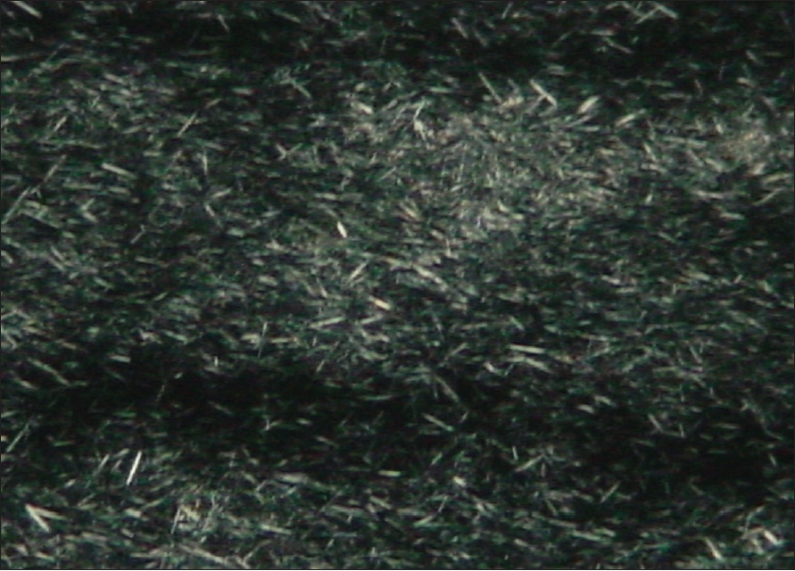
| Figure 4: �Apple green birefringence� of amyloid under polarizing light after Congo red staining |
|
| Figure 4: �Apple green birefringence� of amyloid under polarizing light after Congo red staining |
 |
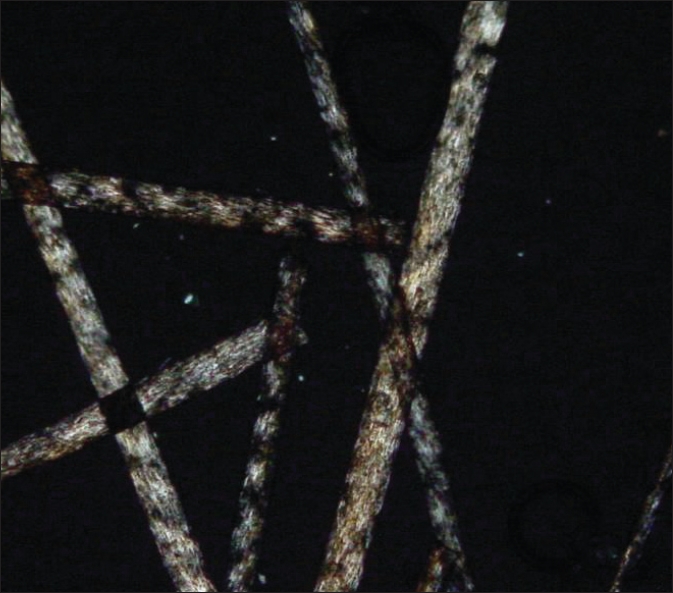
| Figure 3: Needle-shaped monosodium urate crystals in tophaceous gout |
|
| Figure 3: Needle-shaped monosodium urate crystals in tophaceous gout |
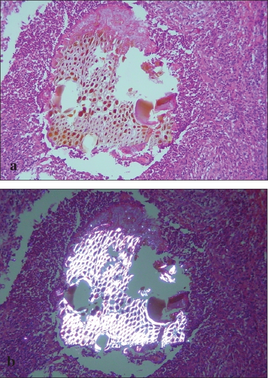 |
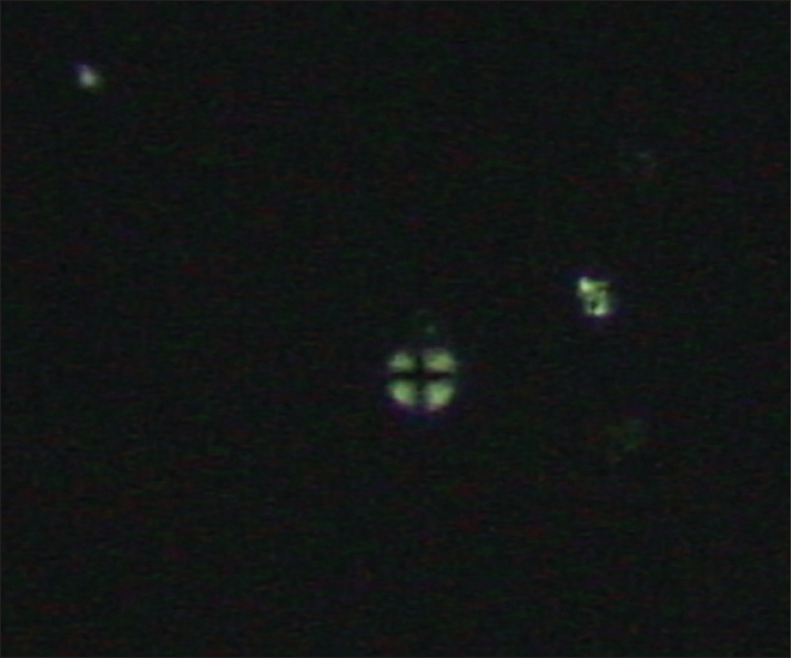
| Figure 2: (a and b) Refractile honeycomb pattern of wooden splinter on polarizing microscopy |
|
| Figure 2: (a and b) Refractile honeycomb pattern of wooden splinter on polarizing microscopy |
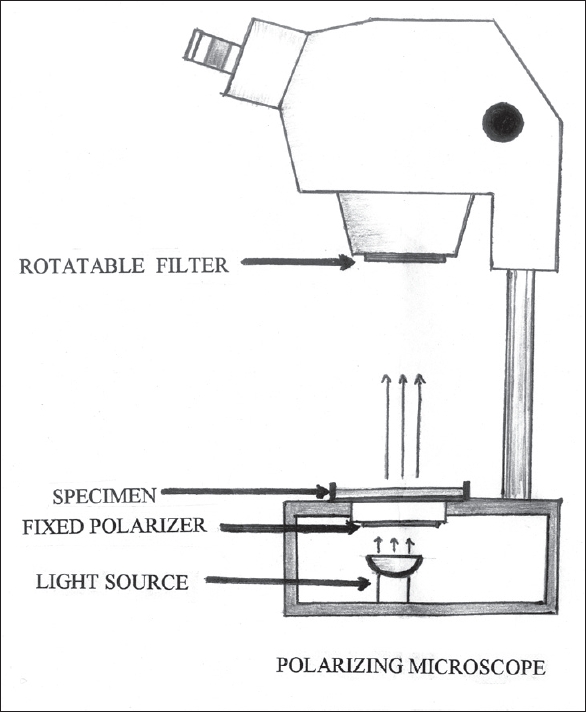 |
| Figure 1: Principle of polarizing microscopy |
|
| Figure 1: Principle of polarizing microscopy |
Polarizing microscopy is the technique of examining histological sections of skin or hair by passing a beam of polarized light. This is an accessory of a light microscope and may not be available with all microscopes.
Principles and technique of polarized microscopy
Polarized light is the light in which all the rays vibrate in one plane. A polarizing microscope has two disk accessories. They are made up of polarizing plastic that allows light vibrating in one plane to pass. One of them is called a polarizer (placed below the condenser). Another similar disc is placed in the top part of the microscope and cuts off all the light vibrating in a perpendicular plane. This disc is called analyzer. The placement of the discs is such that they allow light vibrating in planes perpendicular to each other. Hence when both the disks are in place no light can pass on to the eyepieces. Hence through the eyepieces only dark background is seen unless a doubly refractile object is placed in the path of polarized light, in which case, the doubly refractile object appears illuminated against a dark background [Figure - 1].
Usually organic and inorganic substances with rigid yet repeatable structure like crystals, sand, talc, wood, amyloid and other fibrillary structures are seen on polarized light microscopy. Certain foreign bodies can be detected on routine hematoxylin and eosin staining (H and E) while special stains are required for metabolic or deposition disorders.
Applications in dermatology
These include: foreign body(ies) in the skin, deposition disorders, metabolic disorders, disorders of hair, and genetic disorders.
Foreign body (ies)
On polariscopic examination different foreign bodies have characteristic appearance. They can be found amidst foreign body granulomas (both allergic and non-allergic type) and most commonly within the multinucleated giant cells. Various foreign bodies can be identified on the basis of specific patterns on polarization. Some of the well-described patterns of polarization are mentioned in [Table - 1]. One of the important histopathologic differential diagnoses of silica granuloma is cutaneous sarcoidosis which can be ruled out with polarizing microscopy if it reveals characteristic pattern of doubly refractile spicules of silica. [1] In talc granuloma, Kasper et al demonstrated birefringent talc crystals within nodular granuloma and perivascular histiocytes in the adjacent skin and periprosthetic tissues in a patient having silicone gel-containing prosthetic devices. [2] The ′Maltese cross′ configuration has been described in association with starch granulomas. [3]
Xanthomatosis
Cholesterol esters are doubly refractile and can be demonstrated in conditions like tuberous xanthoma, plane xanthoma, xanthelasma while they are absent in eruptive xanthomas. [4] They can also be demonstrated in non-X-histiocytosis, juvenile xanthogranuloma, erythema elevatum diutinum and dermatofibroma in which deposition of lipids is a secondary phenomenon. To demonstrate the lipids in the sections, tissue should be formalin fixed and frozen sectioned. The minced tissue is either heated or cooled at 2-3°C/min and in the smectic phase, (two-dimensional ordering of cholesterol esters molecules), birefringent crystals of cholesterol esters are identified. [5]
Gout
Gout is a disorder of uric acid metabolism. Severe hyperuricemia is frequently associated with deposits of monosodium urate crystals in the skin leading to ′tophi′ in tophaceous gout. During polarized microscopy, crystals of monosodium urate appear as needle-shaped doubly refractile structures packed in bundles or sheaves [Figure - 3] when alcohol is used for fixation instead of formalin. [6]
Apart from the above metabolic disorders, Savita et al have demonstrated that polarization microscopy is helpful in the identification of various metabolites in the blood i.e. oxalic acid, creatinines, total calcium, glycine. Crystals were isolated, which characterize a type of a metabolic disorder. The fan-type dendrites were indicative of hyperoxaluria; druses and dendrite-like spherulites--hypercreatinemia; oolite spherulities- hypercalcemia; fine-grained particles- hyperglycinemia. [7]
Amyloidosis
Amyloid material shows brick red staining with apple green birefringence under polarizing microscope when stained with Congo red [Figure - 4]. This characteristic staining is due to beta-pleated sheet conformation of polypeptide backbone of amyloid fibrils. In case of macular and lichen amyloidosis it is seen only in the papillary dermis while in systemic amyloidosis it is also deposited in the deeper dermis, subcutaneous tissue and within the walls of the blood vessels. ′Apple green′ birefringence similar to that observed in amyloidosis after Congo red staining has been described in colloid milium. [8]
Hair disorders
Polariscopic examination of the hair in trichothiodystrophy shows characteristic alternate dark and white bands of hair shaft called as ′tiger tail′ appearance which is not visualized on light microscopic examination [Figure - 5].
Genetic disorders
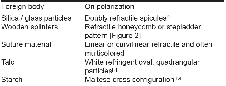
In Fabry′s disease deposits of glycosphingolipids can be demonstrated in the endothelial, perithelial, smooth muscle cells of blood vessels and urinary sediments where they are seen as birefringent ′Maltese crosses′ [Figure - 6].
In normal healthy individuals, the lamins which are structural filaments in the nuclear envelope and nucleus are disorderly oriented and hence can dissipate acute mechanical stress. In Hutchinson-Gilford progeria syndrome, the nuclear lamins have orderly orientations which reduce their deformability to acute mechanical stress. [9] This arrangement or orientation of nuclear lamins can be identified by polarization microscopy.
Thus, polarized microscopy is a simple but useful technique that has varied applications in dermatology making it an essential microscope accessory in the diagnosis of various dermatologic and systemic disorders.
| 1. |
Mowry RG, Sams WM Jr, Caulfield JB. Cutaneous silicone granuloma. Arch Dermatol 1991;127:692-4.
[Google Scholar]
|
| 2. |
Kasper CS, Chandler PJ Jr. Talc deposition in skin and tissues surrounding silicone-gel containing prosthetic devices. Arch Dermatol 1994:130:48-53.
[Google Scholar]
|
| 3. |
Leonard DD. Starch granulomas. Arch Dermatol 1973;107:101-3.
[Google Scholar]
|
| 4. |
Cruz PD Jr, East C, Bergstresser PR. Dermal, subcutaneous and tendon xanthomas: Diagnostic markers for specific lipoprotein disorders. J Am Acad Dermatol 1988;19:95-111.
[Google Scholar]
|
| 5. |
Tall AR, Small DM, Lees RS. Interaction of collagen with the lipids of tendon xanthomata. J Clin Invest 1978;62:836-46.
[Google Scholar]
|
| 6. |
King DF, King LA. The appropriate processing of tophi for microscopy. Am J Dermatopathol 1982;4:239.
[Google Scholar]
|
| 7. |
Savina LV, Pavlishchuk SA, Samsygin VI, Bolotova EV, Gotovtseva LP, Chekmareva SE, et al . Polarization microscopy in diagnosis of metabolic disorders. Klin Lab Diagn 2003;3:11-4.
[Google Scholar]
|
| 8. |
Hashimoto K, Black M. Colloid milium: A final degradation product of actinic elastoid. J Cutan Pathol 1985;12:147-56.
[Google Scholar]
|
| 9. |
Dahl KN, Scaffidi P, Islam MF, Yodh AG, Wilson KL, Misteli T. Distinct structural and mechanical properties of the nuclear lamina in Hutchinson-Gilford progeria syndrome. Proc Natl Acad Sci USA 2006;103:10271-6.
[Google Scholar]
|
Fulltext Views
4,624
PDF downloads
2,157


![[Figure - 1]](#fig_ijdvl_2007_73_3_206_32754_1.jpg)
![[Table - 1]](#tbl_ijdvl_2007_73_3_206_32754_7.jpg)
![[Figure - 3]](#fig_ijdvl_2007_73_3_206_32754_3.jpg)
![[Figure - 4]](#fig_ijdvl_2007_73_3_206_32754_4.jpg)
![[Figure - 5]](#fig_ijdvl_2007_73_3_206_32754_5.jpg)
![[Figure - 6]](#fig_ijdvl_2007_73_3_206_32754_6.jpg)