Translate this page into:
Porokeratosis of mibelli with a mucous membrane lesion
Correspondence Address:
Asok Kumar Gangopadhyay
Department of Dermatology, RKM Seva Pratisthan, Calcutta-700026
India
| How to cite this article: Gangopadhyay AK. Porokeratosis of mibelli with a mucous membrane lesion. Indian J Dermatol Venereol Leprol 1997;63:53-54 |
Abstract
Mucous membrane lesion of porokeratosis is a very vary rare occurrence. Here is a case report of such a case. |
 |
 |
 |
 |
 |
Introduction
Porokeratosis, although the term is a misnomer[1] (since this disorder of keratinistation not necessarily involves the sweat pore always), has got 5 clinical subtypes. They are (i) porokeratosis of Mibelli, (ii) disseminated superficial actinic porokeratosis, (iii) linear porokeratosis, (iv) porokeratosis plantaris palmaris et disseminata, (v) punctate porokeratosis. Oral mucous membrane lesion is very very rerely seen in the Mibelli′s subtype.[2] Porokeratosis of mucous membrane has been mainly reported from Japan and Italy.[3] To the best of my knowledge, this is the first case report of mucous membrane lesion of porokeratosis from India.
Case Report
A young man of 22 years, Hindu by religion, fisherman by profession, presented with multiple circular plaques mainly over the chest wall, neck and one patch over the left lateral aspect of the tongue [Figure - 1] and [Figure - 2]. Ever since the disease started some 8 years back, the lesions were asymptomatic althrough, except the lesion on the tongue which caused little discomfort and slight burning during mastication. The lesions initially appeared as tiny plaques on the chest wall and increased very very incidiously to the present size in a span of 8 years. The lesion on tongue appeared some 6 years back. On examination, there were discrete circular plaques of varying sizes mainly over the chest, neck and medial side of the right arm. The lesions were hyperpigmented with a raised hyperkeratotic border and a bit atropic centre. The plaque on the tongue was about 1 cm in diameter with a slight bluish discrete border. The floor looked greyish and little macerated. The other mucosae were free of any lesion. The systemic examination was non-contributory.
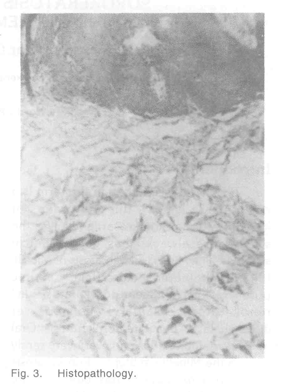
The histology from the edge of the skin lesion showed hyperkeratosis and parakeratotic column (cornoid lamella) in the epidermal invagination. The granular cell layer was absent. There were few chronic inflammatory cells. The histology from the lesion of the tonuge [Figure - 3] showed the parakeratotic column.
Discussion
Poroeratosis is an inherited disorder of keratinisation. An autosomal dominant mode of inheritance with predilection for male sex in now well established. Mibelli, in 1893, coined the term porokeratosis[3] and later on different clinical types were described one by one. Muocus membrane lesions are very very rare and the mucosa of the mouth, nose, glans penis and the conjunctiva are affected. Previously porokeratosis was thought to be a disorder that involves the eccrine sweat pore. But the mucous membrane involvement, where there is no sweat gland, speaks against it. It is now thought that the ring like cornoid lamella, the hallmark of porokeratosis, may be located in the interadnexal epidermis, acro-syringium of eccrine sweat duct or within the infundibulum of pilar apparatus.[4]
| 1. |
Pinkus H, Mehregan AH. A guide to dermato-histopathology, New York:Appleton Century Crafts,1976:491.
[Google Scholar]
|
| 2. |
Mehregan AH, Khalili H, et al. Mibelli's porokeraosis of the face. J Am Acad Dermatol 1980;3:394-6.
[Google Scholar]
|
| 3. |
Mikhali GR, Wertheimer FW. Clinical variants of porokeratosis (Mibelli). Arch Dermatol 1968;98:124-31.
[Google Scholar]
|
| 4. |
Nabai H, Mehregan AH. Porokeratosis of Mibelli. Dermatologica 1979;159:325-31.
[Google Scholar]
|


