Generic selectors
Exact matches only
Search in title
Search in content
Post Type Selectors
Search in posts
Search in pages
Filter by Categories
15th National Conference of the IAOMFP, Chennai, 2006
Abstract
Abstracts from current literature
Acne in India: Guidelines for management - IAA Consensus Document
Addendum
Announcement
Art & Psychiatry
Article
Articles
Association Activities
Association Notes
Award Article
Book Review
Brief Report
Case Analysis
Case Letter
Case Letters
Case Notes
Case Report
Case Reports
Clinical and Laboratory Investigations
Clinical Article
Clinical Studies
Clinical Study
Commentary
Conference Oration
Conference Summary
Continuing Medical Education
Correspondence
Corrigendum
Cosmetic Dermatology
Cosmetology
Current Best Evidence
Current Issue
Current View
Derma Quest
Dermato Surgery
Dermatopathology
Dermatosurgery Specials
Dispensing Pearl
Do you know?
Drug Dialogues
e-IJDVL
Editor Speaks
Editorial
Editorial Remarks
Editorial Report
Editorial Report - 2007
Editorial report for 2004-2005
Errata
Erratum
Focus
Fourth All India Conference Programme
From Our Book Shelf
From the Desk of Chief Editor
General
Get Set for Net
Get set for the net
Guest Article
Guest Editorial
History
How I Manage?
IADVL Announcement
IADVL Announcements
IJDVL Awards
IJDVL AWARDS 2015
IJDVL Awards 2018
IJDVL Awards 2019
IJDVL Awards 2020
IJDVL International Awards 2018
Images in Clinical Practice
Images in Dermatology
In Memorium
Inaugural Address
Index
Knowledge From World Contemporaries
Leprosy Section
Letter in Response to Previous Publication
Letter to Editor
Letter to the Editor
Letter to the Editor - Case Letter
Letter to the Editor - Letter in Response to Published Article
LETTER TO THE EDITOR - LETTERS IN RESPONSE TO PUBLISHED ARTICLES
Letter to the Editor - Observation Letter
Letter to the Editor - Study Letter
Letter to the Editor - Therapy Letter
Letter to the Editor: Articles in Response to Previously Published Articles
Letters in Response to Previous Publication
Letters to the Editor
Letters to the Editor - Letter in Response to Previously Published Articles
Letters to the Editor: Case Letters
Letters to the Editor: Letters in Response to Previously Published Articles
Media and news
Medicolegal Window
Messages
Miscellaneous Letter
Musings
Net Case
Net case report
Net Image
Net Images
Net Letter
Net Quiz
Net Study
New Preparations
News
News & Views
Obituary
Observation Letter
Observation Letters
Oration
Original Article
ORIGINAL CONTRIBUTION
Original Contributions
Pattern of Skin Diseases
Pearls
Pediatric Dermatology
Pediatric Rounds
Perspective
Presedential Address
Presidential Address
Presidents Remarks
Quiz
Recommendations
Regret
Report
Report of chief editor
Report of Hon : Treasurer IADVL
Report of Hon. General Secretary IADVL
Research Methdology
Research Methodology
Resident page
Resident's Page
Resident’s Page
Residents' Corner
Residents' Corner
Residents' Page
Retraction
Review
Review Article
Review Articles
Reviewers 2022
Revision Corner
Self Assessment Programme
SEMINAR
Seminar: Chronic Arsenicosis in India
Seminar: HIV Infection
Short Communication
Short Communications
Short Report
Snippets
Special Article
Specialty Interface
Studies
Study Letter
Study Letters
Supplement-Photoprotection
Supplement-Psoriasis
Symposium - Contact Dermatitis
Symposium - Lasers
Symposium - Pediatric Dermatoses
Symposium - Psoriasis
Symposium - Vesicobullous Disorders
SYMPOSIUM - VITILIGO
Symposium Aesthetic Surgery
Symposium Dermatopathology
Symposium-Hair Disorders
Symposium-Nails Part I
Symposium-Nails-Part II
Systematic Review and Meta-Analysis
Systematic Reviews and Meta-analyses
Systematic Reviews and Meta-analysis
Tables
Technology
Therapeutic Guideline-IADVL
Therapeutic Guidelines
Therapeutic Guidelines - IADVL
Therapeutics
Therapy
Therapy Letter
Therapy Letters
View Point
Viewpoint
What’s new in Dermatology
View/Download PDF

Translate this page into:

Net Letter
ARTICLE IN PRESS
doi:
10.25259/IJDVL_664_2024

Post pemphigus acanthomata: Revisiting an uncommon presentation of pemphigus

Department of Dermatology, Dr BR Ambedkar Medical College, Gandhinagar, KG Halli, Bengaluru, Karnataka, India
Department of Dermatology, Bangalore Medical College and Research Institute, Bengaluru, Karnataka, India.

Corresponding author: Dr. Divya Gupta, Department of Dermatology, Dr BR Ambedkar Medical College, Gandhinagar, KG Halli, Bengaluru, India. divya_gupta@ymail.com

Licence
This is an open-access article distributed under the terms of the Creative Commons Attribution-Non Commercial-Share Alike 4.0 License, which allows others to remix, transform, and build upon the work non-commercially, as long as the author is credited and the new creations are licensed under the identical terms.

How to cite this article: Adithyan P, Loganathan E, Padmanabha SR, Somashekhar S, Reddy JK, Gupta D. Post pemphigus acanthomata: Revisiting an uncommon presentation of pemphigus. Indian J Dermatol Venereol Leprol. doi: 10.25259/IJDVL_664_2024

Dear Editor,

Pemphigus refers to a group of vesiculobullous disorders characterised by the presence of multiple fluid-filled lesions affecting the skin and mucosae. After rupturing, they usually heal with hyper- or hypopigmentation.1 In some cases, verrucous lesions develop at sites of previously healed blisters, this phenomenon is called post pemphigus acanthomata (PPA).2

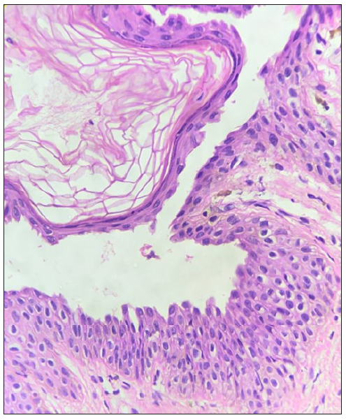
A 22-year-old male patient visited the dermatology outpatient department 12 months ago with complaints of multiple crusted erosions with some fluid-filled flaccid bullae over the face and trunk. Skin biopsy and direct immunofluorescence suggested pemphigus foliaceus. The serum tested positive for anti-desmoglein 1 antibody (anti dsg 1 titres = 184 U/mL). We initiated treatment with dexamethasone pulse therapy along with weekly methotrexate, which effectively controlled the disease. However, after the fourth cycle, he noticed dark elevated lesions over the site of previous erosions along with the reappearance of new superficial blisters. Cutaneous examination revealed multiple hyperpigmented verrucous plaques over the chest, abdomen, back and proximal limbs. Moist erosions with perilesional erythema and positive perilesional Nikolsky sign were seen over the face and trunk [Figures 1a-1c]. There were no other complaints and the general physical examination was unremarkable. Laboratory investigations including haematology and biochemistry panels were within normal limits. Histopathological examination of the verrucous plaque revealed hyperplastic squamous epithelium exhibiting loose hyperkeratosis, acanthosis, papillomatosis and sub-corneal blister formation with acantholytic cells suggestive of post pemphigus acanthomata [Figure 2]. Anti desmoglein-1 antibody titres were elevated [95 U/mL] which indicated a possible relapse of the underlying disease.
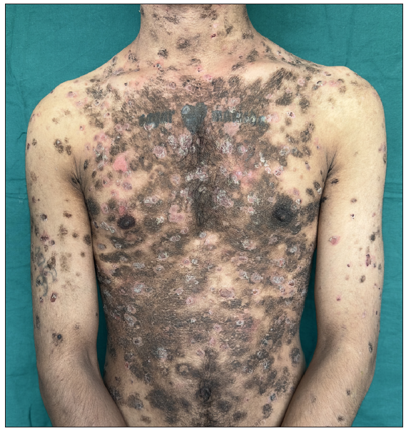
Hyperpigmented verrucous plaques and superficial erosions over chest and abdomen.
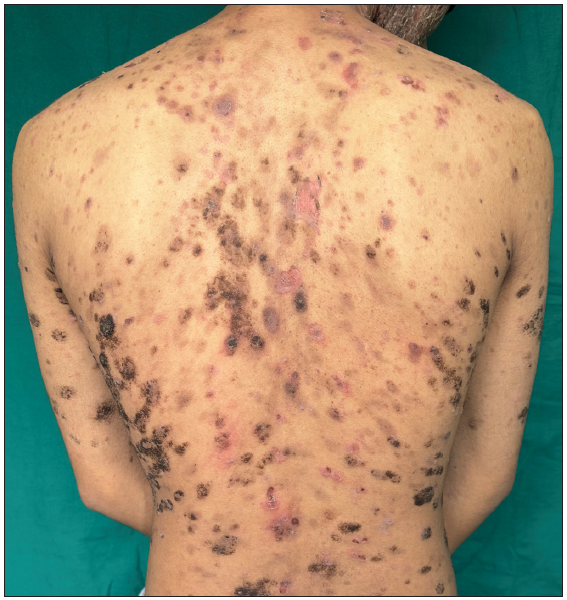
Figure 1a:
Hyperpigmented verrucous plaques and superficial erosions over chest and abdomen.
Hyperpigmented verrucous plaques and superficial erosions over the back.
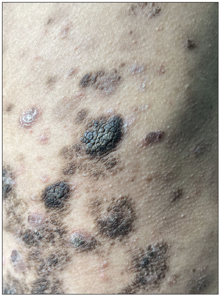
Figure 1b:
Hyperpigmented verrucous plaques and superficial erosions over the back.
Close-up view of the acanthoma.
Figure 1c:
Close-up view of the acanthoma.
Histopathology showing stratum corneum with loose hyperkeratosis, sub-corneal blister with acantholytic cells and irregular acanthosis. (Haematoxylin and Eosin, 400x).
Figure 2:
Histopathology showing stratum corneum with loose hyperkeratosis, sub-corneal blister with acantholytic cells and irregular acanthosis. (Haematoxylin and Eosin, 400x).

PPA is a rare dermatological phenomenon and only a few case reports have been published till date. It typically develops following the resolution of pemphigus lesions, either during remission or after treatment. The trunk is commonly affected. They may arise months to years after the initial pemphigus diagnosis.1,3,4

The exact cause of PPA is unclear, but it possibly involves a complex interplay of autoimmune mechanisms, inflammatory responses, and bacterial colonisation of the skin. Another hypothesis suggests that certain autoantibodies produced during the active phase of pemphigus may continue to stimulate epidermal hyperproliferation even after disease remission, resulting in the development of acanthomata lesions.5

Clinically, PPA lesions present as raised, hyperpigmented plaques with a ‘stuck-on’ appearance, resembling warts or seborrheic keratoses. They may be asymptomatic or associated with mild itching or discomfort.

Clinically these lesions may be confused with seborrheic keratosis, post eczema acanthomata pemphigus vegetans and acantholytic acanthoma.6,7 PPA lesions usually disappear after a period whereas seborrheic keratosis and acantholytic acanthoma lesions are persistent and spontaneous resolution is uncommon. Pemphigus vegetans is characterised by flaccid blisters that erode and subsequently form fungoid vegetations or papillomatous proliferations, especially involving intertriginous areas and scalp, which can be differentiated easily from PPA lesions having truncal predilection.

Histopathologically, PPA lesions demonstrate hyperkeratosis, acanthosis, and papillomatosis along with characteristic intra-epidermal clefting and acantholytic cells. The absence of intra-epidermal clefting is a differentiating histopathological feature from seborrheic keratosis. In acantholytic acanthoma, acantholysis is seen at multiple epidermal levels with occasional focal spongiosis and a variable perivascular lymphohistiocytic infiltrate in the superficial dermis.1,7 Histopathology of pemphigus vegetans is characterised by pseudoepitheliomatous hyperplasia, with numerous eosinophils usually populating the epidermis and dermis producing both eosinophilic spongiosis and eosinophilic pustules. Supra basal split and acantholysis may be focal and not as prominent as seen in PPA.1 Positive direct immunofluorescence of the acanthomata lesions can indicate disease activity in an affected patient.

It is important to differentiate PPA from other skin lesions and provide appropriate management. This often involves close monitoring, as these lesions may resolve spontaneously over time. However, if symptomatic or cosmetically bothersome, treatment options may include topical therapies (e.g., corticosteroids, keratolytics) or systemic immunosuppressive agents, similar to those used for pemphigus vulgaris or pemphigus foliaceus. Since our patient could not afford rituximab and cyclophosphamide was contra-indicated, the case was managed by replacing methotrexate with azathioprine and close monitoring of disease activity.

While PPA typically represents a sign of remission in pemphigus, recent reports have suggested otherwise.1,3,4 In our patient, the phenomenon was accompanied by an increase in the number of blisters, the extension of erosions, the re-appearance of perilesional erythema and positive Nikolsky sign. Taken together, one may suggest that PPA can be seen anytime during the disease course, either during remission or before relapse. Our case underscores the importance of closely monitoring these patients, observing disease activity and tailoring therapy accordingly.

Declaration of patient consent

The authors certify that they have obtained all appropriate patient consent.

Financial support and sponsorship

Nil.

Conflicts of interest

There are no conflicts of interest.

Use of artificial intelligence (AI)-assisted technology for manuscript preparation

The authors confirm that there was no use of artificial intelligence (AI)-assisted technology for assisting in the writing or editing of the manuscript and no images were manipulated using AI.

References

  1. , , , , . Post pemphigus acanthomata: A sign of clinical activity? Int J Dermatol. 1997;36:194-6.
    [CrossRef] [PubMed] [Google Scholar]
  2. , , , , . Post-pemphigus acanthomata presenting as an isolated, hyperkeratotic plaque. Dermatopathology. 2023;10:86-90.
    [CrossRef] [PubMed] [PubMed Central] [Google Scholar]
  3. , , , . Verrucous variant of pemphigus foliaceus. Clin Exp Dermatol. 2019;45:584-5.
    [CrossRef] [PubMed] [Google Scholar]
  4. , , , . Recalcitrant vegetative and nodular scalp lesions on the vertex in pemphigus patients: An immunocompromised district? An Bras Dermatol. 2023;98:102-5.
    [CrossRef] [PubMed] [PubMed Central] [Google Scholar]
  5. , , , . Murray Williams Warts. Indian J Dermatol Venereol and Leprol. 1997;63:107.
    [PubMed] [Google Scholar]
  6. . Acanthomata appearing after eczema. Br J Dermatol. 1956;68:268-71.
    [CrossRef] [PubMed] [Google Scholar]
  7. . Acantholytic acanthoma. J Am Acad Dermatol. 1988;19:783-6.
    [CrossRef] [PubMed] [Google Scholar]

Fulltext Views
988

PDF downloads
693
View/Download PDF
Download Citations
BibTeX
RIS
Show Sections