Generic selectors
Exact matches only
Search in title
Search in content
Post Type Selectors
Search in posts
Search in pages
Filter by Categories
15th National Conference of the IAOMFP, Chennai, 2006
Abstract
Abstracts from current literature
Acne in India: Guidelines for management - IAA Consensus Document
Addendum
Announcement
Art & Psychiatry
Article
Articles
Association Activities
Association Notes
Award Article
Book Review
Brief Report
Case Analysis
Case Letter
Case Letters
Case Notes
Case Report
Case Reports
Clinical and Laboratory Investigations
Clinical Article
Clinical Studies
Clinical Study
Commentary
Conference Oration
Conference Summary
Continuing Medical Education
Correspondence
Corrigendum
Cosmetic Dermatology
Cosmetology
Current Best Evidence
Current Issue
Current View
Derma Quest
Dermato Surgery
Dermatopathology
Dermatosurgery Specials
Dispensing Pearl
Do you know?
Drug Dialogues
e-IJDVL
Editor Speaks
Editorial
Editorial Remarks
Editorial Report
Editorial Report - 2007
Editorial report for 2004-2005
Errata
Erratum
Focus
Fourth All India Conference Programme
From Our Book Shelf
From the Desk of Chief Editor
General
Get Set for Net
Get set for the net
Guest Article
Guest Editorial
History
How I Manage?
IADVL Announcement
IADVL Announcements
IJDVL Awards
IJDVL AWARDS 2015
IJDVL Awards 2018
IJDVL Awards 2019
IJDVL Awards 2020
IJDVL International Awards 2018
Images in Clinical Practice
Images in Dermatology
In Memorium
Inaugural Address
Index
Knowledge From World Contemporaries
Leprosy Section
Letter in Response to Previous Publication
Letter to Editor
Letter to the Editor
Letter to the Editor - Case Letter
Letter to the Editor - Letter in Response to Published Article
LETTER TO THE EDITOR - LETTERS IN RESPONSE TO PUBLISHED ARTICLES
Letter to the Editor - Observation Letter
Letter to the Editor - Study Letter
Letter to the Editor - Therapy Letter
Letter to the Editor: Articles in Response to Previously Published Articles
Letters in Response to Previous Publication
Letters to the Editor
Letters to the Editor - Letter in Response to Previously Published Articles
Letters to the Editor: Case Letters
Letters to the Editor: Letters in Response to Previously Published Articles
Media and news
Medicolegal Window
Messages
Miscellaneous Letter
Musings
Net Case
Net case report
Net Image
Net Images
Net Letter
Net Quiz
Net Study
New Preparations
News
News & Views
Obituary
Observation Letter
Observation Letters
Oration
Original Article
ORIGINAL CONTRIBUTION
Original Contributions
Pattern of Skin Diseases
Pearls
Pediatric Dermatology
Pediatric Rounds
Perspective
Presedential Address
Presidential Address
Presidents Remarks
Quiz
Recommendations
Regret
Report
Report of chief editor
Report of Hon : Treasurer IADVL
Report of Hon. General Secretary IADVL
Research Methdology
Research Methodology
Resident page
Resident's Page
Resident’s Page
Residents' Corner
Residents' Corner
Residents' Page
Retraction
Review
Review Article
Review Articles
Reviewers 2022
Revision Corner
Self Assessment Programme
SEMINAR
Seminar: Chronic Arsenicosis in India
Seminar: HIV Infection
Short Communication
Short Communications
Short Report
Snippets
Special Article
Specialty Interface
Studies
Study Letter
Study Letters
Supplement-Photoprotection
Supplement-Psoriasis
Symposium - Contact Dermatitis
Symposium - Lasers
Symposium - Pediatric Dermatoses
Symposium - Psoriasis
Symposium - Vesicobullous Disorders
SYMPOSIUM - VITILIGO
Symposium Aesthetic Surgery
Symposium Dermatopathology
Symposium-Hair Disorders
Symposium-Nails Part I
Symposium-Nails-Part II
Systematic Review and Meta-Analysis
Systematic Reviews and Meta-analyses
Systematic Reviews and Meta-analysis
Tables
Technology
Therapeutic Guideline-IADVL
Therapeutic Guidelines
Therapeutic Guidelines - IADVL
Therapeutics
Therapy
Therapy Letter
Therapy Letters
View Point
Viewpoint
What’s new in Dermatology
View/Download PDF

Translate this page into:

Letter to the Editor
2015:81:4;400-402
doi: 10.4103/0378-6323.157459
PMID: 25994894

Primary localized cutaneous nodular amyloidosis with bullous lesions

Tutyana Sanusi1 , Yanqiu Li2 , Yue Qian1 , Changzheng Huang1
1 Department of Dermatology, Union Hospital, Tongji Medical College, Huazhong University of Science and Technology, Wuhan, China
2 Zhongshan Hospital, Wuhan, China

Correspondence Address:
Changzheng Huang
Department of Dermatology, Union Hospital, Tongji Medical College, Huazhong University of Science and Technology, Wuhan 430022
China
How to cite this article:
Sanusi T, Li Y, Qian Y, Huang C. Primary localized cutaneous nodular amyloidosis with bullous lesions. Indian J Dermatol Venereol Leprol 2015;81:400-402
Copyright: (C)2015 Indian Journal of Dermatology, Venereology, and Leprology

Sir,

A 43-year-old Chinese female presented with a 3-year history of mildly pruritic bullous skin eruptions on the upper and right lower back. Examination showed flaccid and yellowish-brown bullae surrounded by erythematous plaques on the upper back and hypopigmentation in the center and clear bullae over the right lower back. The bullae were 6 × 3.5 cm in diameter on the upper back and 1 × 1 cm in diameter on the right lower back [Figure - 1]. Mucous membranes and tongue were normal. Systemic examination revealed no abnormality.

Figure 1: (a) Bullous skin eruptions on the upper and right lower back, (b) Large bullae on the upper trunk, flaccid, and yellowish-brown color surrounded by erythematous plaque, (c) Small clear bullae on the right of the lower back

Complete blood count, renal function, liver function and urinalysis were normal. Antinuclear antibody (ANA), extractable nuclear antigen (ENA) screen, qualitative urine porphyrin, serum protein electrophoresis and urine for Bence Jones protein were normal or negative. Roentgenogram and electrocardiogram were normal.

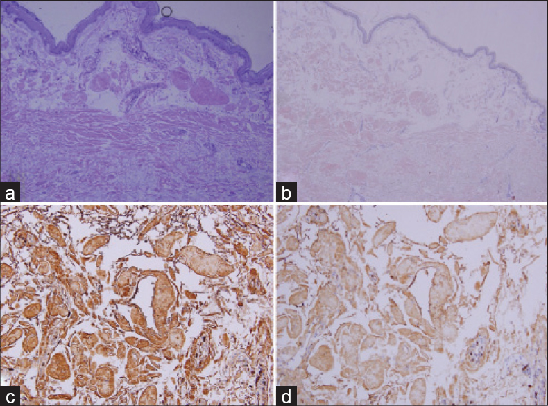
A skin biopsy from the edge of a bulla on the upper back showed subepidermal clefts and mass-like pink colored amorphous deposits in the papillary and reticular dermis which were positive when stained by crystal violet. Congo red staining was also positive and revealed apple-green birefringence with polarized light. There were a few plasma cells around the blood vessels [Figure - 2]. Immunohistochemistry studies showed strong positivity for λ light chain and a comparatively lesser intensity for κ light chain [Figure - 3]. Based on the clinical findings and histologic characteristics of the skin lesions, diagnosis of a primary localized cutaneous nodular amyloidosis (PLCNA) with bullous appearance was made. The lesions were removed by surgical excision.
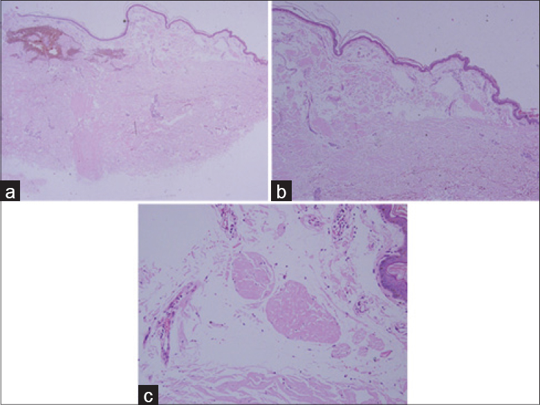
Figure 2: (a and b) Subepidermal clefts and mass-like pink colored amorphous materials deposition in the papillary and reticular dermis (H and E, ×20, ×40), (c) Plasma cells infiltration around the blood vessels (H and E, ×200)
Figure 3: (a) Subepidermal cleft and amyloid deposition in the entire dermis was positive with violet staining (×100) and (b) Congo red staining (×40), also positive for apple-green birefringence with polarized light (not shown). (c) Immunohistochemistry staining showed strong positivity for λ light chain (×200) and (d) less intensity for κ light chain (×200)

Primary localized cutaneous nodular amyloidosis is a rare variant of cutaneous amyloidosis. Approximately 100 cases of nodular cutaneous amyloidosis have been reported in the medical literature. [1] Cutaneous nodular amyloidosis is composed of light-chain immunoglobulins (AL amyloid) derived from a monoclonal expansion of plasma cells. The amyloid infiltrates the entire dermis from the papillary dermis to the subcutis. The amyloid deposits may also be seen in the vessels and nerve sheath. The condition commonly occurs on the legs, feet, trunk, face and genitalia. It presents typically with single or multiple waxy yellow-red nodules. [2] However, the lesions may be indistinguishable from cutaneous lesions of primary systemic amyloidosis. Immunohistochemical analysis of the dermal deposits of amyloid has revealed that the light chains may be λ or κ, or that both may be present. [3]

Bullous lesions of cutaneous amyloidosis are commonly associated with systemic amyloidosis with multiorgan involvement, including primary systemic amyloidosis and myeloma-associated amyloidosis. However, several patients of localized bullous cutaneous amyloidosis without systemic involvement have been described. [4] The bullae can be either intradermal or subepidermal, often induced by trauma or friction which are described as tense painful blisters that are frequently hemorrhagic. [5]

The mechanism of blister formation in cases of cutaneous amyloidosis remains to be described. Ochiai et al.[4] suggested that the amyloid globules might be engulfed through the process of transepidermal elimination initiated by trauma or rubbing which are induced by persistent pruritus that acts as the local precipitating factor for blister formation.

The treatment of primary localized cutaneous nodular amyloidosis includes surgical excision, cryotherapy, electrodesiccation and curettage, intralesional steroid injection and treatment with the CO 2 laser, each with different results. Nevertheless, the lesions are difficult to treat and often recur after treatment. The progression of primary localized cutaneous nodular amyloidosis to systemic amyloidosis has been reported in some case series at the rate of 7-50%. [3] Electron microscopy has shown that the deposits of amyloid, either primary or secondary type, consist of accumulation of rigid non-branching fibrils which are responsible for the Congo Red birefringence seen on polarized light microscopy. [2],[5] Systemic evaluation and long-term follow-up is needed to monitor the progression to systemic amyloidosis. We were able to find only one previous report of this presentation. [6]

References
1.
Santos-Juanes J, Galache C, Curto JR, Astudillo A, Sanchez del Rio J. Nodular primary cutaneous amyloidosis. J Eur Acad Dermatol Venereol 2004;18:224-6.
[Google Scholar]
2.
Fernandez-Flores A. Cutaneous amyloidosis: A concept review. Am J Dermatopathol 2012;34:1-14.
[Google Scholar]
3.
Woollons A, Black MM. Nodular localized primary cutaneous amyloidosis: A long-term follow-up study. Br J Dermatol 2001;145:105-9.
[Google Scholar]
4.
Ochiai T, Morishima T, Hao T, Takayama A. Bullous amyloidosis: The mechanism of blister formation revealed by electron microscopy. J Cutan Pathol 2001;28:407-11.
[Google Scholar]
5.
Lachmann HJ, Hawkins PN. Amyloidosis of the Skin. In: Goldsmith LA, Katz SI, Gilchrest BA, Paller AS, Leffell DJ, Wolff K, editors. Fitzpatrick′s dermatology in general medicine. 8 th ed. New York: McGraw-Hill; 2012. p. 1574-83.
[Google Scholar]
6.
LaChance A, Phelps A, Finch J, Lu J, Elaba Z, Rezuke W, et al. Nodular cutaneous amyloidosis: A bullous variant. Clin Exp Dermatol 2014;39:344-7.
[Google Scholar]

Fulltext Views
2,967

PDF downloads
1,197
Show Sections