Translate this page into:
Progressive symmetric erythrokeratoderma with dermatophytosis
Correspondence Address:
Hong Sang
Zhongshan East Road No. 305, Nanjing, Jiangsu, 210002
China
| How to cite this article: Wu F, Chen J, Li Zh, Hu Zl, Deng L, Sang H. Progressive symmetric erythrokeratoderma with dermatophytosis. Indian J Dermatol Venereol Leprol 2014;80:345-347 |
Sir,
Progressive symmetric erythrokeratoderma (PSEK) or Gottron′s disease is a rare disorder of cornification with an autosomal dominant inheritance. [1] There have been few case reports of this disease superimposed with fungal infection. Here, we present three cases of PSEK complicated with fungal infection.
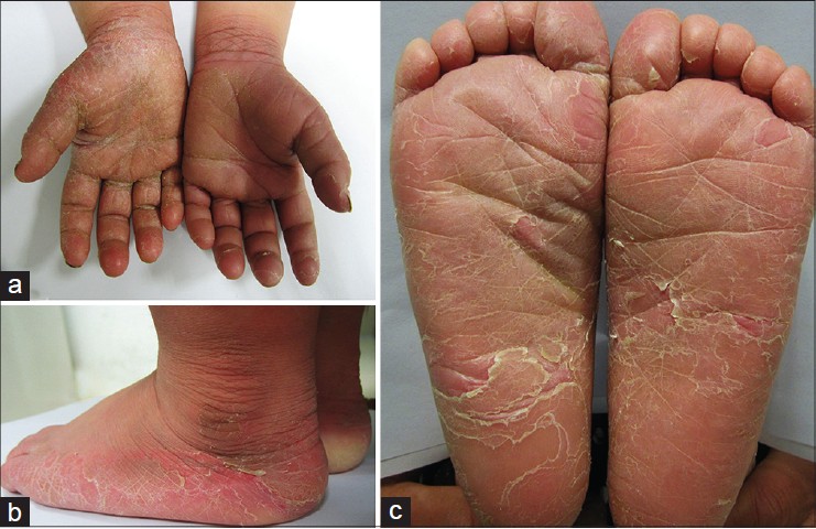
A 4-year-old boy, presented with a history of dryness and erythema over palms and soles since birth. This gradually extended onto the wrists and desquamation developed over both soles and right palm at the age of 2 years. He was otherwise healthy. His first cousin gave a history of similar but milder involvement of both palms.
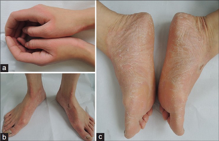
The second case was a 5-year-old girl who presented with a history of scaling and erythema over the palms, dorsa of hands and wrists since the age of 6 months which gradually progressed to involve the soles and dorsa of feet. The child was otherwise healthy and there was no family history of a similar illness.
The third case was a 20-year-old woman who complained of erythematous hyperkeratotic scaly plaques over the palms and soles since 2 years of age and involvement of her toe nails since the age of 12 years. Erythema increased in summers when her hands and feet also became malodorous. Both her father and grandmother were affected by a similar illness.
On clinical examination, all three patients had well- demarcated, symmetrically distributed, hyperkeratotic erythematous plaques on the palms and soles [Figure - 1], [Figure - 2], [Figure - 3]. Several toenails of the second patient′s right foot showed leukonychia while those of the third case also showed yellowish discoloration, thickening, and white spots on the surface [Figure - 2]b and [Figure - 3]b. Direct microscopic examination of KOH mounts of scrapings from the skin lesions and affected nails from all three subjects revealed fungal hyphae. All the fungal cultures yielded white, cottony colonies with a red wine colour on the reverse side, which was identified as Trichophyton rubrum [Figure - 4]. Histopathological examination of the first and the second case showed hyperkeratosis, acanthosis, and a mild superficial perivascular lymphocytic infiltrate with mild spongiosis.
 |
| Figure 1: Well-demarcated, symmetrically distributed hyperkeratotic erythematous plaques on palms, and desquamation on soles, the ridges of the feet and the right palm of case 1 |
 |
| Figure 2: Dry, scaly, well-demarcated plaques over the dorsa of hands and feet; several nail-plates of the right foot showed white spots on the surface in case 2 |
 |
| Figure 3: Erythematous hyperkeratic scaly plaques on the palms and soles, and abnormal toenails of case 3 |
A final diagnosis of progressive symmetrical erythrokeratoderma with dermatophytosis was made for all three cases. The first patient responded well to topical terbinafine cream (0.05%) twice daily and tazarotene gel (0.05%) every night for 1 month. After 1 month, desquamation disappeared, mycological examinations turned negative and keratosis improved, after which the patient was maintained on tazarotene gel 0.05% every night. The second patient was prescribed the same treatment for 1 month but follow up was not available. The third case received the same topical treatment for 1 month with additional oral itraconazole 200 mg twice daily for 1 week. After 1 month, desquamation of feet disappeared and mycological examinations of feet turned negative but those of toenails were positive. The patient received 2 more weeks of itraconazole orally one month apart while continuing tazarotene gel as previously.
Most case reports of PSEK describe nails as being normal. [1],[2] However, in a Chinese family, the prevalence of onychomycosis among PSEK-diseased nails was found to be 20%. [3] To the best of our knowledge, there are no previous reports describing fungal infections in patients of PSEK. The explanation for fungal infection in PSEK is unclear but recurrent fungal infections is one of the characteristics of KID syndrome (keratitis, ichthyosis, deafness) which is one of the erythrokeratoderma syndromes. [3] Transgenic mice expressing a COOH-terminal truncated form of loricrin which is similar to the protein expressed in PSEK exhibited erthrokeratoderma with an epidermal barrier dysfunction. [4] More studies are needed to explore the reasons why all our cases were complicated with fungal infection.
Besides oral retinoids, topical retinoids, keratolytics, steroids and calcipotriol have been reported to be effective in treating this disorder. [5] Considering the coexistence of fungal infection, topical steroids were avoided and antifungal agents and topical retinoids were prescribed. Our cases highlight the importance of mycological examination in patients with PSEK, especially those with scaling and abnormal nails.
| 1. |
Ghorpade A, Ramanan C. Progressive symmetric erythrokeratoderma. Indian J Dermatol Venerol Leprol 1995;61:116-7.
[Google Scholar]
|
| 2. |
Chu DH, Arroyo MP. Progressive and symmetric erythrokeratoderma. Dermatol Online J 2003;9:21.
[Google Scholar]
|
| 3. |
Yan HB, Zhang J, Liang W, Zhang HY, Liu JY. Progressive symmetric erythrokeratoderma: Report of a Chinese family. Indian J Dermatol Venereol 2011;77:597-600.
[Google Scholar]
|
| 4. |
Suga Y, Jarnik M, Attar PS, Lomgley MA, Bundman D, Steven AC, et al. Transgenic mice expressing a mutant form of loricrin reveal the molecular basis of the skin diseases, Vohwinkel syndrome and progressive symmetric erythrokeratoderma. J Cell Biol 2004;151:401-12.
[Google Scholar]
|
| 5. |
Bilgin I, Bozdað KE, Uysal S, Ermete M. Progressive symmetrical erythrokeratoderma- response to topical calcipotriol. J Dermatol Case Rep 2011;5:50-2.
[Google Scholar]
|
Fulltext Views
3,532
PDF downloads
1,499


