Proliferative verrucous leukoplakia
Corresponding author: Dr. Mohamed M. Fawzy, Department of Dermatology and Venereology, Tanta University, Tanta, Egypt. fawzy208222@yahoo.com
-
Received: ,
Accepted: ,
How to cite this article: Fawzy MM, Nofal A, El-Hawary EE. Proliferative verrucous leukoplakia. Indian J Dermatol Venereol Leprol 2021;87:455.
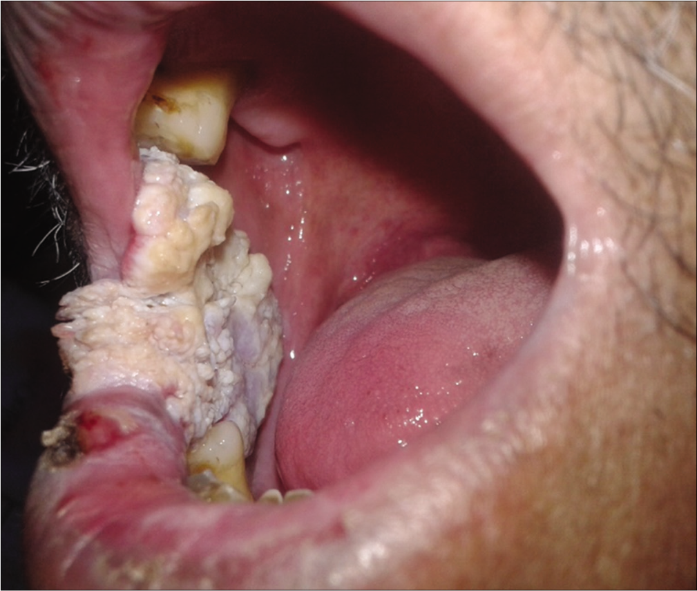
A 65-year-old male came to our hospital for the evaluation of an asymptomatic whitish lesion within the oral cavity that had started 1 year ago. He is a non-smoker with positive serology for hepatitis C. On examination, there was a relatively large (about 5 cm x 3 cm in diameter), white, verrucous plaque over the right inner buccal mucosa [Figure 1]. The clinical differential diagnoses included frictional keratosis, squamous papilloma, verrucous hyperplasia, verrucous carcinoma, squamous cell carcinoma and chronic hyperplastic candidiasis. After obtaining a written informed consent from the patient, skin biopsy was performed which revealed intact mucosal stratified squamous epithelium showing corrugated papillary overgrowths covered by hyperkeratotic acanthotic squamous epithelium. There was interface lymphocytic infiltrate with some areas of basal vacuolar degeneration. No atypia, dyschromatosis, dysplasia, or other malignant changes were detected. These findings support the diagnosis of proliferative verrucous leukoplakia.

- A 65-year-old male with asymptomatic white verrucous plaque on the right buccal mucosa
Declaration of patient consent
The authors certify that they have obtained all appropriate patient consent.
Financial support and sponsorship
Nil.
Conflicts of interest
There are no conflicts of interest.


