Translate this page into:
Pseudolymphoma induced by gold after patch testing: Dermoscopic features
Corresponding author: Dr. Javier Gimeno-Castillo, Department of Dermatology, Hospital Universitario Araba, Vitoria, Spain. javiergimenocastillo@gmail.com
-
Received: ,
Accepted: ,
How to cite this article: Gimeno-Castillo J, Menéndez-Parrón A, Martínez-Aracil AP, Rosés-Gibert P, González-Pérez R. Pseudolymphoma induced by gold after patch testing: Dermoscopic features. Indian J Dermatol Venereol Leprol 2023;89:741-4
Dear Editor,
Late reactions in patch testing are defined as delayed reactions on or after day seven. Allergens such as gold, corticosteroids or paraphenylenediamine can frequently trigger this type of delayed reaction. 1 Among the latter, those caused by gold sodium thiosulfate may be particularly delayed. Moreover, gold sodium thiosulphate has the potential to trigger granulomatous or pseudolymphomatous reactions. Clinical reports of the latter are scarce. 2– 4 Thus, this report highlights a case of pseudolymphoma after patch testing to gold sodium thiosulphate and performs a literature review of previously described cases.
A 64-year-old man presented to the dermatology department with axillary and submental pruritic, erythematous, eczematous plaques for one year. The patient denied a temporal relationship with exposure to any substance. To rule out allergic contact dermatitis, the patient was patch tested using the standard baseline series of the Spanish Contact Dermatitis and Skin Allergy Research Group5 (using as its base the True Test [Smartpractice®, EE. UU] and patching the additional haptens), and corticosteroid-specific series (Chemotechnique Diagnosis®; Vellinge, Sweden). He showed positive reactions to formaldehyde, perfume mix 1 and linalool at 96 hours. These positive results were considered relevant, and the patient was advised to avoid the use of these substances (present mainly in deodorants and perfumes), leading to the resolution of the eczematous lesions. Two months later, a late reaction to gold sodium thiosulfate was observed at the patch test site. The latter was confirmed by a repeat patch testing which showed a positive reaction (++) to gold at 96 hours. The positive reaction to gold sodium thiosulfate was not considered relevant in the present case.
The patient was prescribed topical clobetasol for three weeks. However, he was lost to follow up and returned 12 months later with two erythematous pruriginous, well-demarcated, infiltrated, papules located exactly where both positive reactions to gold sodium thiosulfate had occurred [Figure 1a]. The patient denied any response of his previous lesions to topical therapy with clobetasol after three weeks of treatment. Dermoscopic examination of the latter revealed a telangiectatic background with orangish papules that turned yellow on contact dermoscopy [Figures 1b and c]. A biopsy was done to rule out granulomatous or pseudolymphomatous reactions.
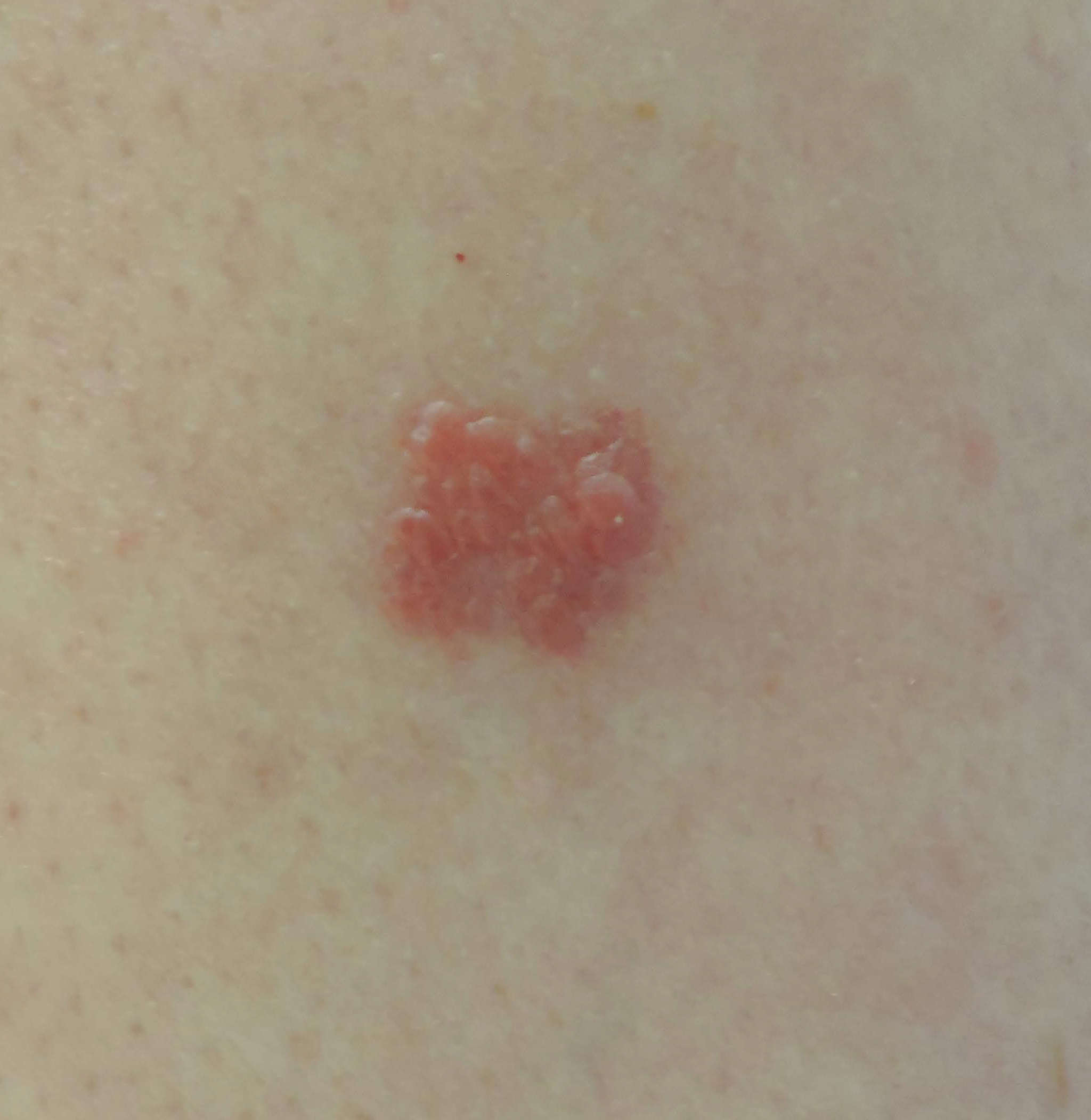
- One of the erythematous pruriginous, well-demarcated papular lesions located on the place where both positive reactions to gold sodium thiosulphate had occurred
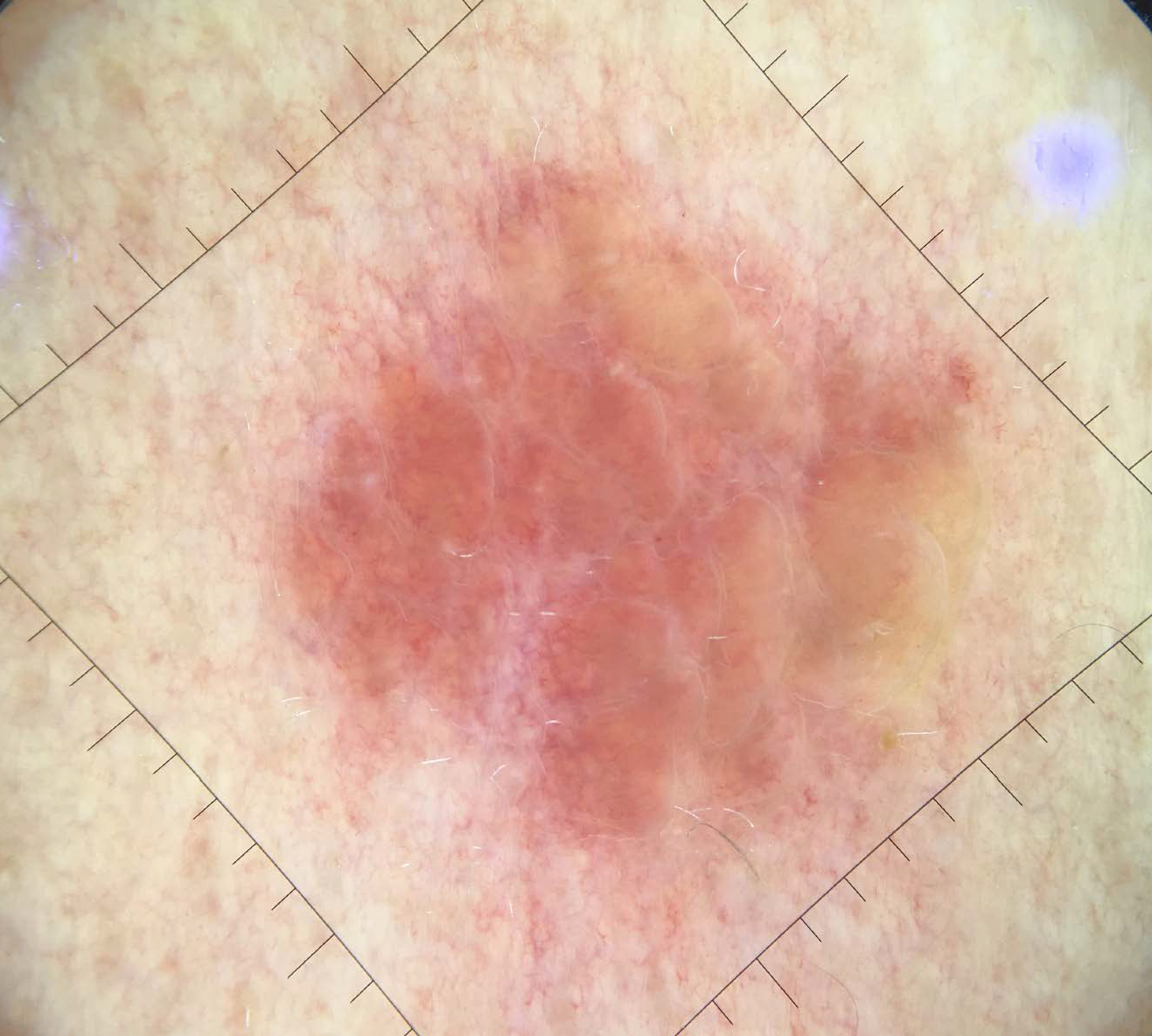
- Dermoscopic features of the lesion: a telangiectatic background along with orangish papules. (Image taken with Handyscope®, Fotofinder) ×10
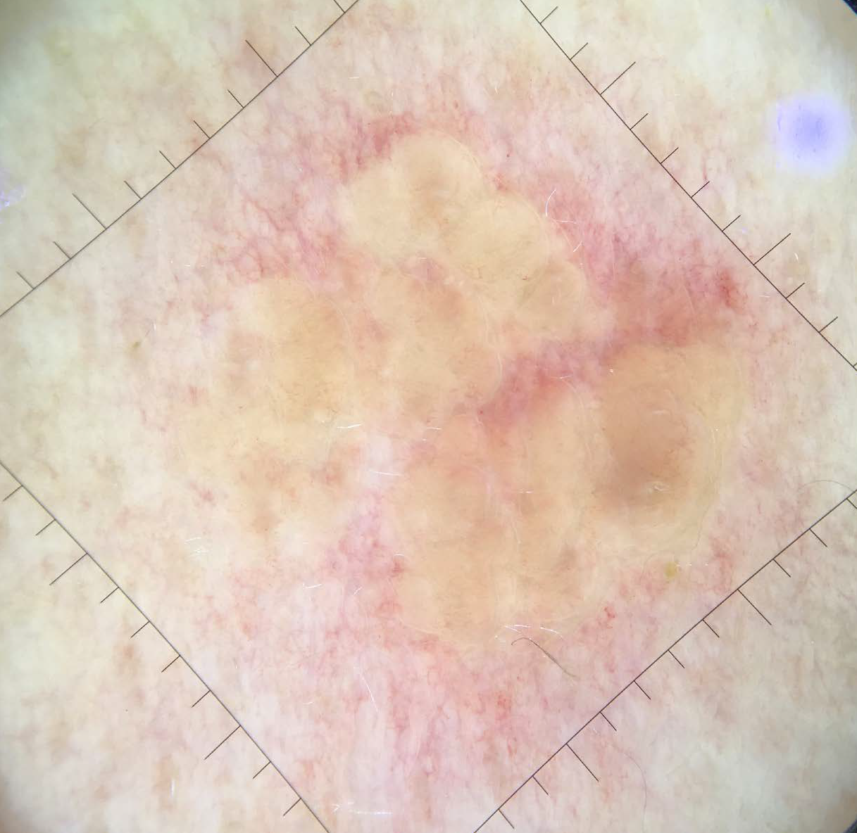
- Yellow discoloration evident on applying contact dermoscopy(Image taken with Handyscope®, Fotofinder) ×10
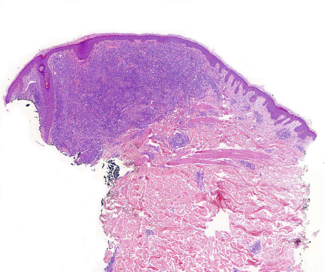
- H&E × 39: A dense dermal lymphohistocytic infiltrate without atypia or epidermotropism
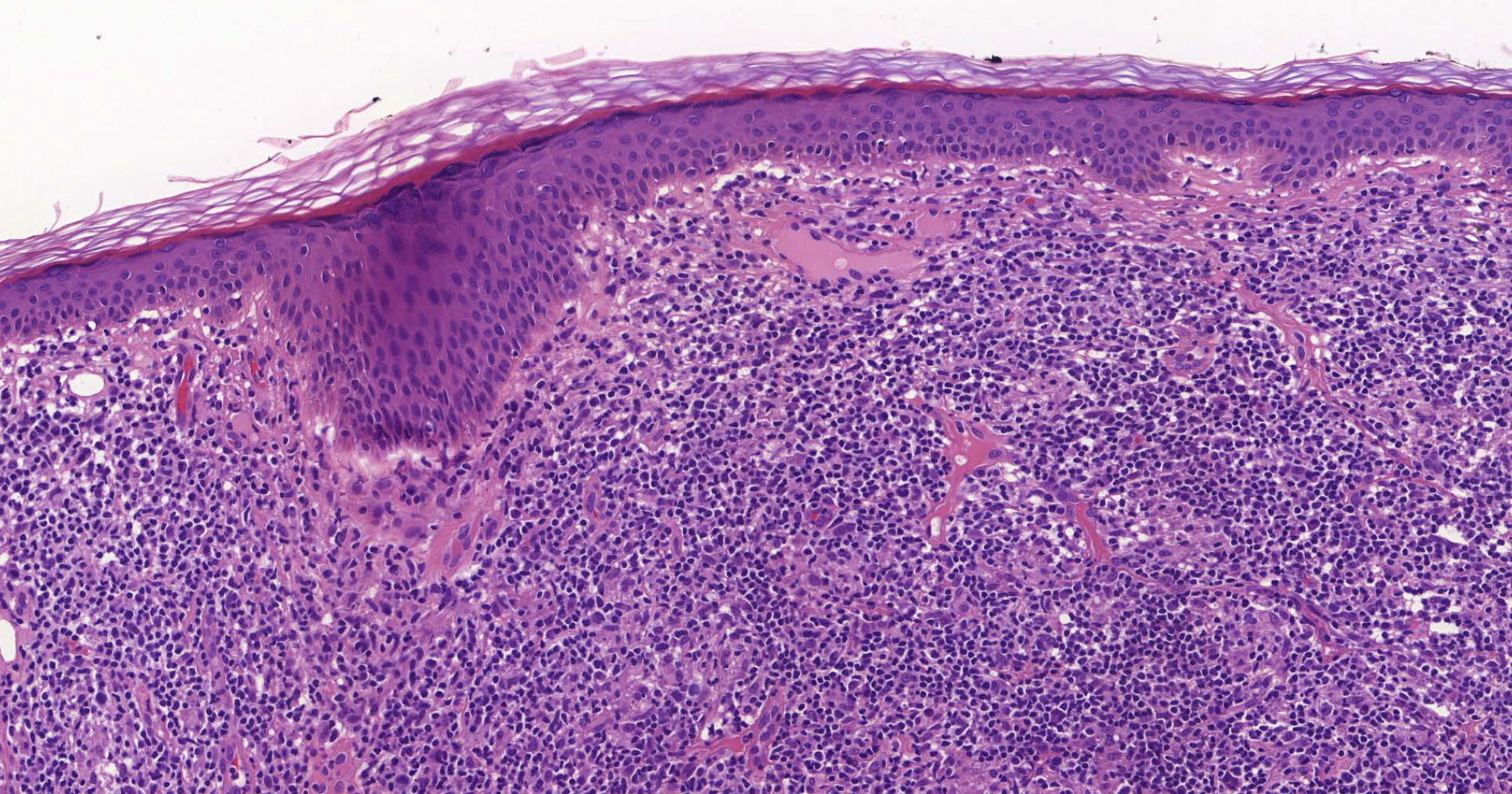
- H&E × 200: Dermal infiltrate primarily composed of lymphocytes and histocytes with numerous plasma cells
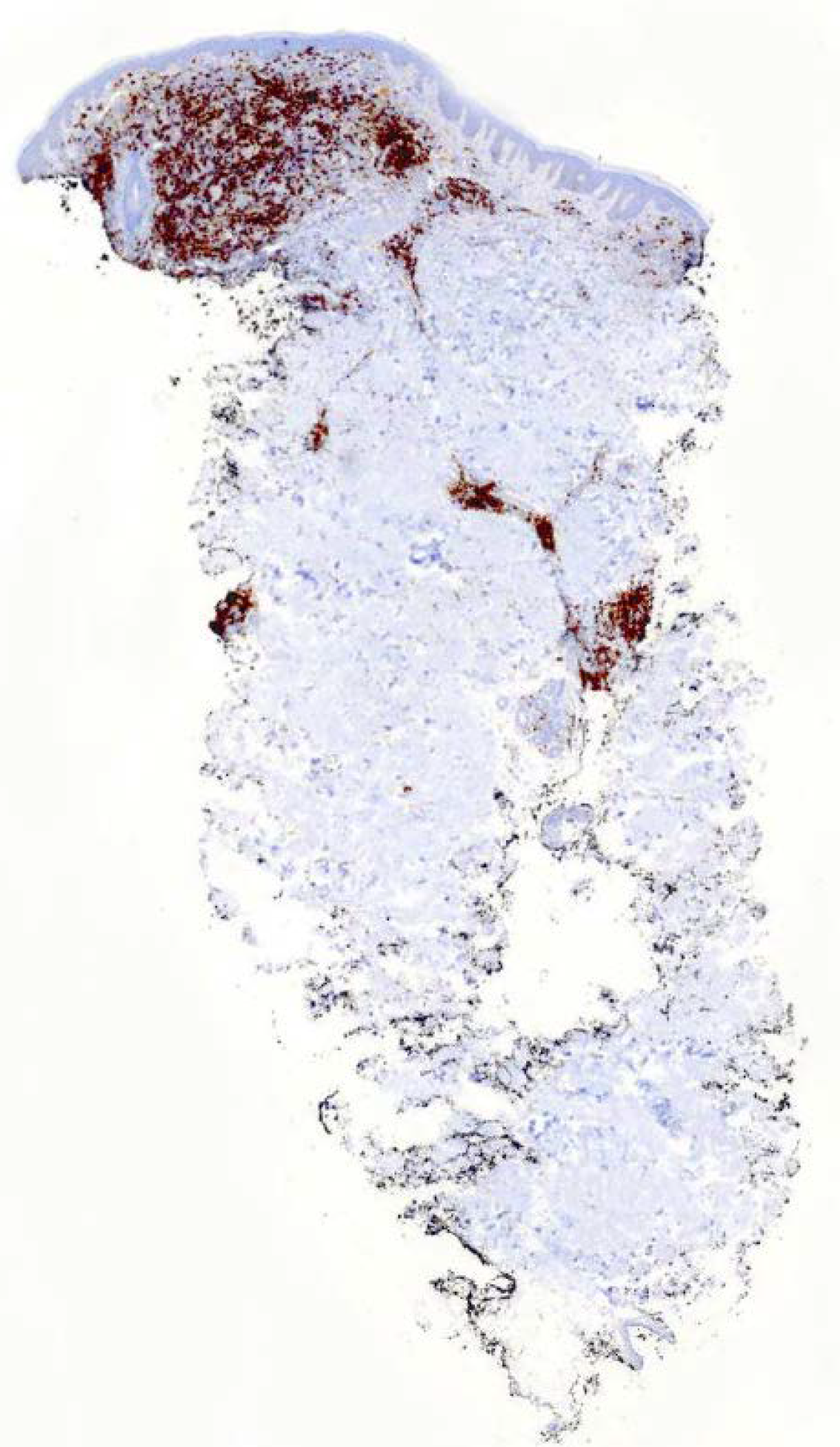
- CD20 × 14 immunohisto chemistry: Cells staining positive for CD-20
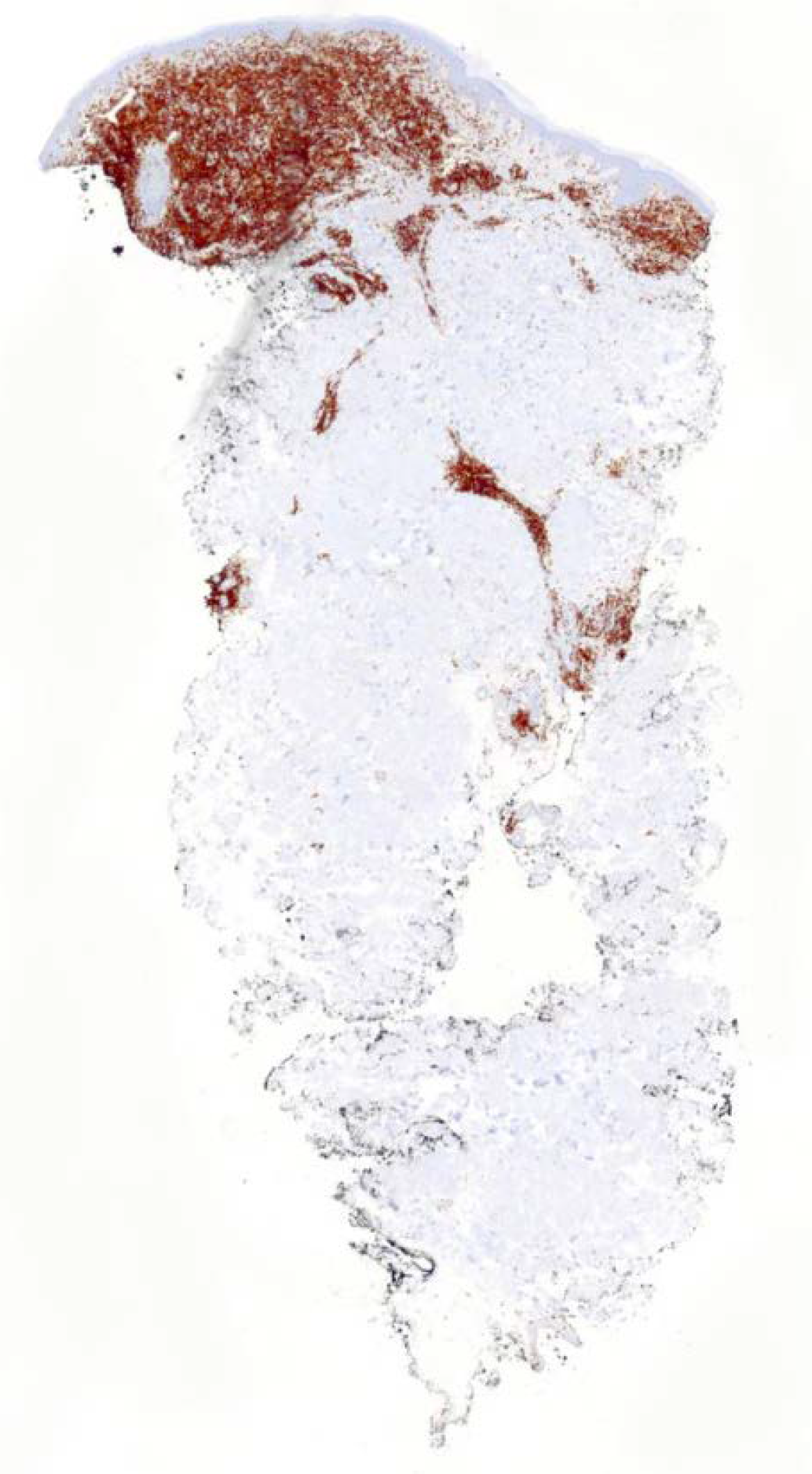
- CD3 × 14 immunohisto chemistry: Cells staining positive for CD-3
Histological examination revealed an unremarkable epidermis. A dense sheet-like lympho-histiocytic infiltration was present, mainly in the superficial and mid-dermis [Figures 2a and b]. Scattered eosinophils, and frequent polytypic plasma cells (Kappa and lambda) were also observed. There was no atypia or granulomas. Immunohistochemically, the infiltrate was composed of a mixture of CD3-positive and CD20-positive B-cells [Figures 2c and d]. These findings were consistent with a histologic diagnosis of pseudolymphoma. Thus, the lesions were treated with intralesional triamcinolone with complete remission after two weeks. No recurrence was reported to date.
Even though positive patch reactions to gold compounds are common, it has been described that percutaneous gold stimulation can induce a pseudolymphomatous change. However, the pathogenesis of this phenomenon remains unclear. The authors believe that this could be explained by an exaggerated localized lymphocytes response after exposure to gold. Previously recorded cases of this condition are summarised in Table 1.
2–4
The present case highlights two pseudolymphomatous lesions after contact with two different gold sodium thiosulfate patches on the same patient.
F: Female, M: Male
Sex
Age
Previous allergy to gold
Time after patch testing
Number of lesions
Clinical manifestation
Symptoms
Dermoscopy
Histology
Treatment
Resolution
Authorship
1
F
66
No
24 months
1
Tiny papules with erythema
Unknown
No
Dense, band-like or nodular infiltrate of mononuclear cells immediately below the epidermis and a small nodular infiltrate of mononuclear cells around eccrine gland structures. Immunohistochemically, there was a mixture of CD3-positive and CD20-positive cells.
Topical steroid
Yes. (Spontaneous)
Kiyohara. T, et al.
2
F
46
No
11 months
1
Well-demarcated round lesion.
Pruritic
No
Nodular dermal infiltrate with suggestion of germinal center formation. Spared epidermis. Immunohistochemically, there was a mixture of CD2, CD3, CD5-positive and CD20-positive cells.
None
Yes. (Spontaneous)
Shpadaruk. V, et al.
3
F
59
No
3 weeks
1
Erythematous plaque
Asymptomatic
No
Dense lymphoid infiltrate, involving the superficial and deep dermis. Immunohistochemically, there was a predominance of CD3-positive cells along with CD4 and CD8 positive cells.
Topical steroid
Yes
García-Arpa. M, et al.
4
F
43
No
4 weeks
1
Persistent reaction (Not specified)
Unknown
No
Dense lymphocytic infiltrate in the dermis, which was predominantly perifollicular. Immunohistochemically, there was a mixture of CD3-positive and CD20-positive cells.
Unknown
Unknown
García-Arpa. M, et al.
5
M
64
No
12 months
2
Papules on an erythematous background.
Pruritic
Telangiectatic background with orangish papules that turned into yellow when contact was applied.
Dense lympho-histocytary infiltrate without atypical cells. Spared epidermis. Immunohistochemically, there was a mixture of CD3-positive and CD20-positive cells.
Intralesional steroids
Yes
Present case.
Previously described dermoscopic findings of pseudo lymphomas i.e. telangiectatic background with orangish papules that turned yellow on contact dermoscopy—could help the recognition of this entity even though it can clinically and dermoscopically mimic late granulomatous reaction or eczema. 6 However, a biopsy is essential to confirm a diagnosis of pseudolymphoma. As reviewed, these lesions usually appear days to weeks after contact with gold sodium thiosulphate and can last for years. They typically present as asymptomatic or mildly pruritic erythematous papules or plaques on an erythematous background at the sites of contact with gold sodium thiosulfate. Histologically a dense lymphoid infiltrate is classically composed of a mixture of CD20-positive and CD3-positive cells. 2–4 In terms of treatment, the response to topical steroids seems inconsistent, however, aggressive options should be avoided considering the indolent behaviour.
Given the variability described in the lag period of development of lesions following contact (from weeks to years) it is likely that this condition remains underdiagnosed. Thus, we suggest that late reactions should be biopsied if they show suggestive clinical and dermoscopic features of pseudolymphoma regardless of their time of evolution.
Declaration of patient consent
The authors certify that they have obtained all appropriate patient consent.
Financial support and sponsorship
Nil.
Conflict of interest
There are no conflicts of interest.
References
- Unusual post-patch testing erythema: A late, granulomatous, non-eczematous reaction to gold sodium thiosulphate. J Eur Acad Dermatol Venereol. 2018;32:e126-7.
- [CrossRef] [PubMed] [Google Scholar]
- Cutaneous pseudolymphoma after patch test to gold sodium thiosulfate in two patients. Contact Dermatitis. 2020;82:322-4.
- [CrossRef] [PubMed] [Google Scholar]
- Pseudolymphoma due to gold sodium thiosulfate. Dermatitis. 2020;31:e1-2.
- [CrossRef] [Google Scholar]
- Small papular pseudolymphoma induced by a patch test for gold. Clin Exp Dermatol. 2020;45:267-9.
- [CrossRef] [PubMed] [Google Scholar]
- The Spanish standard patch test series: 2016 update by the Spanish Contact Dermatitis and Skin Allergy Research Group (GEIDAC) Actas Dermosifiliogr. 2016;107:559-66.
- [CrossRef] [PubMed] [Google Scholar]
- Is dermoscopy useful for the diagnosis of pseudolymphomas? Dermatology. 2021;237:213-6.
- [CrossRef] [PubMed] [Google Scholar]


