Raccoon eye: An ocular presentation in metastatic neuroblastoma
Corresponding author: Dr. Aditya Kumar Gupta, Department of Paediatrics, All India Institute of Medical Sciences, New Delhi, India. adivick@gmail.com
-
Received: ,
Accepted: ,
How to cite this article: Kumar R, Prakash S, Gupta AK. Raccoon eye: An ocular presentation in metastatic neuroblastoma. Indian J Dermatol Venereol Leprol. 2025;91:409-10. doi: 10.25259/IJDVL_1378_2024
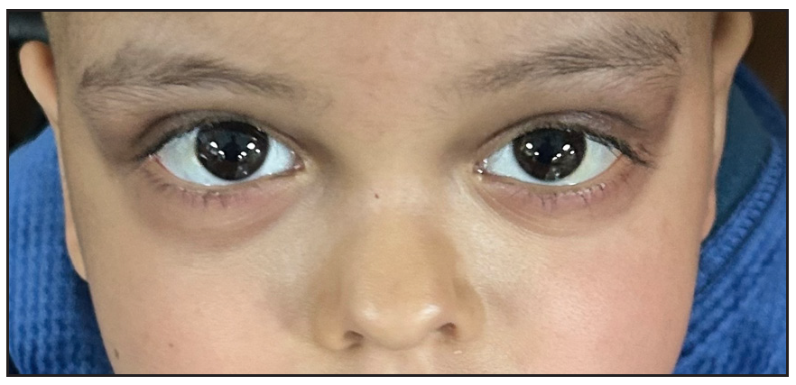
We report the case of a five-year-old girl who presented with progressive painful abdominal distension and bilateral periorbital ecchymosis, subconjunctival haemorrhage, and proptosis (left eye) for two weeks [Figure 1]. The swelling was soft with no limitation of ocular movements. Imaging (ultrasonography, computed tomography) showed a large left suprarenal mass. Bone marrow examination revealed infiltration by small round blue cells, clinching a diagnosis of metastatic neuroblastoma. Post-chemotherapy, there was a striking improvement in the ocular findings [Figure 2].

- Bilateral periorbital ecchymosis, swelling, subconjunctival haemorrhage and proptosis of left eye.

- Normal appearance of the eyes post-chemotherapy for neuroblastoma.
Orbital metastasis is seen in 10–20% of neuroblastoma cases, often presenting with a characteristic ‘raccoon eye’ appearance due to the presence of retrobulbar metastases. Proptosis in neuroblastoma is typically asymmetrical and soft, with a normal range of ocular movements, vis-à-vis leukemic ocular metastases (usually bilateral proptosis) and metastatic sarcoma (firm-to-hard and limited range of ocular movements).
Declaration of patient consent
The authors certify that they have obtained all appropriate patient consent.
Financial support and sponsorship
Nil.
Conflicts of interest
There are no conflicts of interest.
Use of artificial intelligence (AI)-assisted technology for manuscript preparation
The authors confirm that there was no use of AI-assisted technology for assisting in the writing or editing of the manuscript and no images were manipulated using AI.


