Translate this page into:
Raccoon eyes in a case of metastatic neuroblastoma
2 Department of Pediatric Medicine, R.G. Kar Medical College, 1, Khudiram Bose Sarani, Kolkata, West Bengal, India
Correspondence Address:
Sudip Kumar Ghosh
Department of Dermatology, Venereology, and Leprosy, R.G. Kar Medical College, 1, Khudiram Bose Road, Kolkata-700 004, West Bengal
India
| How to cite this article: Ghosh SK, Dutta A, Basu M. Raccoon eyes in a case of metastatic neuroblastoma. Indian J Dermatol Venereol Leprol 2012;78:740-741 |
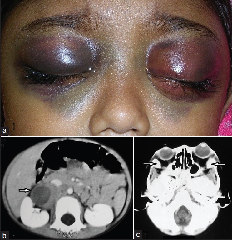
A three-year-old boy presented to us with a history of low-grade intermittent fever for the preceding two months and bruising around his eyes for the last two weeks. He also had a history of loss of appetite and gradual weight loss over the last few months. Examination showed periorbital ecchymoses, bilateral mild proptosis, [Figure - 1]a moderate pallor, and hepatosplenomegaly. There was no other mucocutaneous lesion elsewhere in the body. Ophthalmological evaluation revealed that the vision of his left eye was restricted to perception of light only and he could barely perceive hand movements in the right eye. Ultrasonography of the abdomen showed hepatosplenomegaly and a right-sided adrenal mass. Contrast-enhanced computed tomographic (CT) scan of the abdomen revealed a large mixed-attenuating right-sided adrenal mass with both solid and cystic components, necrotic areas, and foci of calcification [Figure - 1]b. CT scan of the brain showed hyperdense extradural lesions with erosion of the underlying bones. There were extraconal hyperdense lesions in both the orbits with erosions of lateral orbital walls and lateral rectus muscles [Figure - 1]c. Laboratory investigations showed normocytic normochromic anemia and elevated levels of serum lactate dehydrogenase (2-185 IU/l) and urinary vanillylmandelic acid (86.76 mg/day). Bone marrow biopsy demonstrated deposits of round cell tumors suggestive of neuroblastoma (NB). Biopsy from post-caval lymph node showed features of metastatic deposits from NB.
 |
| Figure 1: (a) bilateral periorbital ecchymoses and proptosis (b) Contrast-enhanced CT scan of the abdomen showing a large mixed-attenuating right-sided adrenal mass (marked by arrows) (c) CT scan of the brain showing bilateral orbital metastasis and bony erosion (marked by arrows) |
Based on the clinical features, laboratory investigations, and imaging, a diagnosis of raccoon eyes in a case of metastatic NB was made. His visual evoked potential showed axonal type of bilateral optico-retinal pathway dysfunction (left more than right), with secondary demyelination of the left eye.
Orbital metastases can be seen in up to 20% of children with advanced stage of NB. The periorbital ecchymoses (raccoon eyes) in a case of NB is perhaps related to obstruction of the palpebral vessels (branches of the ophthalmic and facial vessels) by metastatic tissue in and around the orbits. Child abuse, periorbital cellulitis, primary systemic amyloidosis, and blood dyscrasias may be considered in the differential diagnosis of such cases. However, laboratory investigations and imaging have clinched the diagnosis of the present case. Child abuse or traumatic injury must be ruled out in similar cases of periorbital ecchymoses. Signs of injuries may also be found in other areas of the body. In such cases, CT scan might reveal fracture of the skull bones with or without associated intracranial injuries. Periorbital cellulitis usually presents with unilateral erythematous periorbital swelling. Rapid increase in temperature and swelling of tissue may occur. Movement of the extraocular muscles and visual acuity is usually normal and proptosis does not occur in these cases. Primary systemic amyloidosis is a disease of adulthood where, in addition to the raccoon eyes, ecchymoses may be present elsewhere in the body. Macroglossia, sensory and autonomic neuropathy, concomitant renal, cardiac, and hepatic involvement are important features of this condition amongst others. Histopathological examination from abdominal fat pad or rectum is usually diagnostic. Blood dyscrasias usually present with a history of external bleeding. Laboratory evaluation including complete hemogram, platelet function tests, and blood coagulation profile are usually sufficient enough to exclude such cases. Currently, MIBG (meta iodo benzyl guanidine) scan has become an important procedure for staging and defining extent and location of NB. Regarding the management of NB, age of the patient and stage of the tumor along with cytogenetic and molecular features are the most important determinants. Surgery is the main modality of treatment in the management of the low stages of NB. On the other hand, multiple-agent chemotherapy is the conventional therapy for patients with more advanced stages of NB. Commonly used chemotherapeutic agents in NB include cisplatin, carboplatin, cyclophosphamide, etoposide, and doxorubicin amongst others. Autologous stem cell transplantation, surgery, irradiation, and 13-cis-retinoic acid are the other important treatment modalities in high-risk group of NB. Our patient received six cycles of chemotherapy comprising of cisplatin, doxorubicin, etoposide, and ifosfamide along with other supportive measures. He had no evidence of recurrence within a follow-up period of six months and showed significant improvement in the form of general well-being, subsidence of periorbital lesions, and decrease in size of the tumor mass. However, his vision did not improve.
Fulltext Views
14,514
PDF downloads
2,311


