Translate this page into:
Radiotherapy-induced koebnerization in lichen planus
Correspondence Address:
Avinash A Sajgane
87/ 204 A Wing Madhukunj CHS, Nehru Nagar, Kurla East, Mumbai 24, Maharashtra
India
| How to cite this article: Sajgane AA, Dongre AM. Radiotherapy-induced koebnerization in lichen planus. Indian J Dermatol Venereol Leprol 2012;78:665 |
Sir,
Lichen planus (LP) is an inflammatory dermatosis of autoimmune origin, which affects the skin and mucosae. The Koebner phenomenon is the development of isomorphic pathologic lesions in the traumatized uninvolved skin of patients who have cutaneous diseases. These new lesions are clinically and histopathologically identical to those in the diseased skin. [1] This phenomenon has been described in various dermatoses including LP. However, the appearance of LP confined to an irradiated site is extremely rare.
A 40-year-old male presented to our outpatient department with complaint of itchy hyperpigmented lesions on the left foot and right knee. Lesions on the left foot appeared 2 years ago while those on the right knee were of 2-month duration. Two years back he took treatment for skin lesions on the left foot in the form of topical and oral medication with minimal improvement. On detailed history, we came to know that he was a case of non- Hodgkin lymphoma (diffuse large B-cell lymphoma) of right knee joint and had received chemotherapy (CHOP) 6 months back. He had also received external beam radiotherapy with Cobalt 60 in a dose of 45 Gy for 1 month on the right knee joint for lymphoma. After a month of radiotherapy, new itchy hyperpigmented lesions appeared on the right knee.
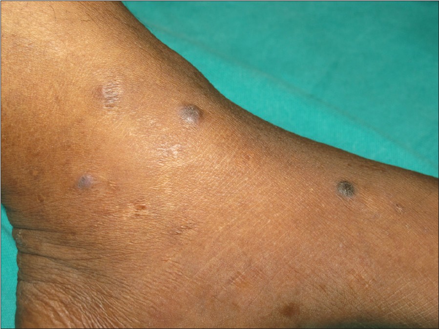
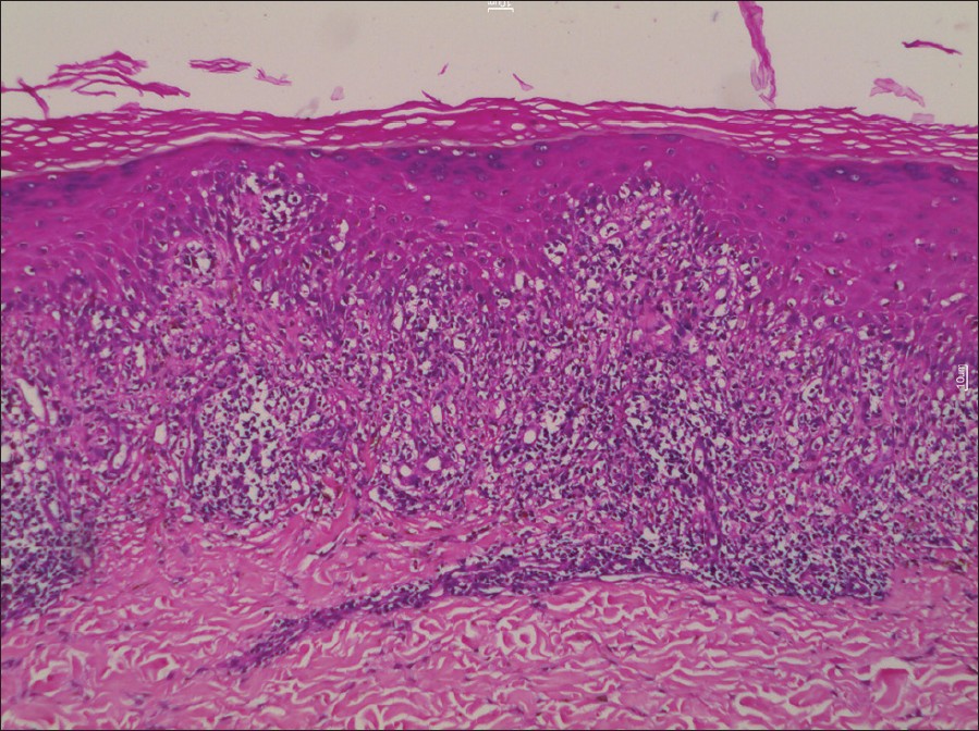
His physical examination was normal except slight swelling of the right knee. Cutaneous examination revealed multiple violaceous, hyperpigmented, shiny papules and plaques on the right knee [Figure - 1]. Violaceous and hyperpigmented papules were also seen on the left foot [Figure - 2]. Lesions on the right knee coincided exactly with the irradiated area. His mucosal examination was normal. Biopsy of papules from the right knee and the left foot showed wedge-shaped hypergranulosis, lichenoid infiltrate consisting of lymphocytes hugging the basal layer and saw toothing of the rete ridges [Figure - 3]. Thus, a diagnosis LP with radiotherapy-induced koebnerization was confirmed.
 |
| Figure 1: Multiple shiny, violaceous, hyperpigmented papules and plaques on the right knee |
 |
| Figure 2: Violaceous and hyperpigmented papules on the left foot |
 |
| Figure 3: Wedge-shaped hypergranulosis, lichenoid infiltrate consisting of lymphocytes hugging the basal layer and saw toothing of the rete ridges (H and E, ×200) |
The Koebner phenomenon is particularly characteristic of psoriasis and LP but occurs in several other dermatoses like lichen nitidus, vitiligo, and lichen sclerosus. The Koebner phenomenon may follow various kinds of trauma which may be mechanical, thermal, chemical, or photo. Various therapies such as Grenz ray therapy, roentgen therapy, and ultraviolet light therapy are known to act as provocative factors. UV light is known to induce lesions of actinic LP. On the other hand, psoralen plus ultraviolet A has been found useful for the treatment of LP. Apart from LP, variety of skin conditions like lichen sclerosus et atrophicus, bullous pemphigoid, pemphigus foliaceus, and pemphigus vulgaris have been reported to be occur following radiotherapy. [2]
New lesions as such can appear in untreated case of LP but in our case as the new lesions occurred exactly at the site of radiotherapy with no new lesion elsewhere on the body, we propose these lesions on the knee to be a result of koebnerization induced by radiotherapy.
Little is known about the mechanism by which LP develops after radiotherapy. Radiotherapy is known to have immunomodulatory properties. Some of the mechanisms which were put forward includes increased expression of proinflammatory molecules such as major histocompatibility complex (MHC), adhesion molecules (ICAM-1 and E-selectin), increased expression of cytokines (IL-1, IL-6, IL-8,TNF-alpha, TGF beta), release of autoantigens or antigenic peptides by damage caused by radiotherapy, and degradation of the basement membrane by increased matrix metalloproteases (MMPs). [3],[4]
LP induced by radiotherapy may be confined to the site of exposure or it may present as generalized eruption. The appearance of LP confined to an irradiated site is very rare and only few cases have been published. [2],[3],[5],[6]
In some of the cases reported in the literature, patients had no prior history of LP and the lesions appeared for the first time after radiotherapy. Only one case reported by Pretel et al. had a history of the presence of similar lesions as in our case. [5] Shurman has suggested the term "isoradiotopic" response to describe the phenomenon of secondary dermatoses arising in radiation fields. [2]
Thus, we present one more rare case of LP where external radiotherapy-induced koebenerization occurred exactly at the site of irradiation and propose that external radiotherapy should be considered as one more factor for the cause of koebnerization in various dermatoses.
| 1. |
Thappa DM. The isomorphic phenomenon of Koebner. Indian J Dermatol Venereol Leprol 2004;70:187-9.
[Google Scholar]
|
| 2. |
Shurman D, Reich HL, James WD. Lichen planus confined to a radiation field: The isoradiotopic response. J Am Acad Dermatol 2004;50:482-3.
[Google Scholar]
|
| 3. |
Sciallis GF, Loprinzi CL, Davis MD. Lichen planus induced by radiotherapy. Br J Dermatol 2005;152:399-401.
[Google Scholar]
|
| 4. |
Morar N, Francis ND. Generalized lichen planus induced by radiotherapy: Shared molecular mechanisms? Clin Exp Dermatol 2009;34:434-5.
[Google Scholar]
|
| 5. |
Pretel M, Espana A. Lichen planus induced by radiotherapy. Clin Exp Dermatol 2007;32:582-3.
[Google Scholar]
|
| 6. |
Kim JH, Krivda SJ. Lichen planus confined to a radiation therapy site. J Am Acad Dermatol 2002;46:604-5.
[Google Scholar]
|
Fulltext Views
3,404
PDF downloads
3,342


