Generic selectors
Exact matches only
Search in title
Search in content
Post Type Selectors
Search in posts
Search in pages
Filter by Categories
15th National Conference of the IAOMFP, Chennai, 2006
Abstract
Abstracts from current literature
Acne in India: Guidelines for management - IAA Consensus Document
Addendum
Announcement
Art & Psychiatry
Article
Articles
Association Activities
Association Notes
Award Article
Book Review
Brief Report
Case Analysis
Case Letter
Case Letters
Case Notes
Case Report
Case Reports
Clinical and Laboratory Investigations
Clinical Article
Clinical Studies
Clinical Study
Commentary
Conference Oration
Conference Summary
Continuing Medical Education
Correspondence
Corrigendum
Cosmetic Dermatology
Cosmetology
Current Best Evidence
Current Issue
Current View
Derma Quest
Dermato Surgery
Dermatopathology
Dermatosurgery Specials
Dispensing Pearl
Do you know?
Drug Dialogues
e-IJDVL
Editor Speaks
Editorial
Editorial Remarks
Editorial Report
Editorial Report - 2007
Editorial report for 2004-2005
Errata
Erratum
Focus
Fourth All India Conference Programme
From Our Book Shelf
From the Desk of Chief Editor
General
Get Set for Net
Get set for the net
Guest Article
Guest Editorial
History
How I Manage?
IADVL Announcement
IADVL Announcements
IJDVL Awards
IJDVL AWARDS 2015
IJDVL Awards 2018
IJDVL Awards 2019
IJDVL Awards 2020
IJDVL International Awards 2018
Images in Clinical Practice
In Memorium
Inaugural Address
Index
Knowledge From World Contemporaries
Leprosy Section
Letter in Response to Previous Publication
Letter to Editor
Letter to the Editor
Letter to the Editor - Case Letter
Letter to the Editor - Letter in Response to Published Article
LETTER TO THE EDITOR - LETTERS IN RESPONSE TO PUBLISHED ARTICLES
Letter to the Editor - Observation Letter
Letter to the Editor - Study Letter
Letter to the Editor - Therapy Letter
Letter to the Editor: Articles in Response to Previously Published Articles
Letters in Response to Previous Publication
Letters to the Editor
Letters to the Editor - Letter in Response to Previously Published Articles
Letters to the Editor: Case Letters
Letters to the Editor: Letters in Response to Previously Published Articles
Medicolegal Window
Messages
Miscellaneous Letter
Musings
Net Case
Net case report
Net Image
Net Images
Net Letter
Net Quiz
Net Study
New Preparations
News
News & Views
Obituary
Observation Letter
Observation Letters
Oration
Original Article
ORIGINAL CONTRIBUTION
Original Contributions
Pattern of Skin Diseases
Pearls
Pediatric Dermatology
Pediatric Rounds
Perspective
Presedential Address
Presidential Address
Presidents Remarks
Quiz
Recommendations
Regret
Report
Report of chief editor
Report of Hon : Treasurer IADVL
Report of Hon. General Secretary IADVL
Research Methdology
Research Methodology
Resident page
Resident's Page
Resident’s Page
Residents' Corner
Residents' Corner
Residents' Page
Retraction
Review
Review Article
Review Articles
Reviewers 2022
Revision Corner
Self Assessment Programme
SEMINAR
Seminar: Chronic Arsenicosis in India
Seminar: HIV Infection
Short Communication
Short Communications
Short Report
Snippets
Special Article
Specialty Interface
Studies
Study Letter
Study Letters
Supplement-Photoprotection
Supplement-Psoriasis
Symposium - Contact Dermatitis
Symposium - Lasers
Symposium - Pediatric Dermatoses
Symposium - Psoriasis
Symposium - Vesicobullous Disorders
SYMPOSIUM - VITILIGO
Symposium Aesthetic Surgery
Symposium Dermatopathology
Symposium-Hair Disorders
Symposium-Nails Part I
Symposium-Nails-Part II
Systematic Review and Meta-Analysis
Systematic Reviews and Meta-analyses
Systematic Reviews and Meta-analysis
Tables
Technology
Therapeutic Guideline-IADVL
Therapeutic Guidelines
Therapeutic Guidelines - IADVL
Therapeutics
Therapy
Therapy Letter
Therapy Letters
View Point
Viewpoint
What’s new in Dermatology
View/Download PDF

Translate this page into:

Net letter
2012:78:5;665-665
doi: 10.4103/0378-6323.100544

Ripple-pattern melanotrichoblastoma arising within nevus sebaceus

Chih-Tsung Hung1 , Chien-Ping Chiang1 , Hong-Wei Gao2 , Wei-Ming Wang1
1 Department of Dermatology, Tri-Service General Hospital, National Defense Medical Center, Taipei, Taiwan
2 Department of Pathology, Tri-Service General Hospital, National Defense Medical Center, Taipei, Taiwan

Correspondence Address:
Wei-Ming Wang
Department of Dermatology, Tri-Service General Hospital, No. 325, Sec. 2, Chenggong Road, Neihu District, Taipei City 114
Taiwan
How to cite this article:
Hung CT, Chiang CP, Gao HW, Wang WM. Ripple-pattern melanotrichoblastoma arising within nevus sebaceus. Indian J Dermatol Venereol Leprol 2012;78:665
Copyright: (C)2012 Indian Journal of Dermatology, Venereology, and Leprology

Sir,

Nevus sebaceus (NS), also known as nevus sebaceus of Jadassohn, is a cutaneous congenital hamartoma. There are several different neoplasms associated with NS. Among them, syringocystadenoma papilliferum and trichoblastoma are the most common benign tumors. [1] Trichoblastoma (TB) is a solitary, non-ulcerated, flesh-colored to bluish-black papule or nodule located mostly on the head and neck. It is derived from the follicular germinative cells with hair papilla-like structures. We reported an extremely rare subtype of TB arising within nevus sebaceus.

A 34-year-old healthy Taiwanese male presented with a dome-shaped, blue-black nodule that erupted from a yellowish hairless plaque on the left side of the temporal scalp. The plaque was present since birth. The patient noticed the pigmented nodule 3 years ago. Physical examination showed a yellowish, verrucous, hairless plague that measured 6.0 cm × 5.0 cm. A dome-shaped, blue-black nodule, 1.0 cm in diameter, was present in the lower half of the plaque [Figure - 1].

Figure 1: A 6.0 cm × 5.0 cm yellowish, verrucous, hairless plaque located on the left temporal scalp. A dome-shaped, blue-black nodule, 1.0 cm in diameter, was present in the lower half of the plaque

Histopathologic examination revealed epidermal papillomatosis and numerous sebaceous glands in the dermis with reduced numbers of hair follicles. A well-circumscribed tumor nodule was found without epidermal connection or clefting artifact between the basaloid tumor islands and the adjacent stroma [Figure - 2]. The nodule was composed of bland basaloid cells arranged in parallel rows of epithelial ribbons, and abundant melanin pigment was also observed. No significant cytologic atypia or abnormal mitoses were found [Figure - 3]. Immunohistochemical staining revealed numerous HMB45-positive dendritic melanocytes in both the pigmented and nonpigmented epithelial cell compartments [Figure - 4]a. Epithelial membrane antigen (EMA) highlighted sebaceous glands of nevus sebaceus. There were no vacuolated EMA-positive sebocytes found in the central blue nodule [Figure - 4]b. We diagnosed the scalp lesion as a rare case of ripple-pattern melanotrichoblastoma arising in a background of congenital nevus sebaceus.

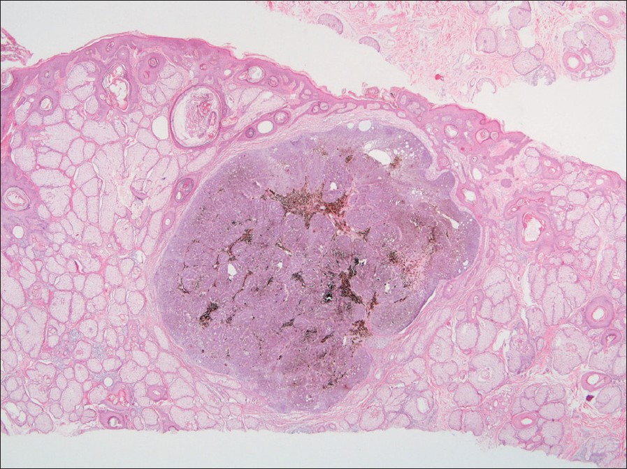
Figure 2: Epidermal papillomatosis and hyperplastic sebaceous glands in the superficial dermis with reduced follicular structures were observed. Besides, a well-demarcated tumor nodule was in the dermis without obvious epithelial stromal retraction cleft (H and E, ×20)
Figure 3: Basaloid cells with elongated cytologically bland nuclei arranged in parallel rows of epithelial ribbons, and abundant melanin pigment was also observed. No high-grade cytologic atypia or mitoses were seen (H and E, ×200)
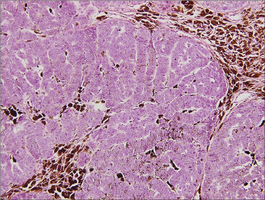
Figure 4: Immunohistochemistry (a) HMB45 (×400): scattered dendritic melanocytes within the tumor nodule (b) EMA (×200): multiple vacuolated positive sebocytes in the sebaceous glands of nevus sebaceus. No sebaceous differentiation in the tumor

TB is a well-circumscribed nodule composed of follicular germinative cells arranged in variably sized nests located in the dermis, sometimes extending to the subcutaneous tissue. In the recent study, the most basaloid neoplasms arising in NS previously diagnosed of basal cell carcinoma have been considered as TB. [2] Pigmented TB is a rare pathological presentation. The pathologic features are not only the presence of basaloid tumor cells arranged in the nodular pattern but also large amounts of melanin deposition within and around the tumor lobules.

Melanotrichoblastoma is a rare subtype of pigmented TB. Besides the deposition of heavy melanin, it is characterized by the presence of abundant dendritic melanocytes within tumor nests [Figure - 4]a. In the literature, only two cases have been reported, but none of them were related to NS. [3],[4] In 1992, Kanitakis et al. [3] presented the first case of a 32-year-old woman with a heavily pigmented nodule on the scalp. The histopathologic findings are compatible to pigmented TB with a well-circumscribed tumor consisting of nodules with uniform basaloid tumor cells, some of them present as the reminiscent of hair follicle germs, and heavy melanin deposition. Besides, the proliferation of dendritic melanocytes were dispersed in the basaloid tumor cells. Due to the resemblance to melanoacanthoma composed of the proliferation of melanocytes and keratinocytes, the author called the tumor melanotrichoblastoma. In 2011, Kim et al. [4] reported the second case of a 51-year-old male with a giant melanotrichoblastoma, 6 cm ΄ 4.3 cm ΄ 3 cm in size, on his back. In our present case, a dome-shaped blue-black nodule, 1 cm in diameter, arose from a yellowish, verrucous, hairless plaque on the left temporal scalp. The histopathologic findings are a smooth-bordered tumor with basaloid cells arranged in the ripple pattern, lots of melanin deposition, and the proliferation of dendritic melanocytes in the background of NS. It is the first reported case of ripple-pattern melanotrichoblastoma arising from a pre-existent NS.

Differential diagnosis would include basal cell carcinoma (BCC); however, the histological findings of BCC within NS include aggregations of basaloid tumor cells that vary markedly in size and shape, limited fibrous stroma, retraction artifact separating tumor and surrounding stroma, cytologic atypia, aberrant mitoses, and the lack of follicular differentiation. [2] In our case, we observed a well-circumscribed dermal tumor nodule without connection to the overlying epidermis and no obvious mitoses or retraction artifact was found [Figure - 2] and [Figure - 3]. Sebaceoma is a benign sebaceous neoplasm, which is composed of basaloid, sebaceous germinative cells, and/or ductal structures. It may associate with nevus sebaceus, [1] and a ripple pattern may be one of the pathologic features. [5] In the present case, no sebaceous differentiation was observed. We further confirmed by the negative immunohistochemical staining of EMA [Figure - 4]b.

References
1.
Cribier B, Scrivener Y, Grosshans E. Tumors arising in nevus sebaceus: A study of 596 cases. J Am Acad Dermatol 2000;42:263-8.
[Google Scholar]
2.
Kaddu S, Schaeppi H, Kerl H, Soyer HP. Basaloid neoplasms in nevus sebaceus. J Cutan Pathol 2000;27:327-37.
[Google Scholar]
3.
Kanitakis J, Brutzkus A, Butnaru AC, Claudy A. Melanotrichoblastoma: Immunohistochemical study of a variant of pigmented trichoblastoma. Am J Dermatopathol 2002;24:498-501.
[Google Scholar]
4.
Kim DW, Lee JH, Kim I. Giant melanotrichoblastoma. Am J Dermatopathol 2011;33:e37-40.
[Google Scholar]
5.
Ansai S, Kimura T. Rippled-pattern sebaceoma: A clinicopathological study. Am J Dermatopathol 2009;31:364-6.
[Google Scholar]

Fulltext Views
1,779

PDF downloads
1,548
Show Sections