Generic selectors
Exact matches only
Search in title
Search in content
Post Type Selectors
Search in posts
Search in pages
Filter by Categories
15th National Conference of the IAOMFP, Chennai, 2006
Abstract
Abstracts from current literature
Acne in India: Guidelines for management - IAA Consensus Document
Addendum
Announcement
Art & Psychiatry
Article
Articles
Association Activities
Association Notes
Award Article
Book Review
Brief Report
Case Analysis
Case Letter
Case Letters
Case Notes
Case Report
Case Reports
Clinical and Laboratory Investigations
Clinical Article
Clinical Studies
Clinical Study
Commentary
Conference Oration
Conference Summary
Continuing Medical Education
Correspondence
Corrigendum
Cosmetic Dermatology
Cosmetology
Current Best Evidence
Current Issue
Current View
Derma Quest
Dermato Surgery
Dermatopathology
Dermatosurgery Specials
Dispensing Pearl
Do you know?
Drug Dialogues
e-IJDVL
Editor Speaks
Editorial
Editorial Remarks
Editorial Report
Editorial Report - 2007
Editorial report for 2004-2005
Errata
Erratum
Focus
Fourth All India Conference Programme
From Our Book Shelf
From the Desk of Chief Editor
General
Get Set for Net
Get set for the net
Guest Article
Guest Editorial
History
How I Manage?
IADVL Announcement
IADVL Announcements
IJDVL Awards
IJDVL AWARDS 2015
IJDVL Awards 2018
IJDVL Awards 2019
IJDVL Awards 2020
IJDVL International Awards 2018
Images in Clinical Practice
Images in Dermatology
In Memorium
Inaugural Address
Index
Knowledge From World Contemporaries
Leprosy Section
Letter in Response to Previous Publication
Letter to Editor
Letter to the Editor
Letter to the Editor - Case Letter
Letter to the Editor - Letter in Response to Published Article
LETTER TO THE EDITOR - LETTERS IN RESPONSE TO PUBLISHED ARTICLES
Letter to the Editor - Observation Letter
Letter to the Editor - Study Letter
Letter to the Editor - Therapy Letter
Letter to the Editor: Articles in Response to Previously Published Articles
Letters in Response to Previous Publication
Letters to the Editor
Letters to the Editor - Letter in Response to Previously Published Articles
Letters to the Editor: Case Letters
Letters to the Editor: Letters in Response to Previously Published Articles
Media and news
Medicolegal Window
Messages
Miscellaneous Letter
Musings
Net Case
Net case report
Net Image
Net Images
Net Letter
Net Quiz
Net Study
New Preparations
News
News & Views
Obituary
Observation Letter
Observation Letters
Oration
Original Article
ORIGINAL CONTRIBUTION
Original Contributions
Pattern of Skin Diseases
Pearls
Pediatric Dermatology
Pediatric Rounds
Perspective
Presedential Address
Presidential Address
Presidents Remarks
Quiz
Recommendations
Regret
Report
Report of chief editor
Report of Hon : Treasurer IADVL
Report of Hon. General Secretary IADVL
Research Methdology
Research Methodology
Resident page
Resident's Page
Resident’s Page
Residents' Corner
Residents' Corner
Residents' Page
Retraction
Review
Review Article
Review Articles
Reviewers 2022
Revision Corner
Self Assessment Programme
SEMINAR
Seminar: Chronic Arsenicosis in India
Seminar: HIV Infection
Short Communication
Short Communications
Short Report
Snippets
Special Article
Specialty Interface
Studies
Study Letter
Study Letters
Supplement-Photoprotection
Supplement-Psoriasis
Symposium - Contact Dermatitis
Symposium - Lasers
Symposium - Pediatric Dermatoses
Symposium - Psoriasis
Symposium - Vesicobullous Disorders
SYMPOSIUM - VITILIGO
Symposium Aesthetic Surgery
Symposium Dermatopathology
Symposium-Hair Disorders
Symposium-Nails Part I
Symposium-Nails-Part II
Systematic Review and Meta-Analysis
Systematic Reviews and Meta-analyses
Systematic Reviews and Meta-analysis
Tables
Technology
Therapeutic Guideline-IADVL
Therapeutic Guidelines
Therapeutic Guidelines - IADVL
Therapeutics
Therapy
Therapy Letter
Therapy Letters
View Point
Viewpoint
What’s new in Dermatology
View/Download PDF

Translate this page into:

Observation Letter
88 (
4
); 541-543
doi:
10.25259/IJDVL_1039_2021
pmid:
35593288

Solitary purpuric plaque in a four-year-old girl: Histopathological diagnostic challenge

Department of Dermatology, TOBB ETU Faculty of Medicine, Yasam cad. No. 5, Baskent University, Ankara, Turkey
Department of Pathology, Reconstructive and Aesthetic Surgery, Baskent University Faculty of Medicine, Baskent University, Ankara, Turkey
Department of Plastic, Reconstructive and Aesthetic Surgery, Baskent University Faculty of Medicine, Baskent University, Ankara, Turkey

Corresponding author: Associate Professor, Pinar Incel Uysal, Department of Dermatology, TOBB ETU Faculty of Medicine, Yasam cad. No. 5, Ankara 06560, Turkey. pinarincel@hotmail.com

Licence
This is an open-access article distributed under the terms of the Creative Commons Attribution-Non Commercial-Share Alike 4.0 License, which allows others to remix, transform, and build upon the work non-commercially, as long as the author is credited and the new creations are licensed under the identical terms.

How to cite this article: Uysal PI, Ayva ES, Tepeoglu M, Uysal AC. Solitary purpuric plaque in a four-year-old girl: Histopathological diagnostic challenge. Indian J Dermatol Venereol Leprol 2022;88:541-3.

Sir,

Lichen aureus with lymphocyte atypia and monoclonal rearrangement is considered as a mimicker of mycosis fungoides. Both disorders; lichen aureus and mycosis fungoides, are rarely recognized in childhood. Here, we describe a healthy girl with a progressive red-brown plaque posing a histopathological challenge.

A four-year-old girl presented with three-week history of asymptomatic eruption on her left axilla. She was otherwise healthy and on no systemic medication. She was participating in a gymnastic class for a few months. The patient had no previous history of infection or exposure to contact allergen. Dermatological examination revealed a solitary reddish plaque with sharp demarcation on her left axilla [Figure 1]. There was no lymphadenopathy or hepatosplenomegaly. Laboratory parameters were within normal limits.
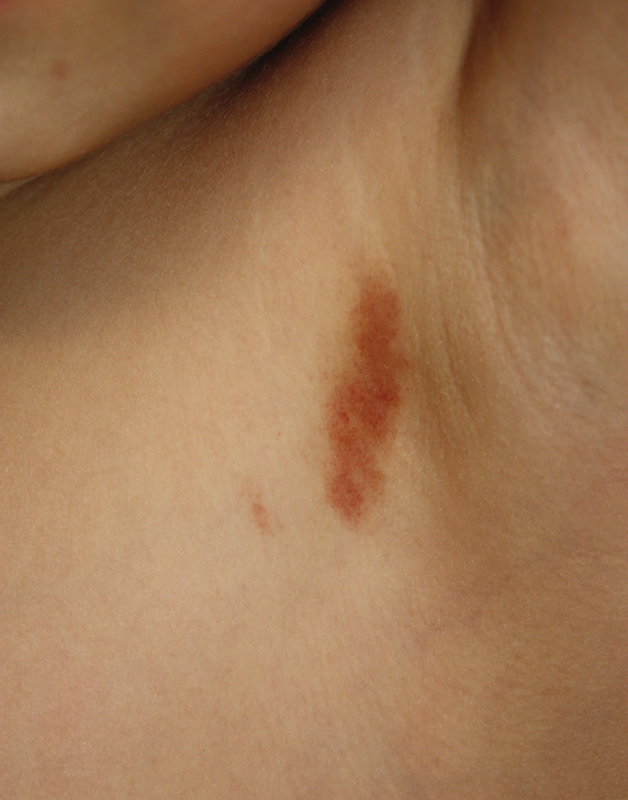
The plaque in left axilla
Figure 1
The plaque in left axilla

Histopathological evaluation of the plaque revealed a moderately dense upper dermal lymphoid infiltrate with intraepidermal lymphocytes tagging the dermo-epidermal junction [Figures 2a and b]. A few intraepidermal lymphocytes were observed. The lymphocytes showed variable nuclear pleomorphism, nuclear membrane irregularity and hyperchromasia with haloes around the nuclei. There were a few clusters of atypical lymphocytes with minimal spongiosis [Figure 2c]. Immunohistochemically, the lymphocytes expressed pan-T-cell markers, CD2, CD3, CD5 and CD7. A substantial reduction in CD7 expression was also observed [Figures 3a and b]. CD8+ phenotype was more prominent than CD4+ phenotype in the lymphoid population [Figures 3c and d]. Monoclonality was detected in the T-cell receptor gene rearrangement study. She was also consulted by a paediatric haematologist. Further detailed clinical and radiological evaluation revealed no abnormality. The patient was treated with local corticosteroids with substantial improvement.
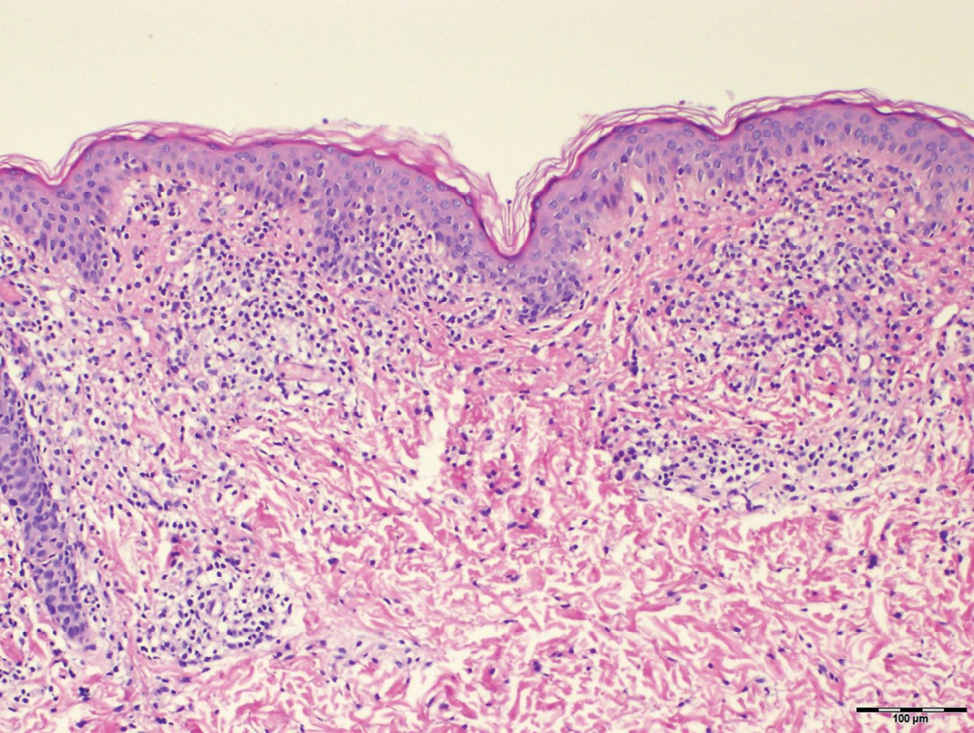
Band-like dermal lymphoid infiltration (hematoxylin and eosin, ×40)
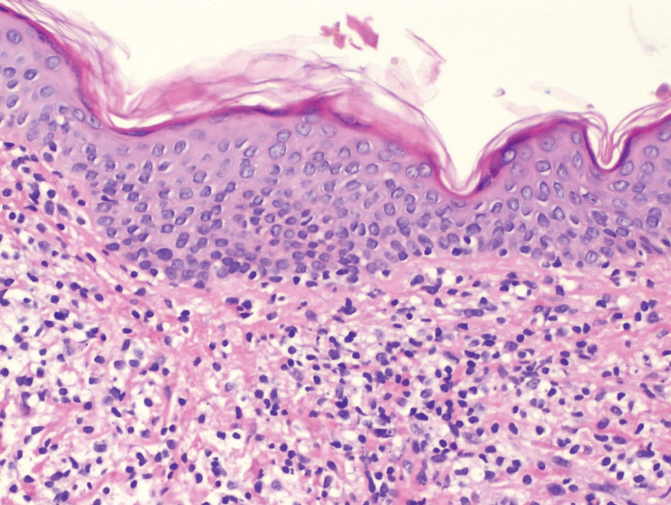
Figure 2 a:
Band-like dermal lymphoid infiltration (hematoxylin and eosin, ×40)
Intraepidermal lymphocytes tagging at the dermo-epidermal junction (hematoxylin and eosin, ×100)
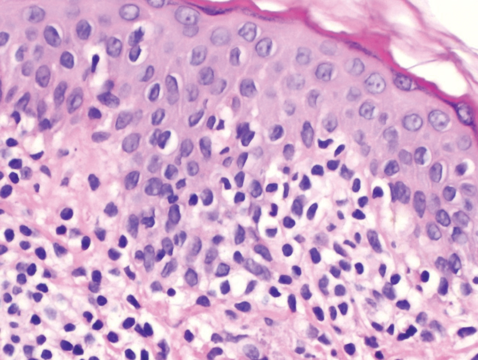
Figure 2b:
Intraepidermal lymphocytes tagging at the dermo-epidermal junction (hematoxylin and eosin, ×100)
Intraepidermal lymphocytes showing variable nuclear pleomorphism with perinuclear haloes (hematoxylin and eosin, ×200)
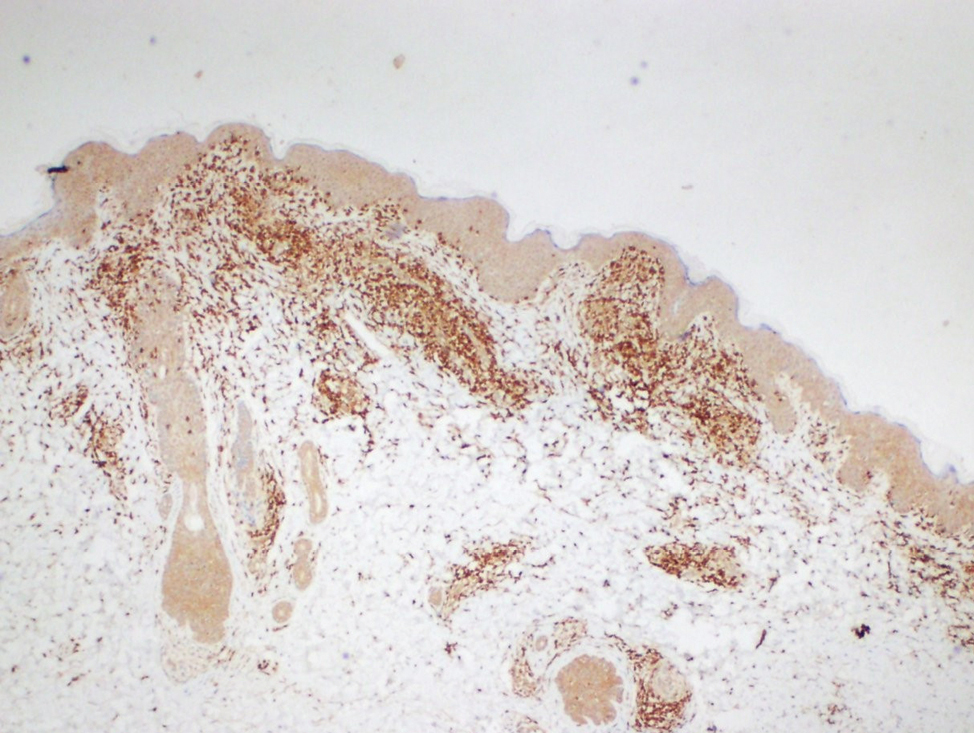
Figure 2c:
Intraepidermal lymphocytes showing variable nuclear pleomorphism with perinuclear haloes (hematoxylin and eosin, ×200)
The lymphocytes immunohistochemically express CD3 which is pan T marker (Diaminobenzidine, ×40)
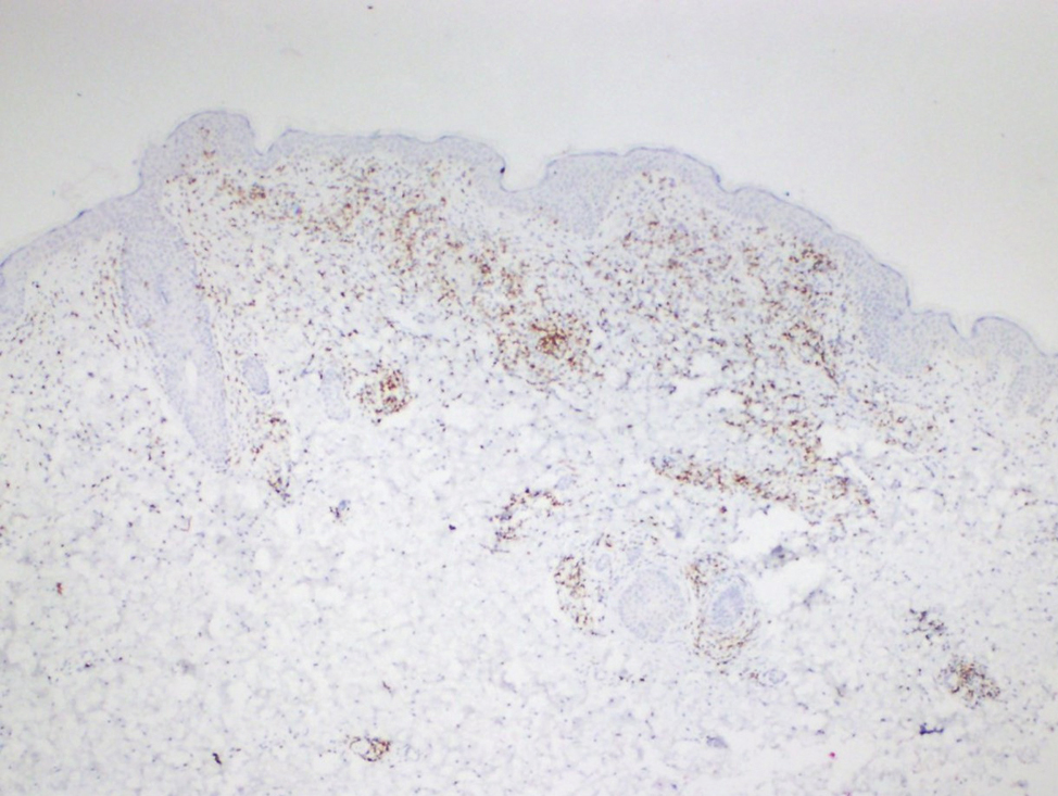
Figure 3a:
The lymphocytes immunohistochemically express CD3 which is pan T marker (Diaminobenzidine, ×40)
Focal loss of CD7 expression (Diaminobenzidine, ×40)
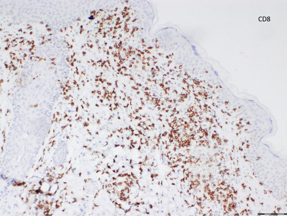
Figure 3b:
Focal loss of CD7 expression (Diaminobenzidine, ×40)
The lymphocytes more profoundly express CD8 expression than CD4 expression (Diaminobenzidine, ×100)
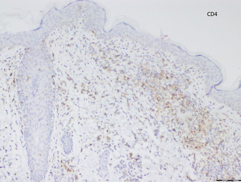
Figure 3c:
The lymphocytes more profoundly express CD8 expression than CD4 expression (Diaminobenzidine, ×100)
Figure 3d:
The lymphocytes more profoundly express CD8 expression than CD4 expression (Diaminobenzidine, ×100)

Lichen aureus and unilesional mycosis fungoides were the main differential diagnoses in our patient. Because of considerable overlap, it is sometimes difficult to differentiate between pigmented purpuric dermatosis (PPD) and PPD-like mycosis fungoides clinically and histologically. It is well recognized that both disorders are much less common in children. Considering the progression of the lesion in a short time, a biopsy was performed on our patient. The presence of atypical lymphocytic infiltrates, microscopic evidence of diminished CD7 expression and clonal rearrangement in T-cell receptor gene arrangement study pointed to the probability of mycosis fungoides.

For many years the linkage between pigmented purpuric dermatosis and mycosis fungoides has been examined in three aspects.1 First, histologically pigmented purpuric dermatosis can mimic mycosis fungoides.2 Among its subtypes, lichen aureus is likely to be the strongest mimicker.3 Currently, pigmented purpuric dermatosis is phenotypically categorized into two types as polyclonal and monoclonal. Besides monoclonality, CD4+ mature T-cell dominance and loss of CD7 expression in pigmented purpuric dermatoses have been reported in the phenotypic and molecular studies.1,4,5 The presence of large cerebriform lymphocytes and Pautrier microabscesses, elevated CD4:CD8 (≥6) ratio and CD4 predominance is generally considered indicators of malignancy. Even so the absence of these findings does not exclude the diagnosis of mycosis fungoides. In our patient CD8 dominance was noted with CD7 loss. Fifteen per cent of mycosis fungoides show a reverse pattern characterized by CD8 dominance on microscopic examination.6 The lesion of our patient was very similar to the patient recently reported by Zubkov et al.7

Secondly, mycosis fungoides can present as PPD-like lesions.8 Autoimmune pathways are likely to be causative in mycosis fungoides related purpura.9 In a large series of patients in whom initial diagnosis was pigmented purpuric dermatosis, mycosis fungoides and suspected mycosis fungoides were detected in 8% (7/87) of patients.10 Notably, among six paediatric patients presenting with PPD-like lesions, one of them was diagnosed as mycosis fungoides. Magro et al. suggest performing T-cell rearrangement analyses in these patients with involvement of large surface areas who are more likely to exhibit monoclonality and loss of CD7 expression.4 Because of presence of CD7 loss, we performed T-cell receptor gene rearrangement study in our patient.

Lastly, mycosis fungoides may develop on preceding pigmented purpuric dermatosis.11,12 According to the literature lichen aureus should be regarded as ‘cutaneous T cell lymphoid dyscrasia’ with potential for progression to cutaneous T-cell lymphoma.4 In the study of Caytemel et al. 2 (1.5%) patients were evolved into mycosis fungoides.10 It is well documented that, only pigmented purpuric dermatosis with T-cell clonotype may progress to mycosis fungoides.4 On the contrary, some authors have proposed that classic lesions of lichen aureus do not progress to cutaneous T-cell lymphoma.13

In conclusion, our patient extraordinarily reflects the fact that the histopathological distinction of lichen aureus and mycosis fungoides can be blurred. Clinically solitary purpuric plaque with partial progression exhibiting exocytosis and nuclear atypia accompanied by CD7 loss, clonal proliferation and lack of characteristic microscopic findings of lichen aureus were strong arguments against the lichen aureus. However, as some authors, we believe that monoclonal lichen aureus belongs to the intermediate stage in the cutaneous T-cell dyscrasia spectrum. A long-term follow-up and serial biopsies are recommended. The question regarding categorizing these patients needs still to be addressed.

Declaration of patient consent

Patient’s consent not required as patient’s identity is not disclosed or compromised.

Financial support and sponsorship

Nil.

Conflict of interest

There are no conflicts of interest.

References

  1. , , . Persistent pigmented purpuric dermatitis and mycosis fungoides: simulant, precursor, or both? A study by light microscopy and molecular methods. Am J Dermatopathol. 1997;19:108.:18.
    [CrossRef] [PubMed] [Google Scholar]
  2. , , , , , , et al. Mimics of cutaneous lymphoma: report of the 2011 Society for Haematopathology/European Association for Haematopathology workshop. Am J Clin Pathol. 2013;139:536.:51.
    [CrossRef] [PubMed] [Google Scholar]
  3. , , , , , . Lichen aureus with pseudolymphomatous infiltrate. J Cutan Pathol. 2021;48:669.:73.
    [CrossRef] [PubMed] [Google Scholar]
  4. , , , , . Pigmented purpuric dermatosis: classification by phenotypic and molecular profiles. Am J Clin Pathol. 2007;128:218.:29.
    [CrossRef] [PubMed] [Google Scholar]
  5. , , , , . Pigmented purpuric dermatosis or mycosis fungoides: A diagnostic dilemma. Indian Dermatol Online J. 2016;7:183.:5.
    [CrossRef] [PubMed] [PubMed Central] [Google Scholar]
  6. , , , , , , et al. Mycosis fungoides: A clinicopathological study of 60 cases from a Tertiary Care Center. Indian J Dermatol. 2020;65:123.:9.
    [CrossRef] [PubMed] [PubMed Central] [Google Scholar]
  7. , , , . Unilesional granulomatous pigmented purpuric dermatosis in a 7-year-old boy. Pediatr Dermatol. 2021;38:506.:7.
    [CrossRef] [PubMed] [Google Scholar]
  8. , , , . Mycosis fungoides presenting as pigmented purpuric dermatitis. Pediatr Dermatol. 2006;23:350.:4.
    [CrossRef] [PubMed] [Google Scholar]
  9. , , , , . Pigmented purpura-like eruption as cutaneous sign of mycosis fungoides with autoimmune purpura. J Am Acad Dermatol. 1999;40:298.:9.
    [CrossRef] [PubMed] [Google Scholar]
  10. , , , , , , et al. Pigmented purpuric dermatosis: Ten years of experience in a tertiary hospital and awareness of mycosis fungoides in differential diagnosis. J Cutan Pathol. 2021;48:611.:6.
    [CrossRef] [PubMed] [Google Scholar]
  11. , , , , . Persistent pigmented purpuric eruption associated with mycosis fungoides: A case report and review of the literature. J Eur Acad Dermatol Venereol. 2001;15:62.:4.
    [CrossRef] [PubMed] [Google Scholar]
  12. , . Progression of pigmented purpura-like eruptions to mycosis fungoides: Report of three cases. J Am Acad Dermatol. 1988;19:25.:31.
    [CrossRef] [PubMed] [Google Scholar]
  13. , , , . Lichen aureus: clinicopathologic features, natural history, and relationship to mycosis fungoides. Arch Dermatol. 2008;144:1169.:73.
    [CrossRef] [PubMed] [Google Scholar]

Fulltext Views
5,462

PDF downloads
2,866
View/Download PDF
Download Citations
BibTeX
RIS
Show Sections