Translate this page into:
Sparing phenomena in dermatology
Correspondence Address:
Jaheersha Pakran
Department of Dermatology, Malabar Institute of Medical Sciences Hospital, Govindapuram P.O. Calicut-673 016, Kerala
India
| How to cite this article: Pakran J. Sparing phenomena in dermatology. Indian J Dermatol Venereol Leprol 2013;79:545-550 |
Introduction
Dermatology being a visual science is adorned with named "signs" and "phenomena." Of much interest today is the sparing phenomena that has accumulated in dermatology over the years. Some of them are anecdotal single case reports, while others have diagnostic or therapeutic significance. A literature search for such "sparing" reports was done by the author and they are listed below in an alphabetical order along with their possible explanations and significance. The author also attempts to untangle the confusion that has risen in connection with these signs.
Alopecia Areata (AA) Sparing Lesional Gray/White Hairs
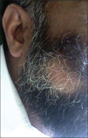
White or graying hair follicles are commonly spared in AA, and regrowing hair shafts are usually white before they become repigmented. A possible explanation can be seen in the hypothesis put forward by Paus et al., [1] They proposed that AA is an autoimmune disorder with two successive waves of inflammatory response against melanogenesis-related proteins generated on hair matrix melanocytes and/or keratinocytes. [1] This results in the loss of only pigmented hairs in the region, with sparing of the inactive melanocytes in white hairs [Figure - 1].
 |
| Figure 1: Alopecia areata of the beard region sparing white hairs |
Anatopic Phenomenon
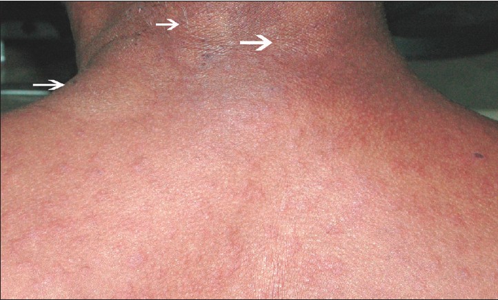
The term is used to describe the modulation of inflammatory response of one dermatoses by another unrelated cutaneous infection at the same site [Figure - 2]. [2] This differs from the reverse isotopic phenomenon in that both the diseases are present simultaneously and one is not healed at the presentation of the other.
 |
| Figure 2: Anatopic phenomenon-Drug hypersensitivity rash to phenytoin sparing tinea versicolor sites (Photo Courtesy: Dr. George Kurien M.D) |
The author has described four cases in which unrelated dermatoses (viral exanthem, acute generalized exanthematous pustulosis, polymorphous light eruption, and irritant contact dermatitis) spared sites affected with tinea versicolor. The authors speculate that the presence of Malassezia spp. may be responsible for this, either by providing a physical barrier or by means of local immunomodulation. A similar case of dithranol-induced erythema sparing tinea versicolor-affected skin was previously reported by Shenoy et al., [3] Two other reports that may also be considered under anatopic phenomena, include the sparing of leprosy patches by ampicillin hypersensitivity rash [4] and dapsone hypersensitivity syndrome. [5] The author proposes that nerve damage in leprosy may affect the microvasculature and local immune response resulting in the non-expression of hypersensitivity reaction. [4]
Atopic Dermatitis Generally Spare the Napkin Area
Despite the fact that atopic individuals are more susceptible to irritant reactions, atopic dermatitis usually spares the napkin area. [6] This helps to differentiate it from infantile seborrheic dermatitis.
Butterfly Sign
This sign describes the butterfly shaped area of sparing observed over the upper central back, corresponding to the zone that is difficult to reach by hands, in conditions with severe generalized pruritus such as primary biliary cirrhosis and atopic dermatitis. [7] The authors infer that this supports the role of scratching in the development of skin lesions of atopic dermatitis.
Deck Chair Sign
The term refers to the distinctive pattern of sparing of the natural skin folds, resembling the slats of a deck chair originally described in papuloerythroderma of Ofuji. But the sign is not specific and has also been reported in Waldenstrom′s macroglobulinemia, adult T-cell leukemia/lymphoma, angioimmunoblastic T-cell lymphoma, drug-induced erythroderma, and mycosis fungoides. [8]
Ear Lobe Sign
The ear-lobe sign is seen in those who develop contact dermatitis to a substance applied with one hand to the skin of the face and neck. On the ipsilateral side of the face, the ear-lobe is spared, whereas, on the contra-lateral side, the ear-lobe is involved. The sign manifests because of the unique combination of the anatomy of the hand and the sweeping movement the hand makes during application. [9]
Epidermal Growth Factor Receptor (EGFR) Inhibitor-Associated Rash Spares Irradiated Areas
There are reports of cancer patients who on treatment with recombinant monoclonal antibody against human EGFR, such as cetuximab and erlotinib, develop skin rashes that spare the previously irradiated areas. [10],[11] It is believed that the development of skin toxicity, which is a major adverse effect of EGFR inhibitors, is due to the inhibition of homodimeric EGFR on normal skin cell surface. The authors propose that this sparing may be either due to decrease in the population of EGFR expressing cells or decrease in the EGFR expression in the irradiated area. This has implications in treatment of recurrent cancers due to predicted unresponsiveness to chemotherapy with EGFR inhibitors.
Ichthyosis Vulgaris Spares Flexures
Ichthyosis vulgaris is characterized by scales over the extensor aspect of limbs and trunk, with characteristic sparing of the flexures. In X-linked recessive ichthyosis, large dirty brown scales cover the extensor and flexor aspects of limbs, but spare the rhomboidal spaces of body folds. The flexural sparing is a useful pointer to differentiate from other types of inherited ichthyosis, where flexures are either involved or predominantly affected. [12]
Irritant Diaper Dermatitis Spares Flexures
The classic description of a primary irritant contact dermatitis is that of a confluent erythema of the convex surfaces in close contact with the diaper, i.e., the buttocks, the genitalia, the lower abdomen, the pubic area, and the upper thighs, with sparing of the deeper parts of the groin flexures. This sparing helps to differentiate it from candidiasis, inverse psoriasis, and intertrigo, where the flexures are predominantly involved. [13]
Islands Of Sparing in Pityriasis Rubra Pilaris (PRP)
PRP is a chronic papulo-squamous disorder of unknown etiology, often characterized by orange-red plaques with keratotic follicular plugging, scalp scaling, and palmoplantar keratoderma. In most cases, sharply demarcated islands of unaffected skin remains a helpful diagnostic sign in PRP. [14]
Isomorphic Non Response/Reverse Koebner Phenomenon
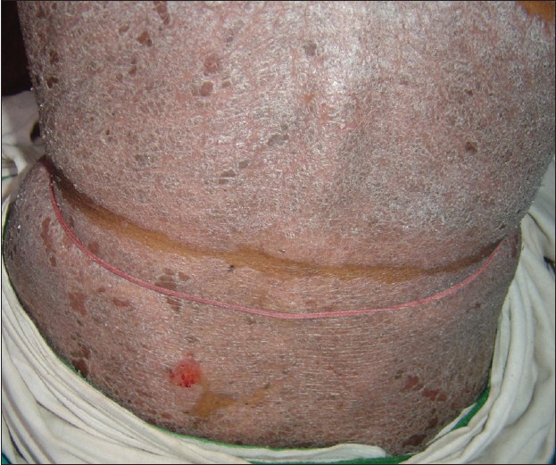
The reverse Koebner response is the nonappearance or disappearance of the lesions of particular dermatoses at the site of injury [Figure - 3]. [15] The exact etiopathogenesis of reverse Koebner phenomenon is poorly understood. It is classically described in psoriasis and has followed trauma by electrodessication, sandpaper abrasion, and surgery. [16] The reason for sparing could be destruction of the dermal papillae within the injured tissue that prevent the epidermal hyperplasia of psoriasis. There are also case reports of this phenomenon occurring in leucocytoclastic vasculitis [17] and vitiligo. [18] In vitiligo "remote reverse Koebner" phenomenon has been reported by Malaker and Dhar, [18] where there was spontaneous repigmentation of the vitiligo patches distant from those treated by autologous punch grafts. The authors hypothesize that fresh cytokines from the donor skin may stimulate the melanocytes at the distant sites after local absorption.
 |
| Figure 3: Reverse Koebner phenomenon-Erythrodermic psoriasis sparing the pressure sites due to chronic use of tight waist band (Photo Courtesy: Dr. Rakhesh SV M.D) |
Isotopic Non-Response
The term "isotopic non-response," was coined by Wolf et al., [19] to describe the absence of an eruption at the site of another unrelated and already healed skin disease. Examples for this response are the case reports of Stevens-Johnson syndrome, erythema multiforme, and erythroderma sparing healed herpes zoster site. [19],[20],[21]
The authors speculate that this may be related to the immune factors and/or to drug metabolism.
A case of generalized contact dermatitis whose lesions spared papules and peri-lesional skin of recent insect bites has been reported under the name of "reverse isotopic phenomenon" by Mansur et al., [22] Though reported under the terminology "reverse isotopic," this case seems to better fit with renbok phenomenon (RP), since both the diseases were active simultaneously and one was not already healed at the presentation of the other. Another case that defies classification to one single group is the patient with cutaneous T-cell lymphoma (CTCL), who developed herpes zoster involving the left T8 dermatome. When his CTCL later became widespread, the previously affected areas of herpes zoster were clinically free of lymphoma. The authors proved the loss of Langerhans cells (LC) in the spared skin using immunohistochemistry, thus pointing towards a role of LC in the epidermotropism in CTCL. [23]
Knuckle Sparing of Acute Cutaneous Lupus Erythematosus (ACLE)
When LE affects the skin of the dorsal hands, erythema and scaling typically spare the knuckles. This is in contrast with dermatomyositis, where the violaceous erythematous papules typically involves the knuckles. [24]
Milian′s Ear Sign
The sign describes the difference between erysipelas and cellulitis of the facial region, where there is involvement of the ear in erysipelas and sparing in cellulitis. This is due to the absence of deeper dermal tissue and subcutaneous fat in the region. [25]
Nasolabial Fold Sparing in the Malar Rash of Systemic Lupus Erythematosus
The "malar rash" of SLE is characterized by well-demarcated erythema and edema over malar areas, connected over the bridge of the nose. [26] It classically spares the nasolabial folds and peri-orbital area in contrast with other facial eruptions like erysipelas and seborrheic dermatitis.
Nose Sign
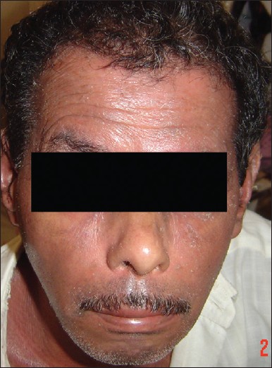
Sparing of the nose and the paranasal area has been described in exfoliative dermatitis [Figure - 4], [27] air-borne contact dermatitis, polymorphic light eruption, severe atopic dermatitis, and PRP. [28] The cause is speculated to be the inability of physical, chemical, or air-borne allergens to lodge at this site, either due to the anatomic peculiarity of nose or from frequent scratching. Another reason cited is the relative vascular insufficiency that prevents the circulating antigens from reaching the skin.
 |
| Figure 4: Nose sign-Erythroderma in a patient sparing the nose and para nasal areas (Photo Courtesy: Dr. Rakhesh SV M.D) |
Paretic Limbs Spared in Rheumatic Diseases
Paretic limb sparing in hemiplegic patients who develop rheumatic diseases such as scleroderma has been reported. The authors propose that sparing could be due to modification of inflammation by the neurological disease perhaps through neuropeptides. [29]
Pityriasis Rosea Spare Sun-Exposed Areas
Costello [30] had observed a number of cases of pityriasis rosea confined to the "bathing trunk" region and other covered areas of the body. The author notes the protective effect of sunlight in preventing certain diseases of the skin.
Pruritic Urticarial Papules and Plaques of Pregnancy (Puppp) Spares Periumbilical Area
Among the pregnancy dermatoses, PUPPP lesions and urticarial pemphigus gestationis (PG) lesions are difficult to differentiate in the early stages. A helpful pointer is that, while PG lesions cluster around the umbilicus, PUPPP lesions uniformly spare the umbilical area. [31]
Psoriasis Often Spare Central Face
The center of the face is relatively spared in psoriasis. Bernhard [32] speculated that ambient UV radiation or some component of sebum may be responsible for this anti-psoriatic activity.
Relapsing Polychondritis Spare Earlobes
Relapsing polychondritis is a rare disease most commonly presenting as inflammation of the cartilage of the ears and nose. Auricular chondritis, with red ears resembling infectious cellulitis, is the most common initial finding. But earlobes are classically spared due to absence of cartilage. [33]
Renbok Phenomenon
The RP was first reported by Happle et al. in 1991 who described four patients with extensive scalp AA showing surprising hair growth within plaques of sebopsoriasis. This phenomenon demonstrates how two distinct autoimmune diseases, psoriasis, and AA interact to result in clinically active psoriasis suppressing AA. It is presumed that the T-helper 17 psoriatic inflammation antagonizes the T-helper 1 AA inflammatory response, presumably through cytokines. [34] This phenomenon was later also found in two patients with genetic mosaicism, where AA spared a nevus flammeus in one case and a congenital nevus in the other. Other case reports that seems to fit in the RP are diltiazem-induced morbilliform drug eruption that spared seborrheic keratoses, [35] widespread maculopapular drug reaction to phenobarbital that spared nevus depigmentosus, [36] guttate psoriasis sparing Becker′s melanosis, [37] and widespread drug reaction that spared psoriatic plaques. [38]
Rosacea Spares Circum-Oral and Peri-Orbital Areas
In rosacea, the circum-oral and peri-orbital areas are typically spared, [26] in contrast with other facial dermatosis like seborrheic dermatitis.
Scrotum Sparing in Tinea Cruris
Tinea cruris forms erythematous, pruritic patches in the groin area that usually spares the scrotum and penis. This helps to differentiate it from candidiasis of the groin that usually involve the scrotum. [39]
Spared Zones in Leprosy
Certain anatomical sites like scalp, groin, axillae, perineum, and a transverse strip of skin over lumbosacral region is considered relatively insusceptible to the development of leprosy. [40] The in-susceptibility is attributed to the relative warmth of these regions, which is unfavorable for Mycobacterium leprae growth, which requires an optimum temperature of 27-30°C.
Sparing Associated with Generalized Rash and Fever
In viral exanthems, there may an accompanying enanthem, whereas, in morbilliform drug rash, oral mucosa is usually spared. In Gianotti-Crosti syndrome, the exanthem spare the trunk in most cases [15] and thus is also called "infantile papular acrodermatitis." In the rash of erythema infectiosum, the blotchy warm edematous plaques that affect the cheeks to form a slapped-cheek appearance, typically spares the nasolabial folds and circum-oral area. [41] The diffuse maculopapular eruption of roseola infantum often spares the face. [42] Scarlet fever is characterized by a generalized rash, but the palms and soles are characteristically spared. In contrast, drug-induced exanthems often spares face but palms and soles may be involved. [43] In staphylococcal scalded-skin syndrome, the rash is usually diffuse and can present as bullous impetigo, scarlatiniform lesions, or diffuse erythema, but the mucous membranes are spared in most patients. [42]
Sunbed Suntan Sacroscapular Sparing Sign
Rowland Payne [44] described this sparing sign in connection with the use of sunbeds. The patients are usually young females having a distinct type of uniform tan on body that spares a palm sized area over sacrum and smaller symmetrical areas over the scapular regions on back. The reason is said to be due to skin blanching by vitropression, while lying on transparent tanning bed surface. Since UVA-induced delayed pigmentation is an oxygen-dependent process, the pressure sites are spared of the tan.
Upper Axillary Area Spared in Pityriasis Versicolor
This is a single case report. Physiological, anatomical, and microbiological factors present in the axillae have been suggested as possible explanations for the sparing. [45]
White Islands in A Sea of Red in Dengue Fever
In cases of dengue fever, individual lesions may coalesce and form generalized confluent erythema and petechiae with characteristic rounded islands of sparing. [46] It is believed to be due to an immune response to the virus. This feature has got importance in the differential diagnosis of viral exanthems.
Vermillion Border Spared in Peri-Oral Dermatitis
Perioral dermatitis is characterized by a papular or papulo-vesicular erythematous skin eruption usually involving the nasolabial folds, upper lip, and the chin, with a clear zone around the vermilion border of the lips. [47] The reason is unknown.
Vermilion Border Spared in an Acrodermatitis Enteropathica (AE)-Like Syndrome
An AE-like syndrome occurred in two nutritionally compromised patients with prolonged catabolic illnesses, [48] in which the vermilion borders of lips were spared. The authors propose that hypozincemia should be considered in the differential diagnosis of widespread AE like rash with vermillion border sparing.
Wilkinson′s Triangle
Photo allergic contact dermatitis spares the skin behind earlobe since it is a photo-protected site. The finding was first described by Wilkinson during the epidemic of photo sensitivity reaction to halogenated salicylinides containing soaps during the 1970′s. He noticed that the eczematous rash affecting the face of photosensitive individuals spared the small area behind the ears, and this area is now known as the Wilkinson′s triangle. [49]
Miscellaneous Single Case Reports of Unknown Significance
A case of oral mucosal pigmentation secondary to treatment with mepacrine sparing the denture bearing area, [50] focal skin toxicity due to methotrexate sparing psoriatic plaques, [51] and sparing of the thumb in Raynaud′s phenomenon [52] are some miscellaneous sparing reports.
Summary
Despite considerable overlap, the confusion hovering over the four common sparing signs can be cleared as follows: Reverse Koebner relates to sparing by physical trauma; isotopic non-response/reverse isotopic response relates to sparing over an already healed dermatosis; Renbok Phenomenon relates to sparing among two concomitant active diseases often with autoimmune etiology; anatopic phenomenon relates to sparing caused by presence of certain infectious diseases of skin.
In conclusion, sparing phenomenon of dermatology seems to command our attention by their sheer number, variety, and clinical implications. Therefore, it may be said that astute clinician is the one who will look, not only for the presence of lesions in the patient but also for the conspicuous absence thereof.
| 1. |
Paus R, Slominski A, Czarnetzki BM. Is alopecia areata an autoimmune-response against melanogenesis-related proteins, exposed by abnormal MHC class I expression in the anagen hair bulb? Yale J Biol Med 1993;66:541-4.
[Google Scholar]
|
| 2. |
Pakran J, Riyaz N. Interesting effect of Malassezia spp. infection on dermatoses of other origins. Int J Dermatol 2011;50:1518-21.
[Google Scholar]
|
| 3. |
Shenoy SD, Wali I, Srinivas CR. Dithranol-induced erythema sparing hypopigmented lesions of pityriasis versicolor. Contact Dermatitis 1993;28:253.
[Google Scholar]
|
| 4. |
Pavithran K. Sparing of leprosy macule in ampicillin hypersensitivity rash. Indian J Lepr 1987;59:309-12.
[Google Scholar]
|
| 5. |
Ng PP, Goh CL. Sparing of tuberculoid leprosy patch in a patient with dapsone hypersensitivity syndrome. J Am Acad Dermatol 1998;39(Pt 1):646-8.
[Google Scholar]
|
| 6. |
Lewis-Jones MS, Charman CR. Atopic dermatitis: Scoring severity and quality of life assessment. In: Harper J, Oranje AP, editors. Textbook of Pediatric Dermatology. 2 nd ed. New Jersey: Blackwell Publishing; 2006. p. 65-78.
[Google Scholar]
|
| 7. |
Kimura T, Miyazawa H. The "butterfly" sign in patients with atopic dermatitis: Evidence for role of scratching in the development of skin manifestations. J Am Acad Dermatol 1989;21(Pt 1):579-80.
[Google Scholar]
|
| 8. |
Bettoli V, Pizzigoni S, Borghi A, Virgili A. Ofuji papulo-erythroderma: A reappraisal of the deck-chair sign. Dermatology 2004;209:1-4.
[Google Scholar]
|
| 9. |
Rotstein E, Rotstein H. The ear-lobe sign: A helpful sign in facial contact dermatitis. Australas J Dermatol 1997;38:215-6.
[Google Scholar]
|
| 10. |
Kanakamedala MR, Packianathan S, Vijayakumar S. Lack of cetuximab induced skin toxicity in a previously irradiated field: Case report and review of the literature. Radiat Oncol 2010;5:38.
[Google Scholar]
|
| 11. |
Chiu HY, Cheng YP, Jee SH, Tsai TF. Erlotinib-induced generalized eczema craquelé and follicular rash sparing the previous radiation field. Clin Exp Dermatol 2012;37:922-4.
[Google Scholar]
|
| 12. |
Thappa DM, editor. Textbook of Dermatology Venereology and Leprology. 3 rd ed. Gurgaon: Elsevier; 2009.p 201-2.
[Google Scholar]
|
| 13. |
William DJ, Timothy GB, Dirk EM. Diaper (Napkin) Dermatitis. In: James WD, Berger TG, Elston DM, editors. Andrew's Diseases of the Skin: Clinical Dermatology. 10 th ed. Canada: WB Saunders; 2006. p. 80.
th ed. Canada: WB Saunders; 2006. p. 80.'>[Google Scholar]
|
| 14. |
Albert MR, Mackool BT. Pityriasis rubra pilaris. Int J Dermatol 1999;38:1-11.
[Google Scholar]
|
| 15. |
Weiss G, Shemer A, Trau H. The Koebner phenomenon: Review of the literature. J Eur Acad Dermatol Venereol 2002;16:241-8.
[Google Scholar]
|
| 16. |
Thappa DM. The isomorphic phenomenon of Koebner. Indian J Dermatol Venereol Leprol 2004;70:187-9.
[Google Scholar]
|
| 17. |
Yadav S, De D, Kanwar AJ. Reverse Koebner phenomenon in leukocytoclastic vasculitis. Indian J Dermatol 2011;56:598-9.
[Google Scholar]
|
| 18. |
Malakar S, Dhar S. Spontaneous repigmentation of vitiligo patches distant from the autologous skin graft sites: A remote reverse Koebner's phenomenon? Dermatology 1998;197:274.
[Google Scholar]
|
| 19. |
Wolf R, Wolf D, Ruocco E, Brunetti G, Ruocco V. Wolf's isotopic response. Clin Dermatol 2011;29:237-40.
[Google Scholar]
|
| 20. |
Tenea D. Carbamazepine-induced Stevens-Johnson syndrome sparing the skin previously affected by herpes zoster infection in a patient with systemic lupus erythematosus: A reverse isotopic phenomenon. Case Rep Dermatol 2010;2:140-5.
[Google Scholar]
|
| 21. |
Park H, Kang Y, Lee U. Erythema multiforme sparing regressing herpes zoster lesion: "Reverse Isotopic Phenomenon?" J Am Acad Dermatol 2008;58(Suppl 2):AB40.
[Google Scholar]
|
| 22. |
Mansur AT, Aydingöz IE. Reverse isotopic response: A rarely reported phenomenon. Int J Dermatol 2009;48:783-4.
[Google Scholar]
|
| 23. |
Twersky JM, Nordlund JJ. Cutaneous T-cell lymphoma sparing resolving dermatomal herpes zoster lesions: An unusual phenomenon and implications for pathophysiology. J Am Acad Dermatol 2004;51:123-6.
[Google Scholar]
|
| 24. |
Costner M, Sontheimer RD. Lupus Erythematosus. In: Wolff K, Goldsmith LA, Katz SI, Gilchrest BA, Paller AS, Leffell DJ, editors. Fitzpatrick's Dermatology In General Medicine. Vol. 2. 7 th ed. New York: Mcgrawhill; 2008. p. 1520.
th ed. New York: Mcgrawhill; 2008. p. 1520.'>[Google Scholar]
|
| 25. |
Madke B, Nayak C. Eponymous signs in dermatology. Indian Dermatol Online J 2012;3:159-65.
[Google Scholar]
|
| 26. |
Datta I, Casanas B, Vincent AL, Greene JN. The red face: Erysipelas versus, parvovirus B19, SLE, and rosacea. Asian Biomed 2009;3:681-8.
[Google Scholar]
|
| 27. |
Pavithran K. Nose sign of exfoliative dermatitis. Indian J Dermatol Venereol Leprol 1988;54:42.
[Google Scholar]
|
| 28. |
Agarwal S, Khullar R, Kalla G, Malhotra YK. Nose sign of exfoliative dermatitis: A possible mechanism. Arch Dermatol 1992;128:704.
[Google Scholar]
|
| 29. |
Sethi S, Sequeira W. Sparing effect of hemiplegia on scleroderma. Ann Rheum Dis 1990;49:999-1000.
[Google Scholar]
|
| 30. |
Costello MJ. Pityriasis rosea sparing tanned areas of the skin (bathing Trunk Distribution). Arch Dermatol 1938;38:75-6.
[Google Scholar]
|
| 31. |
Bremmer M, Driscoll MS, Colgan R. The skin disorders of pregnancy: A family physician's guide. J Fam Pract 2010;59:89-96.
[Google Scholar]
|
| 32. |
Bernhard JD. Facial sparing in psoriasis. Int J Dermatol 1983;22:291-2.
[Google Scholar]
|
| 33. |
Rapini RP, Warner NB. Relapsing polychondritis. Clin Dermatol 2006;24:482-5.
[Google Scholar]
|
| 34. |
Harris JE, Seykora JT, Lee RA. Renbok phenomenon and contact sensitization in a patient with alopecia universalis. Arch Dermatol 2010;146:422-5.
[Google Scholar]
|
| 35. |
Heymann WR. Diltiazem-induced drug eruption sparing seborrheic keratoses. Cutis 2000;66:129-30.
[Google Scholar]
|
| 36. |
Naik RP, Srinivas CR, Das PC. Generalized drug reaction sparing nevus depigmentosus. Arch Dermatol 1986;122:509-10.
[Google Scholar]
|
| 37. |
Weiss RM, Schulz EJ. Guttate psoriasis sparing Becker's melanosis-A case report. Dermatologica 1990;180:160-2.
[Google Scholar]
|
| 38. |
Krivanek J, Doyle J, Paver K, Kossard S. A case report on the psoriatic sparing of a drug reaction. Australas J Dermatol 1975;16:20-1.
[Google Scholar]
|
| 39. |
Vander Straten MR, Hossain MA, Ghannoum MA. Cutaneous infections dermatophytosis, onychomycosis, and tinea versicolor. Infect Dis Clin North Am 2003;17:87-112.
[Google Scholar]
|
| 40. |
Kaur I, Indira D, Dogra S, Sharma VK, Das A, Kumar B. "Relatively spared zones" in leprosy: A clinicopathological study of 500 patients. Int J Lepr Other Mycobact Dis 2003;71:227-30.
[Google Scholar]
|
| 41. |
Scott LA, Stone MS. Viral exanthems. Dermatol Online J 2003;9:4.
[Google Scholar]
|
| 42. |
Mckinnon HD Jr, Howard T. Evaluating the febrile patient with a rash. Am Fam Physician 2000;62:804-16.
[Google Scholar]
|
| 43. |
Ely JW, Seabury Stone M. The generalized rash: Part I. Differential diagnosis. Am Fam Physician 2010;81:726-34.
[Google Scholar]
|
| 44. |
Rowland Payne CM. The sunbed suntan sacroscapular sparing sign. Dermatology 1995;190:172.
[Google Scholar]
|
| 45. |
Aljabre SH. Sparing of the upper axillary area in pityriasis versicolor. Rev Iberoam Micol 2005;22:167-8.
[Google Scholar]
|
| 46. |
Thomas EA, John M, Kanish B. Mucocutaneous manifestations of dengue fever. Indian J Dermatol 2010;55:79-85.
[Google Scholar]
|
| 47. |
Jen I. Perioral dermatitis. Can Fam Physician 1976;22:43-4.
[Google Scholar]
|
| 48. |
Carr PM, Wilkin JK, Rosen T. Sparing of the vermilion border in an acrodermatitis enteropathica-like syndrome. Cutis 1983;31:82-3.
[Google Scholar]
|
| 49. |
White IR. Phototoxic and photoallergic reactions. In: Rycroft RJ, Menné T, Frosch PJ, Lepoittevin JP, editors. Textbook of Contact Dermatitis. 3 rd ed. Berlin: Springer; 2001. p. 529.
[Google Scholar]
|
| 50. |
Martin TJ, Sharp I. Oral mucosal pigmentation secondary to treatment with mepacrine, with sparing of the denture bearing area. Br J Oral Maxillofac Surg 2004;42:351-3.
[Google Scholar]
|
| 51. |
Truchuelo T, Alcántara J, Moreno C, Vano-Galván S, Jaén P. Focal skin toxicity related to methotrexate sparing psoriatic plaques. Dermatol Online J 2010;16:16.
[Google Scholar]
|
| 52. |
Chikura B, Moore TL, Manning JB, Vail A, Herrick AL. Sparing of the thumb in Raynaud's phenomenon. Rheumatology (Oxford) 2008;47:219-21.
[Google Scholar]
|
Fulltext Views
34,806
PDF downloads
6,498


