Spontaneous ecchymoses and conjunctival deposits in primary systemic amyloidosis
Corresponding author: Dr. Vishal Gupta, Department of Dermatology and Venereology, All India Institute of Medical Sciences, New Delhi, India. doctor.vishalgupta@gmail.com
-
Received: ,
Accepted: ,
How to cite this article: Mehta N, Khan MD, Gupta V. Spontaneous ecchymoses and conjunctival deposits in primary systemic amyloidosis. Indian J Dermatol Venereol Leprol. 2024;90:680. doi: 10.25259/IJDVL_427_2023.

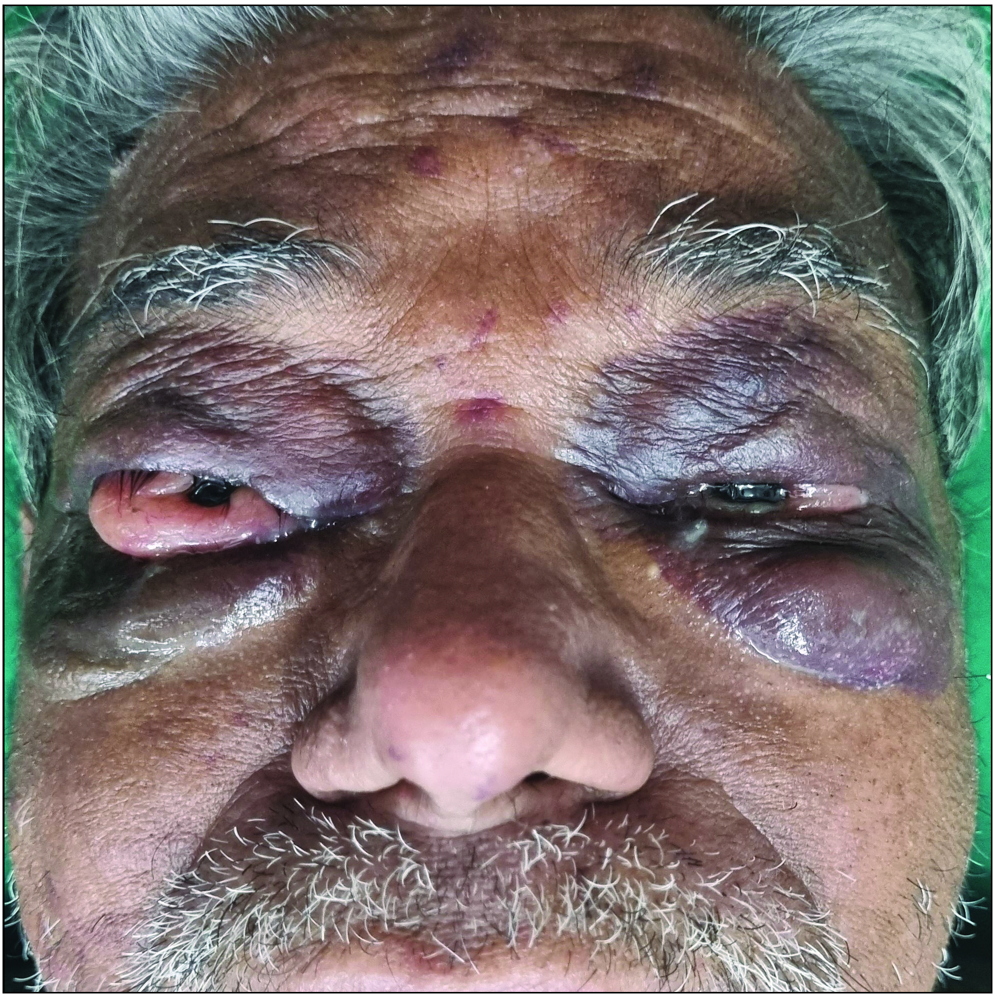
- Bilateral periorbital purpura and fleshy conjunctival growths.

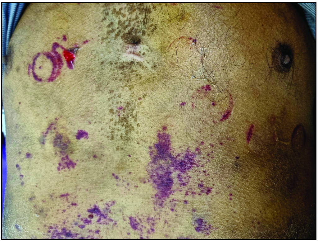
- Purpura on the chest at the site of ECG electrodes and upper abdomen. Note the skin peeling over a purpura on the right chest.
A 55-year-old man was referred to us for spontaneous bruising and increased skin fragility for 5 years. He had periorbital ecchymoses and puffiness [Figure 1] and purpura on the chest at the site of ECG electrodes [Figure 2]. In addition, there were fleshy conjunctival growths in bilateral eyes that showed amyloid deposits on histopathology. Serum electrophoresis revealed an IgG-kappa restricted M-band (2.1 g/dL) and an elevated kappa/lambda ratio (2.32, normal <1.65), while bone marrow biopsy showed 15% kappa-restricted monoclonal plasma cells. A diagnosis of primary systemic light chain (AL) amyloidosis with plasma cell dyscrasia was made and the patient was referred to the haematology department.
Characteristic dermatologic manifestations may be seen in about 40% of AL amyloidosis and may even be the presenting feature leading to diagnosis. Easy bruising of skin occurs due to amyloid infiltrating and weakening the blood vessels. Other changes include waxy nodules and plaques, skin thickening and increased skin fragility. Amyloid deposits in the conjunctiva are quite rare. These have been more commonly described to be an isolated finding, but rarely can be a part of systemic amyloidosis as well, as seen in our patient.
Declaration of patient consent
The authors certify that they have obtained all appropriate patient consent.
Financial support and sponsorship
Nil.
Conflicts of interest
There are no conflicts of interest.
Use of artificial intelligence (AI)-assisted technology for manuscript preparation
The authors confirm that there was no use of artificial intelligence (AI)-assisted technology for assisting in the writing or editing of the manuscript and no images were manipulated using AI.


