Translate this page into:
Subcutaneous fat necrosis of the newborn mimicking generalized lymphadenopathy
2 Department of Paediatrics, Manipal Hospital, Bangalore, India
3 Department of Paediatric surgery, Manipal Hospital, Bangalore, India
Correspondence Address:
Sanjay A Pai
Manipal Hospital, Airport Road, Bangalore - 560017
India
| How to cite this article: Pai SA, Nagesh K, Radhakrishnan C N. Subcutaneous fat necrosis of the newborn mimicking generalized lymphadenopathy. Indian J Dermatol Venereol Leprol 2007;73:357-358 |
 |
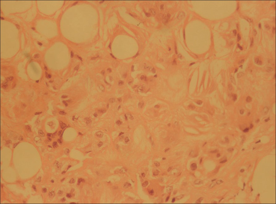
| Figure 1: Subcutaneous fat showing giant cells and needle shaped crystals (H and E, X20) |
 |
| Figure 1: Subcutaneous fat showing giant cells and needle shaped crystals (H and E, X20) |
Sir,
A one and a half month old boy presented with multiple swellings on the body. He had been born prematurely at 34 weeks, by caesarian section of a mother who had had pregnancy-induced hypertension. There was severe hypoxia at birth and he had a seizure on day one. On day two, he developed unconjugated hyperbilirubinemia, for which he was treated with phototherapy. He also developed thrombocytopenia and was given packed red cells. There was no history of steroid intake. The swellings were noticed 10 days after birth, had gradually increased in size and were bilaterally in the neck, axillae and inguinal area. They ranged from 1.5 to 2 cm and were mobile, firm and non-tender. There was also a 3 cm, firm, tender epigastric swelling. His hemoglobin was 8.5 gm% and a TORCH screening was negative. The clinical diagnosis was generalized lymphadenopathy of unknown cause. A biopsy of the axillary swelling, believed to be a lymph node was performed; this showed adipose tissue containing a lobular panniculitis with foci of fat necrosis. Multiple macrophages, foreign body giant cells and lymphoid cells were present. Many needle-shaped crystals in a radial arrangement were seen in the cytoplasm of the macrophages [Figure - 1]. The histologic features were those of subcutaneous fat necrosis. Evaluation of serum calcium was advised but the parents chose to get the child discharged and the patient was lost to followup.
Subcutaneous fat necrosis is characterized by rubbery firm, mobile nodules and erythematous violaceous plaques over the trunk, arms, buttocks, thighs and cheeks. These lesions appear in neonates who have had fetal distress. The child is usually afebrile and appears well. Most cases of subcutaneous fat necrosis are self-limiting. [1],[2] Thrombocytopenia is a common association with subcutaneous fat necrosis and anemia, as in our patient, has also been recorded. [1],[3] Neonatal stress due to difficult delivery and hypothermia are believed to be aetiologic factors. [1],[2] Recognition of the condition is important because a small but significant percentage of cases proceed to a hypercalcemia. When associated with hypercalcemia, seizures, failure to thrive, weight loss, irritability, apathy, hypotonia and even mortality can result. [1],[2],[4]
Treatment consists of analgesics. Hypercalcemia must be treated aggressively with fluid loading, calcium wasting diuretics and low calcium/ vitamin D diet. Serum Calcium must be monitored for several weeks. [1],[2],[4] The differential diagnosis includes erythema nodosum and sclerema neonatorum. [5] These conditions have different morphologies and are unlikely to be misdiagnosed by a pathologist. Erythema nodosum is a septal panniculitis, with little fat necrosis of the lobules and no crystals. Children with sclerema neonatorum are severely ill and though the skin biopsy shows crystals, there is no inflammatory infiltrate or fat necrosis. Poststeroid panniculitis is a condition which is morphologically similar but is preceded by cessation of hydro corticosteroid treatment. [5] In our patient, the unusual sites of the nodules led to the clinical suspicion of generalized lymphadenopathy, which is in itself a rare condition in children. Subcutaneous fat necrosis, an uncommon entity, must be kept in the differential diagnosis of multiple cutaneous nodules in premature children.
| 1. |
Pride, H. Subcutaneous fat necrosis of the newborn. http://www.emedicine.com/derm/topic410.htm [accessed, 16 March 2006]
[Google Scholar]
|
| 2. |
Schulzke S, Buchner S, Fahnenstich H. Subcutaneous fat necrosis of the newborn. Swiss Med Wkly, 2005; 19:135:122-123.
[Google Scholar]
|
| 3. |
Varan B, Gurakan B, Ozbek N, Emir S. Subcutaneous fat necrosis of the newborn associated with anemia. Pediatr Dermatol, 1999;16:381-383.
[Google Scholar]
|
| 4. |
Thomsen RJ. Subcutaneous fat necrosis of the newborn and idiopathic hypercalcemia. Report of a case. Arch Dermatol 1980;116:1155-1158.
[Google Scholar]
|
| 5. |
Maize JC, Burgdorf W, Hurt MA, LeBoit PE, Metcalf JS, Smith T, et al . Cutaneous pathology. Philadelphia: Churchill Livingstone 1998.p.337-8.
[Google Scholar]
|
Fulltext Views
1,273
PDF downloads
1,405


