Translate this page into:
Subcutaneous phaeohyphomycoses caused by Exophiala xenobiotica in an immunocompromised patient: A case report
Corresponding author: Dr. Arathy Menon Anandabhavan, Department of Dermatology and Venereology, Govt Medical College, Kannur, Pariyaram, Kannur, India. arathymenonvstar@gmail.com
-
Received: ,
Accepted: ,
How to cite this article: Anandabhavan AM, Anoop T, Narayanan DP, Payyappilly RJ. Subcutaneous phaeohyphomycoses caused by Exophiala xenobiotica in an immunocompromised patient: A case report. Indian J Dermatol Venereol Leprol. 2024;90:266. doi: 10.25259/IJDVL_259_2022
Dear Editor,
Phaeohyphomycosisis is caused by dematiaceous fungi, which has melanin in the fungal cell wall. The morphology of these varies from septate hyphae, pseudohyphae and yeast. Ajello et al. coined the term “phaeohyphomycosis.” The genera commonly seen in India are Exophiala, Phialophora, Cladosporium, Curvularia, Fonsecaea and Alternaria.1 Exophiala species exists as opportunistic pathogen causing subcutaneous or even fatal disseminated infection especially in immunocompromised patients. Exposed areas of the body are more prone to these infections.
A 42-year-old woman presented to the Government medical college, Kannur, Kerala with multiple cystic swellings of right forearm of 1-year duration. There was no history of trauma. She had a history of episodes of purulent discharge from lesions without any systemic symptoms. She was on oral azathioprine for panuveitis for the last 4 years. Cutaneous examination revealed multiple skin-coloured, non-tender, firm to soft cystic subcutaneous nodules of varying sizes on the extensor aspect of the right forearm [Figure 1a]. The differential diagnoses considered were subcutaneous mycoses and foreign body granuloma.
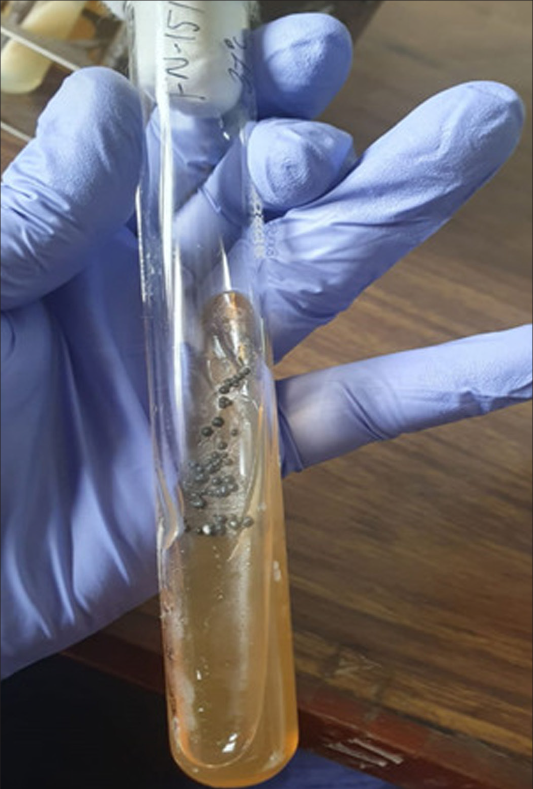
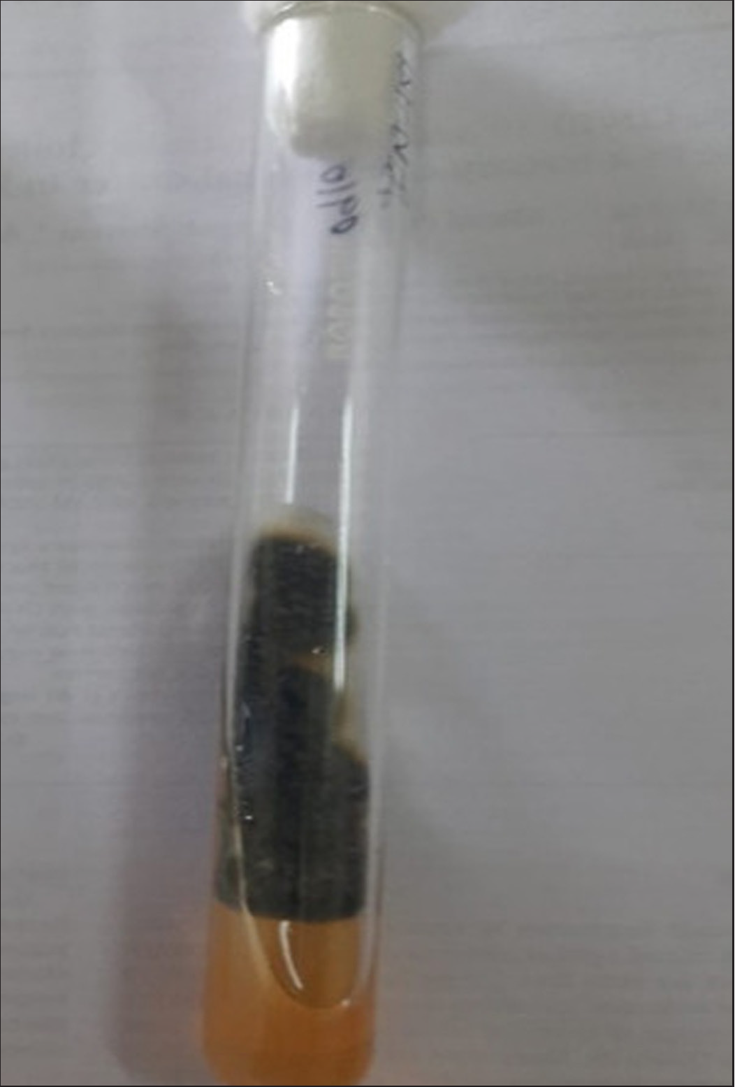
Fine-needle aspiration of the content with potassium hydroxide (KOH) mount and gram staining showed branching septate hyphae [Figures 1b and 1c]. Fungal culture in sabouraud agar showed slow-growing yeast-like colonies with ovoid to elliptical budding cells, olivaceous grey coloured, with aerial mycelium [Figures 2a and 2b]. Slide culture and staining with lactophenol cotton blue mount showed short-branched annelophores with multiple sessile oval conidia [Figure 2c].

- Multiple firm to soft cystic subcutaneous nodules of varying sizes on the extensor aspect of right forearm (black arrows)
![KOH mount from aspirate showing branched hyphae [10x] (black arrow)](/content/126/2024/90/2/img/IJDVL-259-2022-g001b.png)
- KOH mount from aspirate showing branched hyphae [10x] (black arrow)
![Gram-positive branching fungal filaments from the aspirate [100x] (black arrows)](/content/126/2024/90/2/img/IJDVL-259-2022-g001c.png)
- Gram-positive branching fungal filaments from the aspirate [100x] (black arrows)

- Culture on Sabouraud agar with mucoid colonies after 7 days

- Culture on Sabouraud agar with velvety growth on 14th day
![Lactophenol cotton blue mount showing conidiophores with supporting hyphae [40x]](/content/126/2024/90/2/img/IJDVL-259-2022-g002c.png)
- Lactophenol cotton blue mount showing conidiophores with supporting hyphae [40x]
Exophiala xenobiotica was identified by mass spectrometry with VITEK® MS matrix-assisted laser desorption ionisation time-of-flight technology. Histopathological examination of the biopsy specimen on haematoxylin and eosin stain showed cystic lesion in the dermis with cyst wall composed of polygranulomatous inflammation and necrotic debris at the centre, with few pigmented fungal hyphae seen along the cyst wall [Figures 3a and 3b]. Gomori methenamine silver stain showed silver-stained fungal hyphae and Masson-Fontana stain highlighted the melanin pigments of phaeohyphomycosis [Figures 3c and 3d]. Antifungal sensitivity testing was not done due to the lack of facility. The lesions were surgically excised. Because the patient had a mild elevation in liver enzymes, oral itraconazole was started at a dose of 100 mg twice daily. Amphotericin B was not given due to financial constraints. Azathioprine could not be stopped as ophthalmologist advised to continue the drug in view of panuveitis. Patient was closely monitored for signs of dissemination during each follow up. After 6 months, while on itraconazole, she got a relapse at the same site and was empirically started on oral terbinafine 250 mg twice daily for 1 month. Unfortunately, the patient was lost to follow-up after a few visits.
![Cystic lesion in the dermis with cyst wall, granulomatous inflammation and necrotic debris at the centre [H&E, 10x] (black arrows)](/content/126/2024/90/2/img/IJDVL-259-2022-g003a.png)
- Cystic lesion in the dermis with cyst wall, granulomatous inflammation and necrotic debris at the centre [H&E, 10x] (black arrows)
![Granulomatous inflammation and necrotic debris at the centre with few pigmented fungal hyphae [H&E, 40x] (black arrows)](/content/126/2024/90/2/img/IJDVL-259-2022-g003b.png)
- Granulomatous inflammation and necrotic debris at the centre with few pigmented fungal hyphae [H&E, 40x] (black arrows)
![Gomori methenamine silver stain showing silver-stained fungal hyphae [40x] (black arrows)](/content/126/2024/90/2/img/IJDVL-259-2022-g003c.png)
- Gomori methenamine silver stain showing silver-stained fungal hyphae [40x] (black arrows)
![Masson-Fontana stain highlighting the melanin pigments of phaeohyphomycosis [40x] (black arrows)](/content/126/2024/90/2/img/IJDVL-259-2022-g003d.png)
- Masson-Fontana stain highlighting the melanin pigments of phaeohyphomycosis [40x] (black arrows)
The clinical presentation of phaeohyphomycosis varies from superficial forms such as Tinea nigra and black piedra, cutaneous forms such as scytalidiosis and subcutaneous mycotic cyst to systemic phaeohyphomycosis in an immunocompromised host. The mycotic cyst is characterised by asymptomatic presentation after traumatic implantation, the usual sites being the extremities.
A very few case reports are available from published literature about phaeohyphomycosis caused by Exophiala xenobiotica species. The most common clinical presentations among the cases reported were subcutaneous phaeohyphomycosis, followed by invasive or disseminated infection. Exophiala xenobiotica is usually isolated from habitats rich in monocyclic aromatic hydrocarbons.2 According to the study conducted by Singh et al. and based on the reports retrieved from the national culture collection of pathogenic fungi (NCCPF), Chandigarh, India, 23 cases due to Exophiala species have been reported. Exophiala jeanselmei was the most common species identified. Exophiala xenobiotica was identified only in a single case.3 We were unable to find any other reports of phaeohyphomycosis caused by Exophiala xenobiotica from India. Among the cases reported in India till 2015, the species of Exophiala isolated were, Exophiala spinifera, Exophiala jeanselmei and Exophiala dermatitidis.4 Systemic and disseminated cases of these fungi can take a fatal course in young and otherwise-healthy individuals.
A panel of experts of the European fungal infections study group from the European society of clinical microbiology and infectious diseases has done a data review and prepared guidelines for the diagnosis and management of infections caused by melanised fungi.5 Melanised fungi are usually susceptible to azoles such as itraconazole and voriconazole followed by amphotericin B. The early clinical suspicion of phaeohyphomycosis in immunocompromised patients is extremely important, as immunosuppression is the major risk factor for the systemic spread of the disease, causing severe and fatal outcomes.
Declaration of patient consent
Patient’s consent is not required as patient’s identity is not disclosed or compromised.
Financial support and sponsorship
Nil.
Conflicts of interest
There are no conflicts of interest.
References
- Three rare cases of cutaneous phaeohyphomycosis. Indian J Plast Surg. 2016;49:271-4.
- [CrossRef] [PubMed] [PubMed Central] [Google Scholar]
- Isolation and screening of black fungi as degraders of volatile aromatic hydrocarbons. Mycopathologia. 2013;175:369-79.
- [CrossRef] [PubMed] [Google Scholar]
- Clinical spectrum, molecular characterization, antifungal susceptibility testing of Exophiala spp. from India and description of a novel Exophiala species, E. arunalokei sp. nov. Front Cell Infect Microbiol. 2021;11:686120.
- [CrossRef] [PubMed] [PubMed Central] [Google Scholar]
- Phaeohyphomycosis due to Exophialajeanselmei: An emerging pathogen in India—Case report and review. Mycopathologia. 2016;181:279-84.
- [CrossRef] [PubMed] [Google Scholar]
- ESCMID and ECMM joint clinical guidelines for the diagnosis and management of systemic phaeohyphomycosis: diseases caused by black fungi. Clin Microbiol Infect. 2014;20:47-75.
- [CrossRef] [PubMed] [Google Scholar]


