Translate this page into:
Supraventine sparing of salt-and-pepper pigmentation in a patient with systemic sclerosis
Corresponding Author: Dr. Vishal Gupta, Department of Dermatology and Venereology, All India Institute of Medical Sciences, New Delhi, India. doctor.vishalgupta@gmail.com
-
Received: ,
Accepted: ,
How to cite this article: Sindhuja T, Danish M, Gupta V. Supraventine sparing of salt-and-pepper pigmentation in a patient with systemic sclerosis. Indian J Dermatol Venereol Leprol 2022;88:812-3.
Sir,
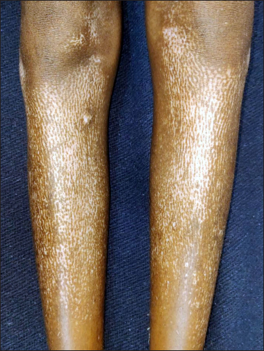
A 13-year-old Indian girl presented with a nine-month history of progressive binding down of the skin on the face, trunk and distal extremities including fingers. There were depigmented macules interspersed with areas of normal pigmentation around the follicles suggestive of salt-and-pepper pigmentation, over the forehead and legs [Figure 1]. There was a striking sparing of depigmentation along the course of the superficial veins on her forehead and frontal scalp [Figure 2]. She also had Raynaud’s phenomenon, dysphagia to solid food, exertional dyspnea and symmetric proximal muscle weakness. Nail fold capillaroscopy demonstrated a few giant capillaries. Laboratory investigations were significant for a positive indirect immunofluorescence test for antinuclear antibody (4+, 1:100, fine speckled pattern), a positive line immunoblot assay for Scl-70 antibodies and an elevated serum creatine kinase (1441 U/L; normal 55–135 U/L). Anti-centromere, antimitochondrial M2, Jo-1, PM-Scl, Mi-2 and Ku antibodies were negative. Pulmonary function tests showed a restrictive pattern (forced vital capacity 78%, forced expiratory volume in 1 second 66%) and high resolution computerized tomographic scan of the chest revealed basal and subpleural predominant reticular opacities and traction bronchiectasis suggestive of interstitial lung disease. Barium swallow showed poor oesophageal motility and 2-D echocardiography showed a large pericardial effusion without an impending cardiac tamponade. Based on these clinical and investigative findings, a diagnosis of diffuse cutaneous systemic sclerosis with inflammatory myopathy was made. During the hospital stay, she was treated with intravenous dexamethasone (50 mg) pulse for three days, daily nifedipine 10 mg/day, torsemide 5 mg/day, sildenafil 10 mg thrice a day and mosapride 5 mg twice a day, along with cold protection measures. There was symptomatic improvement in muscle weakness and a reduction in the size of the pericardial effusion on repeat 2-D echocardiography and the patient was discharged in a stable condition. Unfortunately, she passed away two weeks later at home and the cause of death could not be ascertained.

- Salt-and-pepper pigmentation over bilateral legs

- Sparing of depigmentation over the course of superficial veins within areas of salt-and-pepper pigmentation on the forehead and scalp
Salt-and-pepper pigmentation is a characteristic pigmentary change in patients with systemic sclerosis and is particularly common in patients with darker skin phototypes with a prevalence ranging from 51 to 94%.1 Patients with systemic sclerosis have an abnormal temperature response and the perifollicular sparing of the salt-and-pepper depigmentation is thought to be due to the rich capillary network around the hair follicles warming the perifollicular skin. Preservation of normal pigment overlying the superficial veins within the areas of salt-and-pepper pigmentation is quite rare and may be attributed to the warm skin overlying superficial veins.2
The thermographic examination has previously demonstrated a temperature difference of 0.5–1.5° C in the pigmented areas over the vessels compared to the surrounding depigmented skin.2
We came across only one earlier report of three patients with this distinctive pigmentary change; all had progressive systemic sclerosis, two of whom also had active myositis like our case.2 However, the clinical significance of this observation as a clue to more severe disease needs to be further explored.
Pigment retention overlying superficial veins within areas of depigmentation has rarely been seen in certain other dermatoses as well, such as vitiligo, dermatitides and dermatophytic infections.3 It is hypothesized that a significant inflammatory response may cause vasodilatation leading to increased vascular permeability and endothelial cell activation. Endothelial injury and subclinical-to-mild phlebitis may result in basal epidermal hyperpigmentation and dermal pigment incontinence.3
In addition, serpentine supravenous hyperpigmentation is also known to occur following intravenous infusion of certain chemotherapy agents, including 5-fluorouracil, docetaxel, cyclophosphamide, bleomycin and vinca alkaloids.3
Declaration of patient consent
The authors certify that they have obtained all appropriate patient consent.
Financial support and sponsorship
Nil.
Conflicts of interest
Dr. Vishal Gupta is the Deputy Editor of the journal.
References
- Salt-and-pepper appearance: A cutaneous clue for the diagnosis of systemic sclerosis. Indian J Dermatol. 2012;57:412-3.
- [CrossRef] [PubMed] [Google Scholar]
- A new skin manifestation of progressive systemic sclerosis. J Am Acad Dermatol. 1984;11:265-8.
- [CrossRef] [PubMed] [Google Scholar]
- Serpentine supravenous hyperpigmentation – A rare cutaneous manifestation in non-neoplastic conditions. J Skin Sex Transm Dis. 2019;1:97-100.
- [CrossRef] [Google Scholar]


