Generic selectors
Exact matches only
Search in title
Search in content
Post Type Selectors
Search in posts
Search in pages
Filter by Categories
15th National Conference of the IAOMFP, Chennai, 2006
Abstract
Abstracts from current literature
Acne in India: Guidelines for management - IAA Consensus Document
Addendum
Announcement
Art & Psychiatry
Article
Articles
Association Activities
Association Notes
Award Article
Book Review
Brief Report
Case Analysis
Case Letter
Case Letters
Case Notes
Case Report
Case Reports
Clinical and Laboratory Investigations
Clinical Article
Clinical Studies
Clinical Study
Commentary
Conference Oration
Conference Summary
Continuing Medical Education
Correspondence
Corrigendum
Cosmetic Dermatology
Cosmetology
Current Best Evidence
Current Issue
Current View
Derma Quest
Dermato Surgery
Dermatopathology
Dermatosurgery Specials
Dispensing Pearl
Do you know?
Drug Dialogues
e-IJDVL
Editor Speaks
Editorial
Editorial Remarks
Editorial Report
Editorial Report - 2007
Editorial report for 2004-2005
Errata
Erratum
Focus
Fourth All India Conference Programme
From Our Book Shelf
From the Desk of Chief Editor
General
Get Set for Net
Get set for the net
Guest Article
Guest Editorial
History
How I Manage?
IADVL Announcement
IADVL Announcements
IJDVL Awards
IJDVL AWARDS 2015
IJDVL Awards 2018
IJDVL Awards 2019
IJDVL Awards 2020
IJDVL International Awards 2018
Images in Clinical Practice
Images in Dermatology
In Memorium
Inaugural Address
Index
Knowledge From World Contemporaries
Leprosy Section
Letter in Response to Previous Publication
Letter to Editor
Letter to the Editor
Letter to the Editor - Case Letter
Letter to the Editor - Letter in Response to Published Article
LETTER TO THE EDITOR - LETTERS IN RESPONSE TO PUBLISHED ARTICLES
Letter to the Editor - Observation Letter
Letter to the Editor - Study Letter
Letter to the Editor - Therapy Letter
Letter to the Editor: Articles in Response to Previously Published Articles
Letters in Response to Previous Publication
Letters to the Editor
Letters to the Editor - Letter in Response to Previously Published Articles
Letters to the Editor: Case Letters
Letters to the Editor: Letters in Response to Previously Published Articles
Media and news
Medicolegal Window
Messages
Miscellaneous Letter
Musings
Net Case
Net case report
Net Image
Net Images
Net Letter
Net Quiz
Net Study
New Preparations
News
News & Views
Obituary
Observation Letter
Observation Letters
Oration
Original Article
ORIGINAL CONTRIBUTION
Original Contributions
Pattern of Skin Diseases
Pearls
Pediatric Dermatology
Pediatric Rounds
Perspective
Presedential Address
Presidential Address
Presidents Remarks
Quiz
Recommendations
Regret
Report
Report of chief editor
Report of Hon : Treasurer IADVL
Report of Hon. General Secretary IADVL
Research Methdology
Research Methodology
Resident page
Resident's Page
Resident’s Page
Residents' Corner
Residents' Corner
Residents' Page
Retraction
Review
Review Article
Review Articles
Reviewers 2022
Revision Corner
Self Assessment Programme
SEMINAR
Seminar: Chronic Arsenicosis in India
Seminar: HIV Infection
Short Communication
Short Communications
Short Report
Snippets
Special Article
Specialty Interface
Studies
Study Letter
Study Letters
Supplement-Photoprotection
Supplement-Psoriasis
Symposium - Contact Dermatitis
Symposium - Lasers
Symposium - Pediatric Dermatoses
Symposium - Psoriasis
Symposium - Vesicobullous Disorders
SYMPOSIUM - VITILIGO
Symposium Aesthetic Surgery
Symposium Dermatopathology
Symposium-Hair Disorders
Symposium-Nails Part I
Symposium-Nails-Part II
Systematic Review and Meta-Analysis
Systematic Reviews and Meta-analyses
Systematic Reviews and Meta-analysis
Tables
Technology
Therapeutic Guideline-IADVL
Therapeutic Guidelines
Therapeutic Guidelines - IADVL
Therapeutics
Therapy
Therapy Letter
Therapy Letters
View Point
Viewpoint
What’s new in Dermatology
View/Download PDF

Translate this page into:

Observation Letter
ARTICLE IN PRESS
doi:
10.25259/IJDVL_1093_2024

Synovitis, acne, pustulosis, hyperostosis, osteitis (SAPHO) syndrome with co-existing ulcerative and superficial granulomatous variants of pyoderma gangrenosum

Department of Dermatology, Venereology and Leprosy, King George’s Medical University, Lucknow, India
Department of Dermatology, Venereology and Leprosy, Hind Institute of Medical Sciences, Sitapur Road, India
Department of Nuclear Medicine, Dr Ram Manohar Lohia Institute of Medical Sciences, Lucknow, India
Department of Pathology, Dr. Ram Manohar Lohia Institute of Medical Sciences, Lucknow, India
Department of Rheumatology, King George’s Medical University, Lucknow, India.

Corresponding author: Dr. Swastika Suvirya, Department of Dermatology, Venereology and Leprosy, King George’s Medical University, Lucknow, India. swastika.p@gmail.com

Licence
This is an open-access article distributed under the terms of the Creative Commons Attribution-Non Commercial-Share Alike 4.0 License, which allows others to remix, transform, and build upon the work non-commercially, as long as the author is credited and the new creations are licensed under the identical terms.

How to cite this article: Verma P, Shukla P, Deswal S, Malhotra KP, Kumar P, Suvirya S. Synovitis, acne, pustulosis, hyperostosis, osteitis (SAPHO) syndrome with co-existing ulcerative and superficial granulomatous variants of pyoderma gangrenosum. Indian J Dermatol Venereol Leprol. doi: 10.25259/IJDVL_1093_2024

Dear Editor,

SAPHO syndrome is a rare seronegative spondyloarthropathy that stands for synovitis, acne, palmoplantar pustulosis, hyperostosis and osteitis. Palmoplantar pustulosis is the most common associated cutaneous feature, but around 16% of adults never manifest cutaneous symptoms.1

A young boy in his late teens presented to the dermatology outpatient department with multiple painful ulcerative lesions over bilateral cheeks and legs for the past two years. He also recalled a history of episodic pain over multiple joints since the age of 14 years. There was no history of morning stiffness in joints, bowel disturbances or other systemic complaints. Patient was prescribed non-steroidal anti-inflammatory drugs (NSAIDs), isotretinoin, and antibiotics from various physicians, without relief.

On examination, erythematous plaques of approximately 8 × 6 cm size were present on both cheeks with an overlying yellowish crust and serosanguinous discharge [Figure 1a]. A tender indurated ulcer of size 10 × 6 cm with undermined edges was present on the lateral aspect of the right leg, covered with haemorrhagic crust and serosanguinous discharge, with the surrounding skin showing violaceous discoloration [Figure 1b]. Multiple rounds to oval depressed cribriform scars were noted on all four limbs.
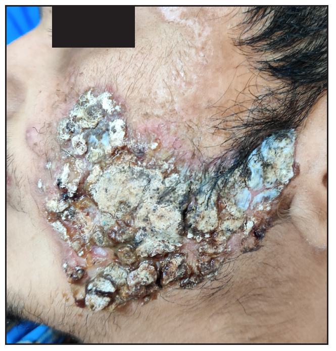
Erythematous plaque over the left cheek with areas of yellow-black crusting and scarring.
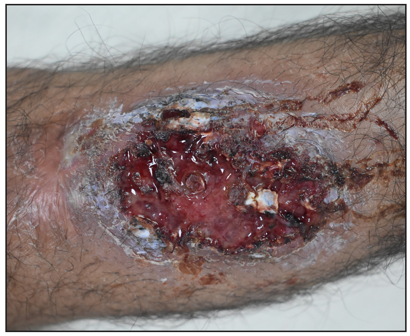
Figure 1a:
Erythematous plaque over the left cheek with areas of yellow-black crusting and scarring.
An ulcer over the right leg with haemorrhagic crust and serosanguinous discharge. Surrounding peripheral skin showed violaceous discoloration.
Figure 1b:
An ulcer over the right leg with haemorrhagic crust and serosanguinous discharge. Surrounding peripheral skin showed violaceous discoloration.

There was tenderness at the bilateral knee, ankle and right sternoclavicular joint. Medial end of the right clavicle appeared enlarged as compared to the left [Supplementary Figure 1]. Routine laboratory investigations did not reveal any abnormality. Erythrocyte sedimentation rate was 64 mm/hr (Normal range < 10 mm/hr for men). Anti-nuclear antibodies were positive (2+, fine speckled). Anti-double stranded deoxyribonucleic acid (Anti-dsDNA) antibody, rheumatoid factor, Copmlement levels C3 and C4, anti-myeloperoxidase and anti-proteinase-3 antibodies were negative. Biopsy from facial plaque showed deep dermal ulcer with intense mixed dermal inflammation and ill-defined granulomas [Figure 1c]. Biopsy from leg lesion showed epidermal ulceration with mixed dermal inflammation and perivascular lymphocytic infiltrate [Figure 1d]. Direct immunofluorescence (DIF), bacterial, mycobacterial, and fungal cultures were negative. Ultrasound whole abdomen and colonoscopy were within normal limits. X-rays of hip joints, bilateral elbow, knee and chest revealed no abnormality.
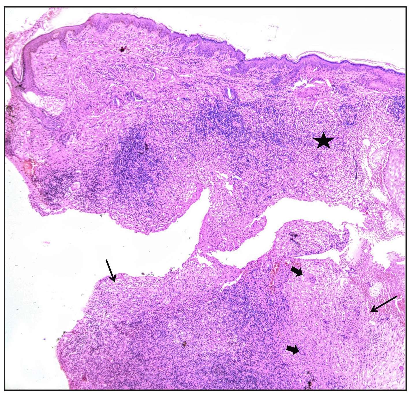
Biopsy from face showing intense mixed inflammation in superficial dermis (asterisk) and deep dermal granulomas (black arrows) with giant cells (block arrows) (Haematoxylin & eosin, 50x.)
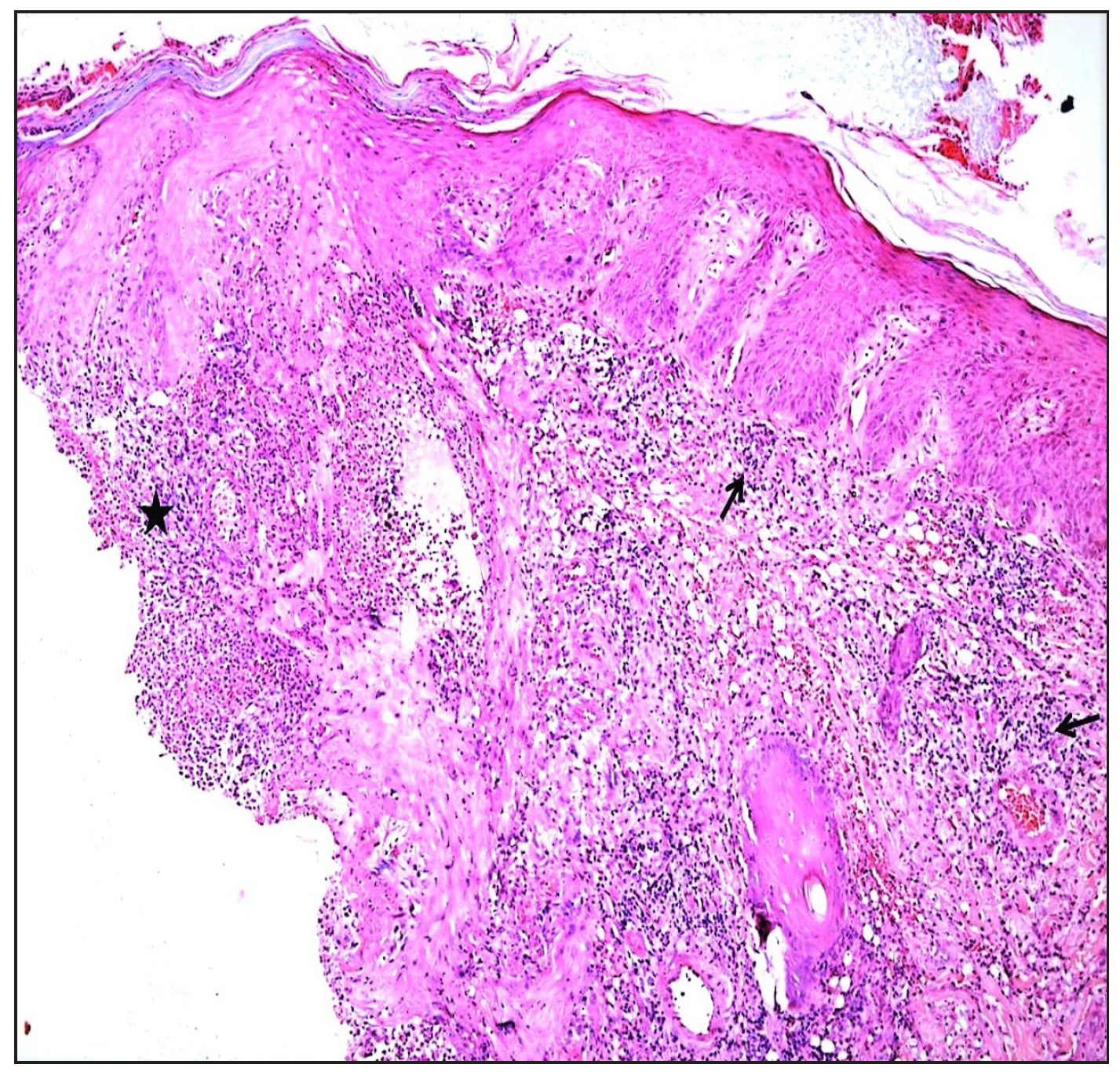
Figure 1c:
Biopsy from face showing intense mixed inflammation in superficial dermis (asterisk) and deep dermal granulomas (black arrows) with giant cells (block arrows) (Haematoxylin & eosin, 50x.)
Biopsy from leg plaque showing ulcer edge (asterisk) and lymphocytic vasculitis with infiltrate around dermal vessels (black arrows) (Haematoxylin & eosin, 200x).
Figure 1d:
Biopsy from leg plaque showing ulcer edge (asterisk) and lymphocytic vasculitis with infiltrate around dermal vessels (black arrows) (Haematoxylin & eosin, 200x).

Tc-99m methylene diphosphonate (MDP) whole body bone scan performed in the anterior and posterior view revealed increased radiotracer uptake in bilateral shoulders (right>left), left elbow, bilateral knee and ankle joints. Right clavicle was also expanded along with increased tracer uptake [Figure 2a]. The scan findings were consistent with osteitis of the clavicle and inflammation at the above joints. Reconstructed three-dimensional (3D) computed tomography (CT) image of bones of the anterior chest wall showed hyperostosis of the bony and sternal end of the right clavicle [Figure 2b]. Synovial fluid aspirate from the knee joint did not show any growth on culture, suggestive of the aseptic nature of inflammation.
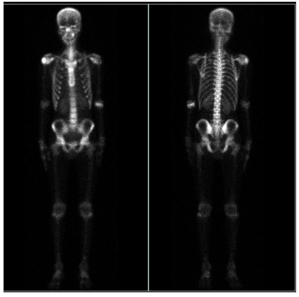
99mTc MDP (Methyl Diphosphonate) bone scan anterior and posterior images showing increased radiotracer uptake in bilateral shoulders (right> left), left elbow, bilateral knee and bilateral ankle joint. The right clavicle appears expanded with a mild increase in tracer uptake.
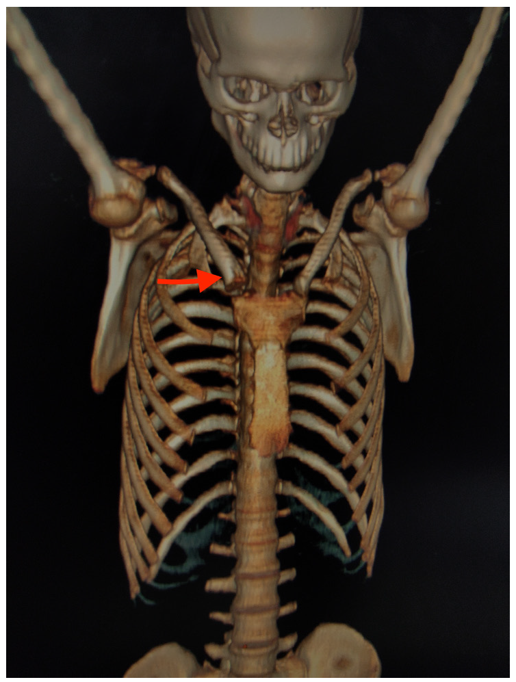
Figure 2a:
99mTc MDP (Methyl Diphosphonate) bone scan anterior and posterior images showing increased radiotracer uptake in bilateral shoulders (right> left), left elbow, bilateral knee and bilateral ankle joint. The right clavicle appears expanded with a mild increase in tracer uptake.
Reconstructed 3D CT image of bones of the anterior chest wall showed hyperostosis of bony and sternal end of right clavicle (red arrow).
Figure 2b:
Reconstructed 3D CT image of bones of the anterior chest wall showed hyperostosis of bony and sternal end of right clavicle (red arrow).

A final diagnosis of SAPHO syndrome2 was established along with co-existing superficial granulomatous (face) and ulcerative variants (legs) of pyoderma gangrenosum (PG) in consultation with a rheumatologist. Patient was administered infliximab 5mg/kg IV at weeks 0, 2, 6 and 14. On induction of infliximab therapy, he showed promising responses in both cutaneous and bony symptoms. Patient is presently in remission one month after the last dose of infliximab therapy [Figure 3a,b]. Dermatological disorders, infrequently reported with SAPHO syndrome, include acne conglobata, acne fulminans, pustular psoriasis, subcorneal pustular dermatosis, hidradenitis suppurativa, psoriasis vulgaris, linear IgA (immunoglobulin A) disease, Behçet’s disease, Sweet syndrome and very rarely, PG (pyoderma gangrenosum).3 PG is an uncommon, chronic, recurrent neutrophilic dermatosis that requires clinicopathological correlation for diagnosis. With four different subtypes, ulcerative PG is the most common variant. Superficial granulomatous PG, also known as the vegetative subtype, is infrequent and very rarely reported.4
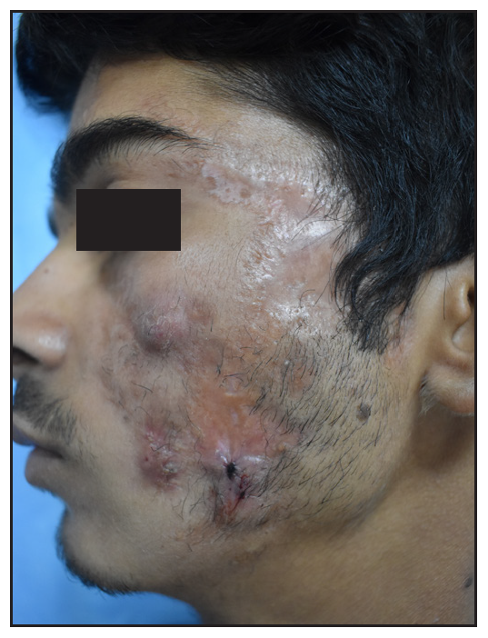
Scarring over right cheek post-treatment.
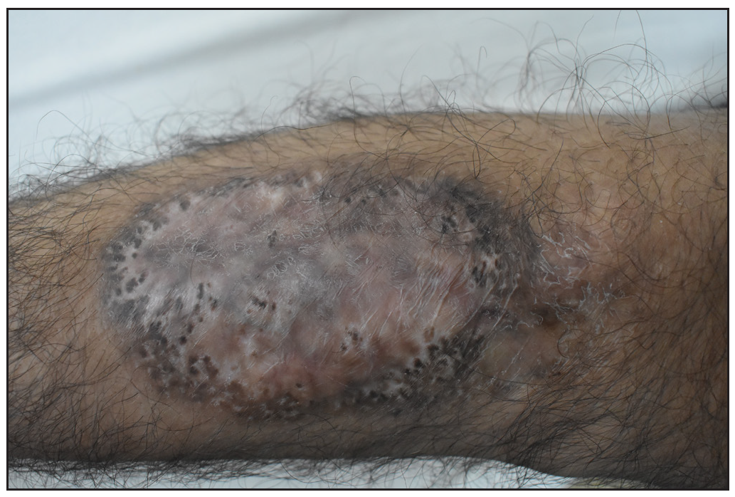
Figure 3a:
Scarring over right cheek post-treatment.
Healed ulcer with cribriform scarring over the right leg post-treatment.
Figure 3b:
Healed ulcer with cribriform scarring over the right leg post-treatment.

So far, only a very few cases of SAPHO syndrome with PG have been described, and none with two subtypes of PG simultaneously co-existing with SAPHO.5,6 The pathogenesis of SAPHO remains unclear, but similarities with auto-inflammatory disorders and involvement of cytokines like interleukin-1-beta (IL-1β) and tumour necrosis factor-alpha (TNFα) have been observed. These findings suggest that PG and SAPHO may represent different manifestations of a single auto-inflammatory spectrum.7 Treatment options include NSAIDs and intra-articular corticosteroids for arthritis; bisphosphonates for bone involvement; and systemic corticosteroids or other immunosuppressive medications, TNFα inhibitors and IL-1antagonists for both skin and bone disease.5

In the above case, simultaneous presence of PG with arthritis served as a diagnostic dilemma. The differentials for the same have been illustrated in Table 1. Distinctive radiographic features of the osteoarticular lesions formed the cornerstone for diagnosis in the index case. Despite being rare, it would be a good practice to keep an eye out for this condition when dealing with a case of neutrophilic dermatoses or a seronegative spondyloarthropathy.

Table 1: Differential diagnosis for Pyoderma Gangrenosum (PG) and PG like ulcer with associated arthritis
S.no. A) PG lesions with arthritis
Condition Clinical findings
Laboratory findings Imaging
Cutaneous Osteoarticular Others
1. Vasculitis
  • Cutaneous ulcers

  • Palpable purpura

  • Arthralgia

  • Mono/oligoarthritis and joint swelling

  • Other systemic symptoms

  • Positive autoantibodies (ANA, ANCA)

  • Histology and DIF findings consistent with vasculitis

  • Histology PG

X-ray joints: Meniscus horns, intra-articular bodies of Pellagri-Shtaydi and Hoff, Baker cysts, tendovaginitis, subchondral sclerosis and osteocystosis
2. *Inflammatory arthritides:
a) RA (Rheumatoid arthritis)
  • PG

  • Rheumatoid nodules, Felty syndrome, rheumatoid vasculitis

  • Joint pain (palindromic rheumatism), morning stiffness, swelling, deformities

  • MCP, PIP and MTP joints.

  • CTS

  • Positive RF, anti-CCP antibodies.

  • HPE s/o PG

X-ray joints: Periarticular osteopenia, uniform joint space loss, bone erosions and soft tissue swelling
b) PsA1 (Psoriatic arthritis)
  • PG

  • Psoriatic lesions

  • Nail changes of psoriasis

  • Enthesitis, dactylitis, tenosynovitis, sacroilitis

  • DIP joints

  • Uveitis

  • Negative RF

  • HPE of skin showing psoriatic changes

  • HPE s/o PG

X-ray joints: Periarticular soft tissue swelling, marginal erosion and pencil-in-cup deformity
3. *Autoinflammatory syndromes
a) PAPA syndrome7 [Pyoderma Gangrenosum, Acne, and Pyogenic arthritis syndrome]
  • PG

  • Acne

  • Recurrent sterile pyogenic arthritis

-
  • Sterile synovial fluid analysis with neutrophil predominance

  • HPE s/o PG

X-ray joints: Effusion without erosions

Ultrasound:Soft tissue swelling

b) PAPASH syndrome7 [Pyoderma gangrenosum, Pyogenic arthritis, Acne, and Suppurative hidradenitis syndrome]
  • PG

  • Acne

  • Suppurative hidradenitis

  • Pyogenic arthritis

-
  • Negative RF and ANA

  • HPE s/o PG

X-ray joints:Similar to PAPA
c) SAPHO syndrome2
  • Synovitis

  • Acne

  • Pustulosis

  • PG rarely

  • Hyperostosis, osteitis, back pain, tenderness over sternoclavicular joint

-
  • Elevated complements C3, C4 and alkaline phosphatase

  • HPE s/o PG

X-ray joints:Hyperostosis, sclerosis, osteitis, osteolysis and periosteal reaction

Bone scintigraphy: Increased uptake in affected areas

4. *IBD {Inflammatory bowel disease} (e.g. Crohn’s disease, ulcerative colitis)
  • PG

  • Erythema Nodosum

  • Joint pain, swelling

  • Mono/polyarticular

  • Axial arthropathy, dactylitis, enthesopathy

  • Abdominal pain

  • Diarrhoea

  • Weight loss

  • Elevated fecal calprotectin

  • Positive p-ANCA and ASCA

  • HPE s/o PG

Radiograph:Axial spondyloarthritis and non-erosive peripheral arthritis

Barium studies and CT findings: IBD

5. *SLE (Systemic lupus erythematosus)
  • PG rarely

  • Malar rash, photosensitivity, oral ulcer

  • Migratory, symmetrical, polyarticular, arthralgia/arthritis

  • Systemic manifestations.

  • Positive ANA, anti-dsDNA, anti-Smith antibodies

  • HPE s/o PG

X-ray joints: Non-erosive, non-deforming arthritis

MRI: effusions, synovial and/or tenosynovial hypertrophy and vascularity

6. *Behçet’s disease
  • PG

  • Oral and genital aphthae

  • EN

  • Arthralgia

  • Oligoarthritis

  • Cardiac, neurological and ocular symptoms

  • Positive pathergy

  • Histology suggestive of PG

  • HLA-B51 positivity

X-ray joints: Non-erosive, non-deforming arthritis commonly involving knee, wrist, ankle, elbow
B) PG like ulcers with arthritis
1. *Infectious diseases
  • Localised erythema, swelling, warmth

  • Painful necrotic ulcers

  • Pain, swelling, limited range of motion, limping

  • Fever, chills, weight loss

  • Positive cultures of synovial fluid, bone or tissue

X-ray joints: Effusion, joint space narrowing, joint destruction

MRI in TB arthritis: #Phemister triad

2. *Malignancy (e.g., Mycosis fungoides, leukemia)
  • Haemorrhagic necrotic ulcer with advancing border composed of multiloculated bullae

  • Arthralgia

  • Symmetrical polyarthritis

  • Lymphadenopathy

  • HPE findings of malignancy

  • Cytological examination of PBS and bone marrow showing malignant cells

X-ray hands:Synovitis, erosive and deforming polyarthritis, joint space narrowing, periarticular osteoporosis
Inflammatory markers (ESR and CRP) are elevated

#Phemister triad: marginal erosion, periarticular osteoporosis and joint space narrowing.

PG: Pyoderma gangrenosum; ANA: anti-nuclear antibodies; ANCA: anti-neutrophil cytoplasmic antibodies; DIF: Direct immunofluorescence; s/o: suggestive of; RA: Rheumatoid arthritis; MCP: Metacarpophalangeal; PIP: Proximal interphalangeal; MTP: Metatarsophalangeal; CTS: Carpel tunnel syndrome; RF: Rheumatoid factor; anti-CCP: anti-cyclic citrullinated peptide; PsA: Psoriatic arthritis; DIP: Distal interphalangeal; PAPA: Pyogenic arthritis, Pyoderma gangrenosum, and Acne; PAPASH: Pyogenic arthritis, Pyoderma gangrenosum, Acne, and Hidradenitis suppurativa; SAPHO: Synovitis, Acne, Pustulosis, Hyperostosis, and Osteitis; IBD: Inflammatory bowel disease; ASCA: anti-saccharomyces cerevisiae antibodies; SLE: Systemic lupus erythematosus; MRI: Magnetic resonance imaging; HLA-B51: Human leukocyte antigen-B51

Declaration of patient consent

The authors certify that they have obtained all appropriate patient consent.

Financial support and sponsorship

Nil.

Conflicts of interest

There are no conflicts of interest.

Use of artificial intelligence (AI)-assisted technology for manuscript preparation

The authors confirm that there was no use of AI-assisted technology for assisting in the writing or editing of the manuscript and no images were manipulated using AI.

References

  1. , , , , , . The SAPHO syndrome. Semin Arthritis Rheum. 2012;42:254-65.
    [CrossRef] [PubMed] [Google Scholar]
  2. . SAPHO syndrome: A review. J Child Orthop. 2015;9:19-27.
    [CrossRef] [PubMed] [PubMed Central] [Google Scholar]
  3. , , . Dermatology (4th ed). China: Elsevier; . p. :468-70.
  4. , , , , , , et al. Superficial granulomatous pyoderma gangrenosum involving the face: A case series of five patients and a review of the literature [Formula: see text] J Cutan Med Surg. 2021;25:371-6.
    [CrossRef] [PubMed] [Google Scholar]
  5. , , , , , . Synovitis, acne, pustulosis, hyperostosis, osteitis (SAPHO) syndrome associated with pyoderma gangrenosum: Report of two patients. Int J Dermatol. 2023;62:e186-9.
    [CrossRef] [PubMed] [Google Scholar]
  6. , , , . SAPHO syndrome associated with hidradenitis suppurativa and pyoderma gangrenosum successfully treated with adalimumab and methotrexate: A case report and review of the literature. Int J Dermatol. 2018;57:10-8.
    [CrossRef] [PubMed] [Google Scholar]
  7. , , , . Pyoderma gangrenosum and its syndromic forms: Evidence for a link with autoinflammation. Br J Dermatol. 2016;175:882-91.
    [CrossRef] [PubMed] [Google Scholar]

Fulltext Views
585

PDF downloads
18
View/Download PDF
Download Citations
BibTeX
RIS
Show Sections