
Translate this page into:
The dermatological evolution of the first case of monkeypox reported in India
Corresponding author: Dr. Anuja Elizabeth George, Department of Dermatology & Venereology, Government Medical College, Trivandrum, Kerala, India. aegeor4@yahoo.in
-
Received: ,
Accepted: ,
How to cite this article: George AE, Heera AB, Harikrishnan H, Reghukumar A, Varghese SA, Chandran R, et al. The dermatological evolution of the first case of monkeypox reported in India. Indian J Dermatol Venereol Leprol 2023;89:578–81.
Monkeypox is a zoonotic disease caused by the monkeypox virus.1 After the first human case was reported in 1970,2 only minor outbreaks have occurred till the recent global outbreak in May 2022, raising concern due to its resemblance to the deadly disease smallpox.
It is in this backdrop, that a 35-year-old man who returned to Kerala, India, from UAE with a one-week history of fever and myalgia; followed by the development of painless genital lesions; painful erosions on the lips and tip of the tongue; and difficulty in swallowing, was seen in the Department of Dermatology & Venereology, Government Medical College, Trivandrum, Kerala. He noticed the genital lesions and oral ulcers on the 2nd day of fever with mildly pruritic raised lesions on the body. The patient agreed to a history of condom-protected heterosexual peno-vaginal and also oro-vaginal contact, one week prior to the onset of symptoms. His partner had skin lesions on her thigh at the time of sexual contact and had a prior fever for which she was under investigation for suspected monkeypox. On examination, he had upper lip oedema with extensive painful, discrete and confluent, well-defined erosions with yellowish slough, some having crusting, on the centre of the upper lip, angles of the mouth and the tip and sides of the tongue [Figure 1], along with features of follicular tonsillitis. There were multiple well-defined, discrete skin-coloured, as well as yellowish, umbilicated papulo-vesicles with an erythematous base on his forehead, ears, chest, shoulders, arms, hands and palms [Figure 2]. Two papules with erosions were present on the shaft of the penis [Figure 3]. There were only less than 20 lesions in total. Cervical lymph nodes were enlarged non-tender, firm and non-matted.
Samples of the patient’s blood, urine, nasopharyngeal swab and swabs from the roof, exudate and floor of skin lesions in three different sites, were sent on the same day for monkeypox Polymerase Chain Reaction (PCR), all of which tested positive. By the 10th day of symptom onset (day 3 of hospitalisation), the lesions evolved into pustules [Figures 4a and b]. PCR of the swabs from the pustules, and the scarified base of the papules on the trunk; blood; urine and nasopharyngeal swabs again tested positive for monkeypox.
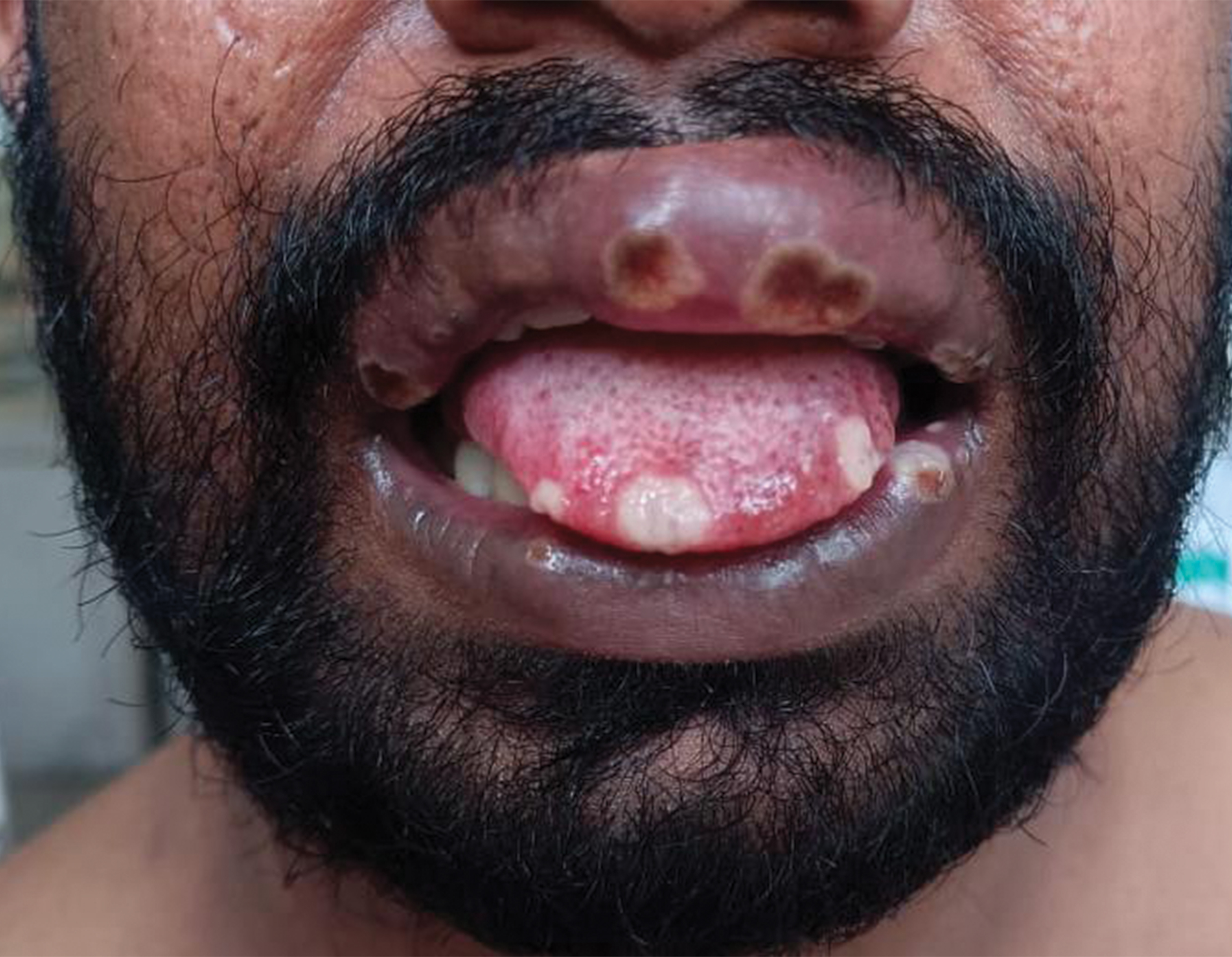
- Monkeypox. Day-7. Upper lip oedema with well-defined erosions with slough
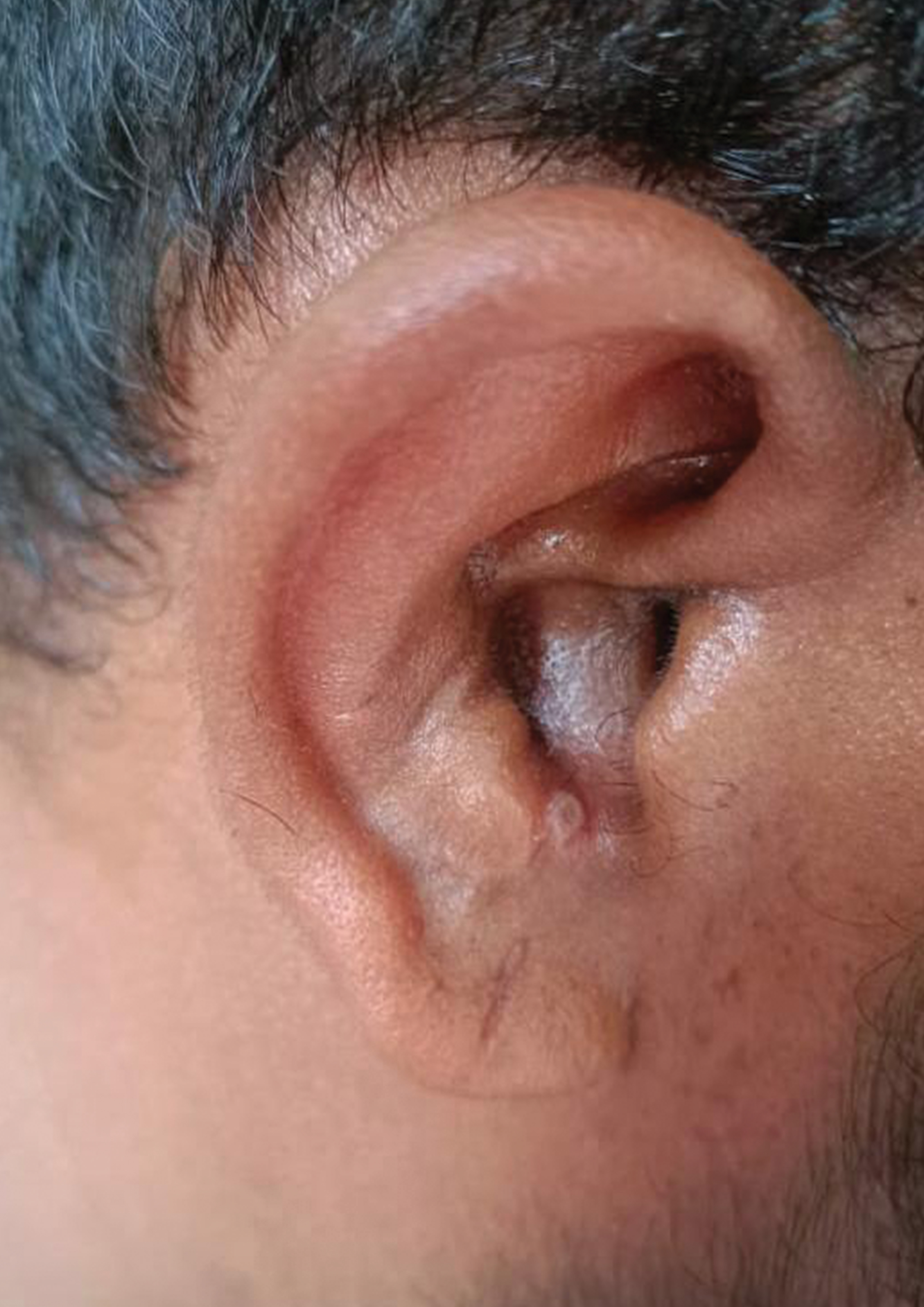
- Monkeypox. Day-7. Umbilicated papulo-vesicle
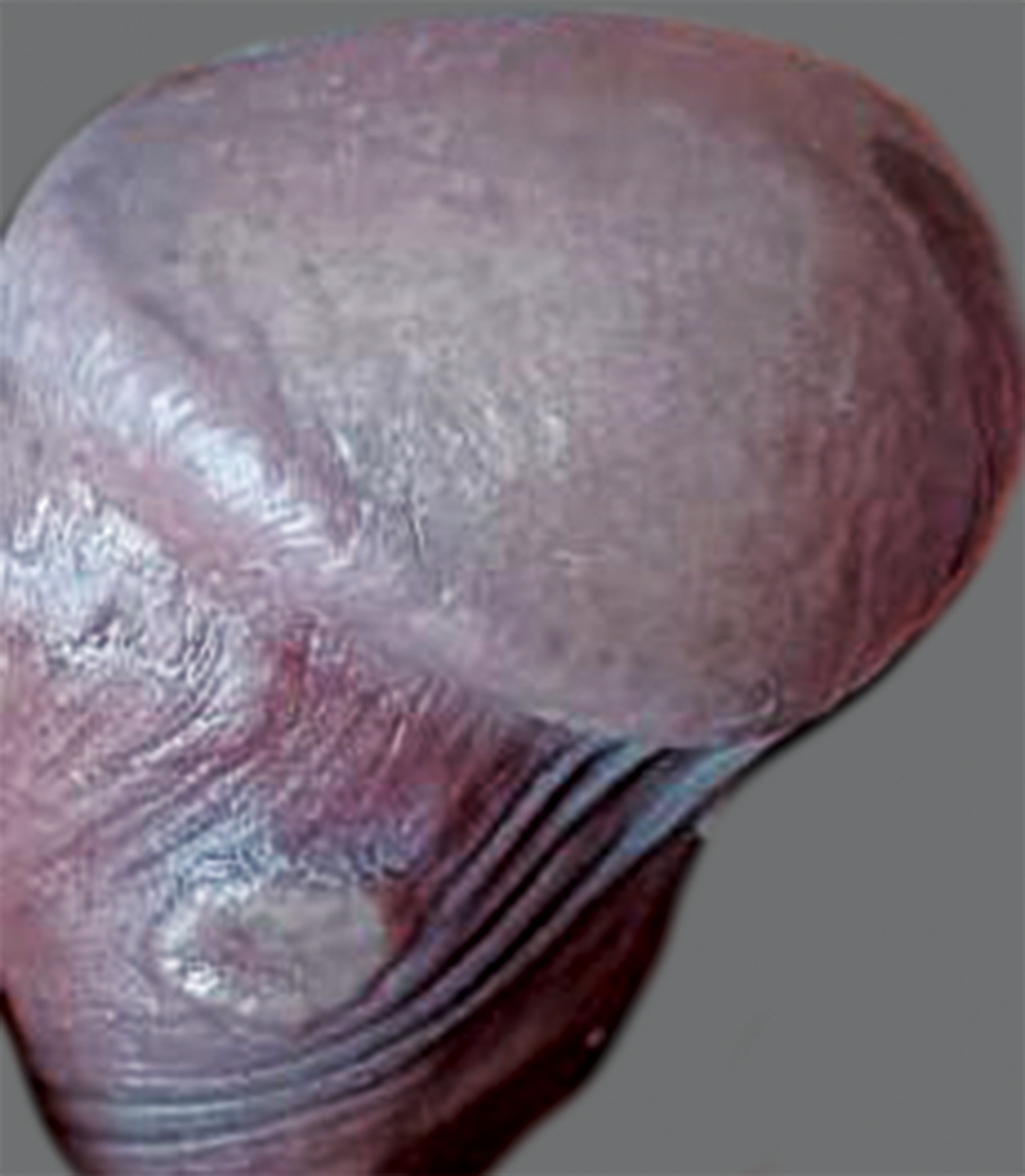
- Monkeypox. Day-7. Papule with erosion on shaft of penis
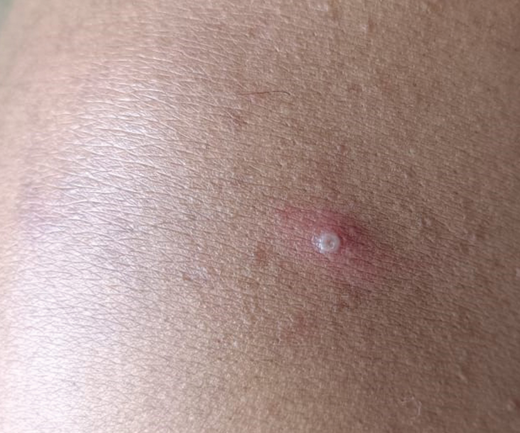
- Monkeypox. Day-10. Pustule on shoulder with central umbilication
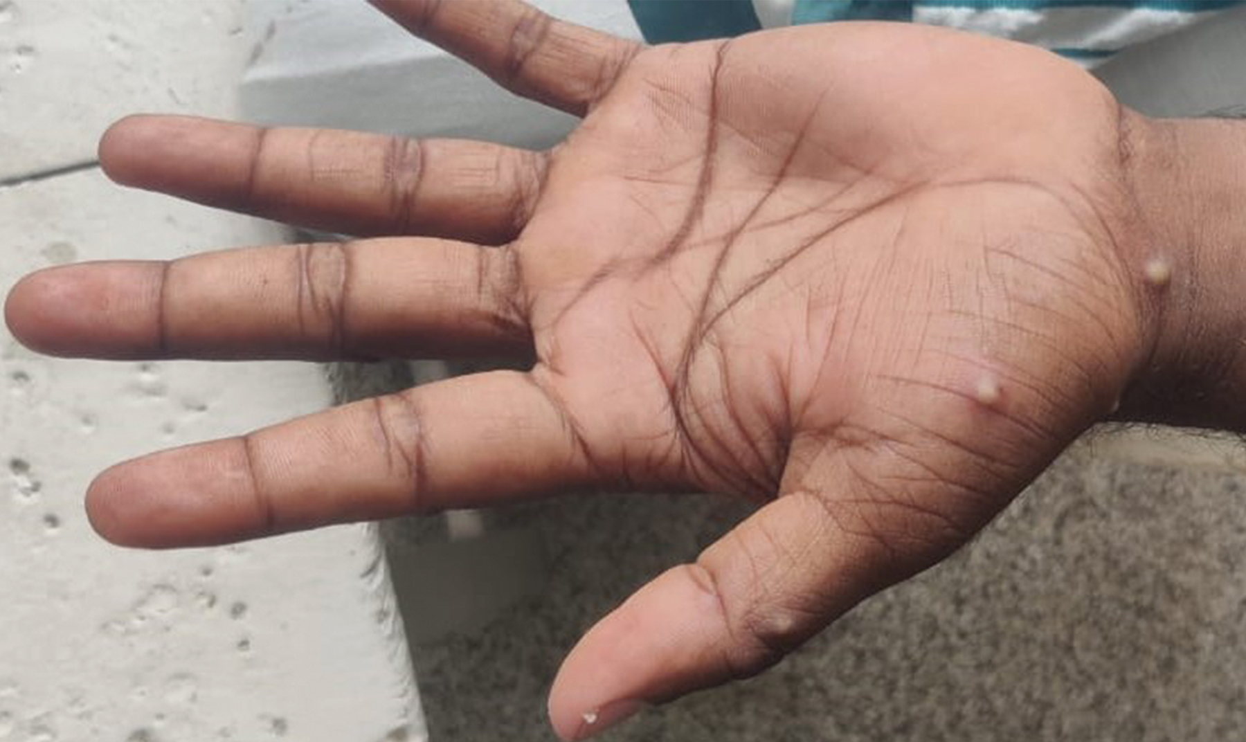
- Monkeypox. Day-10. Pustules on palm
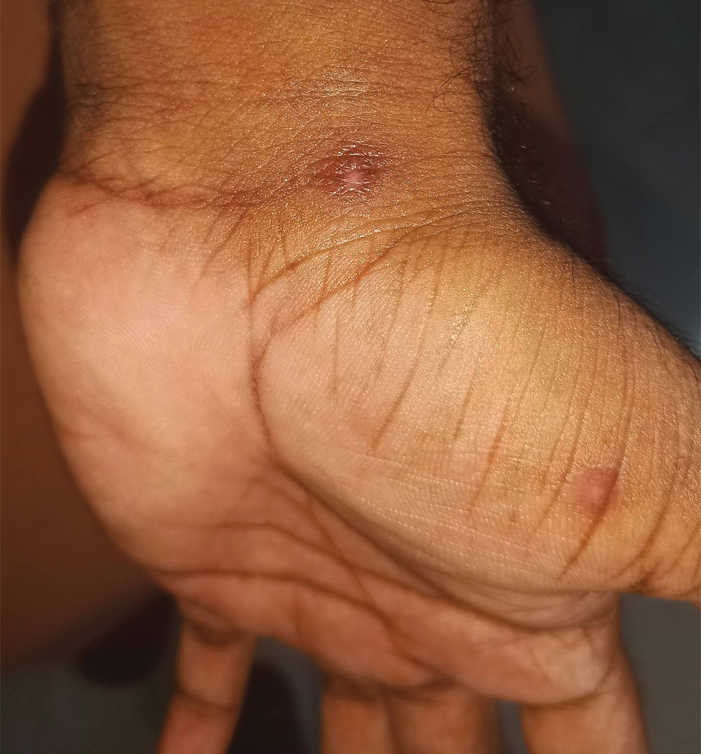
- Monkeypox. Day-23. Healed lesion on palm, with central atrophy and hypopigmentation; peripheral hyperpigmentation
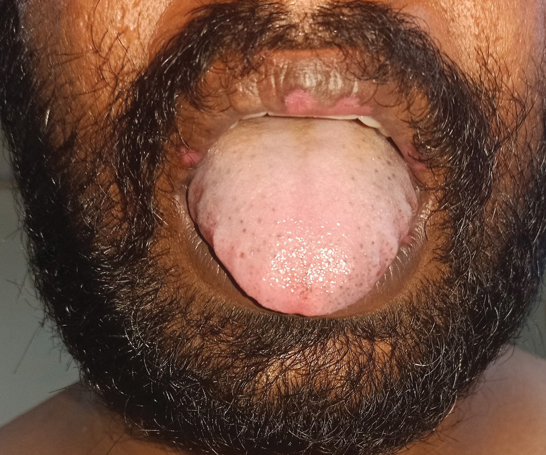
- Monkeypox. Day-23. Healed lesions on lips with central atrophy and hypopigmentation; peripheral hyperpigmentation
Incidentally, the patient also turned positive for Herpes Simplex Virus (HSV) (1 and 2) Immunoglobulin IgG and IgM. During the course of the hospital stay, he was also detected to have transaminitis; elevated levels of D-dimer and inflammatory markers C-Reactive Protein (CRP), Lactate Dehydrogenase (LDH) & Erythrocyte Sedimentation Rate (ESR). The test values and their changes on the various days of hospitalisation are shown in Table 1.
Tests
D-1 in hospital
D-3 in hospital
D-5 in hospital
D-7 in hospital
D-11 in hospital
D-16 in hospital
D-18 in hospital
D-8 of onset
D-10 of onset
D-12of onset
D-14 of onset
D-18 of onset
D-23 of onset
D-25 of onset
PCR
Positive
Positive
Positive
Negative
Negative
SGOT
48
76
42
SGPT
109
133
132
ESR
9
120
120
82
60
D-dimer (µg/mL)
3.52
1.27
CRP (mg/dl)
Positive
2.22
LDH (IU/L)
1024
394
HSV 1/2 IgM (RU/mL)
2.74
HSV 1/2 IgG (RU/mL)
147.2
By the 14th day (day 7 of hospitalisation), lesions crusted and those on the lips had started to heal. In addition to samples as before, PCR was done from the crust on the lips. All samples remained positive, except that of the lip. On the 18th day (day 11 of hospitalisation), the crusts had fallen off, and lesions had almost re-epithelialised with residual pigmentary changes. PCR repeated on all samples became negative. By the 23rd day (day 16 of hospitalisation), all lesions had healed well, leaving minimal central atrophy, hypopigmentation and peripheral hyperpigmentation [Figures 5a and b]. PCR of all samples was repeated for confirmation to ensure that the patient was no longer infectious to others before he was discharged from the hospital and it was found to be negative. He did not have any systemic involvement or complications.
The patient was accompanied by his mother and brother-in-law in the same vehicle to the hospital. Being close contacts, they were also isolated in the hospital and their blood, urine and nasopharyngeal swabs were sent for PCR for monkeypox, and it was negative.
The patient was managed conservatively with antibiotics (amoxycillin 500 mg + clavulanic acid 125 mg, thrice daily for 5 days) for follicular tonsillitis, acyclovir (400 mg thrice daily for 5 days) and analgesics, in addition to supportive therapy which included regular psychological counselling also. He was discharged on the 25th day after the onset of symptoms (18th day of hospitalisation).
Discussion
The monkeypox virus (genus: Orthopoxvirus, family: Poxviridae),1 has two genetic clades, the West African and Central African.3 The current strain is now said to belong to a new clade 3, of the West African strain, which is milder and less fatal (<1%) as per Centre for Infectious Disease Research and Policy (CIDRAP News of Jun 24, 2022). There have been only about 10 fatalities since January 2022. Although first discovered in monkeys in a research facility in 1958 in Copenhagen, Denmark,4 which gave it its name, the largest animal reservoirs of the virus have been found in rodents, including squirrels and giant pouched rats, both of which are hunted for food.3 The first human case was documented in 1970 in an infant from The Democratic Republic of Congo,2 while smallpox which is caused by a related virus, was declared eradicated by WHO in 1980 as a result of vaccinations. Smallpox vaccination confers 85% immunity against monkeypox too,5 but the waning herd immunity due to the discontinuation of smallpox vaccination 42 years ago, could be a reason for the resurgence of monkeypox.6 The virus spreads by close skin or mucosal contact with an infected person (touching, kissing, cuddling, sex), or through aerosols, contaminated fomites (beddings, towels, utensils) or by handling an infected animal or its meat. (CDC)
The incubation period of monkeypox is 5–21 days,7 (average 8.1 days), after which a prodrome of fever, headache, myalgia, chills, nasal congestion, cough and fatigue occur, along with lymphadenopathy, a feature which differentiates it from smallpox. Prodrome is followed by small oral mucosal papules or erosions and skin lesions over the face which extend centrifugally to the extremities. Over the next four weeks, the rashes evolve through macular, papular, vesicular and pustular phases in 1–2 day increments, being monomorphic at each stage. All lesional changes are synchronous and the pustular phase heals in 5–7 days with crusting. In most patients, the scabs fall off in 7–14 days and patients are considered to cease being infective at this point.8 Monkeypox can spread to others from the time of onset of symptoms till the crust has fallen off, the rash has fully healed and a new layer of skin has formed.9 Extracutaneous manifestations like secondary infection of skin and/or soft-tissue, pneumonitis, encephalitis, eye involvement and blindness, foetal transmission in infected pregnant women, etc., are possible complications.10 Atypical presentations without prodrome, onset of lesions in genitalia,11 perianal lesions associated with proctitis,9 pleomorphism of skin lesions or even absence of skin lesions etc can also occur.12 In the current outbreak, monkeypox has been found more in males having sex with males and had onset of lesions on genitalia.11
The oral mucosal lesions of herpes simplex present as 1–2 mm blisters that rapidly break down and coalesce to form small painful shallow irregular ulcers covered by a yellowish-grey pseudomembrane and surrounded by an erythematous halo.13 The co-existence of herpes simplex infection could explain the extensive painful oral lesions in this patient.
Lab diagnosis is crucial to differentiate monkeypox from smallpox infection. Real-time PCR is the gold standard for diagnosis and this patient tested positive in all the samples sent, throughout the stages from maculopapular to crust and became negative from the stage when the crust fell off and re-epithelialisation occurred. This means that the patient has to be in the infective state for a diagnosis.14 Conventional tests such as viral isolation from a clinical specimen, electron microscopy, and immunohistochemistry are valid techniques but require advanced technical skills and training and a sophisticated laboratory.14
The current treatments for human monkeypox include oral tecovirimat, brincidofovir, cidofovir and vaccinia immune globulin-VIGIV.15 The vaccines available are JYNNEOS and ACAM2000, both live vaccines. JYNNEOS is administered subcutaneously, 0.5 mL, two doses four weeks apart, including as post-exposure prophylaxis.16 ACAM2000 is given as a single 0.0025 mL percutaneous dose.16
The extensive oral involvement seen in this patient is not a common feature in the ongoing monkeypox outbreak and could be due to the concurrent HSV infection. This patient is being reported as the first confirmed case of monkeypox in India and is unique with respect to the shorter duration and rapid clearance of lesions.
Declaration of patient consent
The authors certify that they have obtained all appropriate patient consent.
Financial support and sponsorship
Nil.
Conflict of interest
There are no conflicts of interest.
References
- Genomic history of human monkey pox infections in the Central African Republic between 2001 and 2018. Sci Rep. 2021;11:13085.
- [CrossRef] [PubMed] [Google Scholar]
- Human monkeypox: Epidemiologic and clinical characteristics, Diagnosis, and prevention. Infect Dis Clin North Am. 2019;33:1027-43.
- [CrossRef] [PubMed] [Google Scholar]
- A review of experimental and natural infections of animals with monkeypox virus between 1958 and 2012. Future Virol. 2013;8:129-57.
- [CrossRef] [PubMed] [Google Scholar]
- Major increase in human monkeypox incidence 30 years after smallpox vaccination campaigns cease in the Democratic Republic of Congo. Proc Natl Acad Sci U S A. 2010;107:16262-7.
- [CrossRef] [PubMed] [Google Scholar]
- The changing epidemiology of human monkeypox—A potential threat? A systematic review. PLoSNegl Trop Dis. 2022;16:e0010141.
- [CrossRef] [PubMed] [Google Scholar]
- Monkeypox, Who.int. 2022 [cited 12 October 2022] Available from: https://www.who.int/news-room/fact-sheets/detail/monkeypox
- Monkeypox virus and insights into its immunomodulatory proteins. Immunol Rev. 2008;225:96-113.
- [CrossRef] [PubMed] [Google Scholar]
- 2022 Clinical Recognition [online] Available at: https://www.cdc.gov/poxvirus/monkeypox/clinicians/clinical-recognition.html [Accessed 12 October 2022]
- Human monkeypox: An emerging zoonosis. Lancet Infect Dis. 2004;4:15-25.
- [CrossRef] [PubMed] [Google Scholar]
- Global human monkeypox outbreak: Atypical presentation demanding urgent public health action. Lancet Microbe. 2022;3:e554-5.
- [CrossRef] [PubMed] [Google Scholar]
- Monkeypox: An emerging global threat during the COVID-19 pandemic. J Microbiol Immunol Infect. 2022;55:787-94.
- [CrossRef] [PubMed] [Google Scholar]
- Herpes Simplex Virus Type 1 infection: overview on relevant clinico-pathological features: HSV-1 literature review. J Oral Pathol Med. 2007;37:107-21.
- [CrossRef] [PubMed] [Google Scholar]
- Detection of monkeypox virus with real-time PCR assays. J Clin Virol. 2006;36:194-203.
- [CrossRef] [PubMed] [Google Scholar]
- Clinical features and management of human monkeypox: Aretrospective observational study in the UK. Lancet Infect Dis. 2022;22:1153-62.
- [CrossRef] [PubMed] [Google Scholar]
- Interim Clinical Considerations for use of JYNNEOS and ACAM2000. Vaccines during the 2022 U.S. Monkeypox Outbreak. Updated October 19, 2022
- [Google Scholar]


