Translate this page into:
Translating tissue expression of STAT 1, 3 and 6 in prurigo nodularis to clinical efficacy of oral tofacitinib – A prospective single-arm investigational study
Corresponding author: Dr. Kabir Sardana, Department of Dermatology, Atal Bihari Vajpayee Institute of Medical Sciences & Dr. Ram Manohar Lohia Hospital, New Delhi, Delhi, India. kabirijdvl@gmail.com
-
Received: ,
Accepted: ,
How to cite this article: Sardana K, Mathachan SR, Muddebihal A, Agrawal D, Ahuja A. Translating tissue expression of STAT 1, 3 and 6 in prurigo nodularis to clinical efficacy of oral tofacitinib – A prospective single-arm investigational study. Indian J Dermatol Venereol Leprol. doi: 10.25259/IJDVL_1017_2024
Abstract
Background
Interleukin (IL)-4, IL-13, IL-17, IL-22 and IL-3 are overexpressed in prurigo nodularis (PN). They mediate their action via the Janus Kinase (JAK) Signal transducer and activator of transcription (STAT) pathway.
Objectives
Our aim was to study the expression of tissue STAT1, STAT3, and STAT6, as well as the efficacy of the JAK-STAT inhibitor, tofacitinib, in PN.
Methods
A prospective study was conducted in a tertiary care hospital. Patients with PN were recruited after excluding secondary causes. Pruritus was graded using Pruritus Grading System Score (PGSS). All cases underwent histological assessment using immunohistochemical markers for STAT1, STAT3, and STAT6 in both lesional and perilesional skin. Tofacitinib was initiated at a dose of 5 mg twice daily or 11 mg once daily and then tapered to a maintenance dose. The final PGSS at the time of data evaluation, as well as the occurrence of remissions and relapses, was assessed.
Results
The majority of the 17 patients included in the study had moderate to severe disease. Immunohistochemical analysis revealed marked tissue expression of STAT6 in 13 and STAT3 in 10 patients, while STAT1 expression was seen in only 4 patients [p < 0.05], suggesting a Th2/Th17 tissue response. The mean onset of action of tofacitinib was 11.2 ± 6.44 days and the mean duration of treatment was 5.6 ± 2.2 months. A significant reduction in PGSS was noted after treatment (66.1%, P value 0.0004). Fourteen of the patients maintained remission on low-dose therapy (5 mg OD or A/D) while one patient experienced a relapse. No serious adverse effects were noted.
Limitation
We could not study the tissue cytokines and the expression of STATs after achieving clinical response on oral tofacitinib.
Conclusion
The efficacy of tofacitinib in PN is based on its inhibitory effect on Th2 and Th17 cytokines, which is dependent on STAT6 and STAT3.
Keywords
Prurigo nodularis
JAK
STAT
Tofacitinib
efficacy
Th2
Th17
JAK inhibitor
dupilumab
cytokine
Introduction
Prurigo nodularis (PN) is characterised by an immunological interplay among T helper (Th) 2, Th17, Th22, and Th1 cells, along with dysregulation of fibroblastic biology and neural dysfunction.1 T helper subtypes are assessed by the tissue expression of signal transducer and activator of transcription (STAT) proteins, a part of the Janus kinase (JAK)/STAT signalling pathway. STAT proteins mediate the actions of immune cells, including STAT1 and STAT4 (Th1), STAT5 and STAT6 (Th2), and STAT3 and STAT5 (Th17).2 Notably, cytokines such as IL-4, IL-13, IL-17, IL-22, and IL-31, which are up-regulated in PN, act via the JAK/STAT pathway.3,4
STAT1, STAT3, and STAT6 are all implicated in the pathophysiology of PN, with a dominance of STAT3 and STAT6,5,6 suggesting that the Th2/Th17 pathway and its related cytokines play a significant role in PN. The marked neural proliferation observed in PN is a consequence of certain cytokines, specifically IL-4, IL-13, and IL-31.4,6 The role of STAT receptors in PN, particularly in the context of concurrent use of JAK inhibitors, has not been previously investigated. In this study, we assessed the expression of STAT1, STAT3, and STAT6 in biopsies of PN lesions and evaluated the efficacy of oral tofacitinib.
Methods
Settings and Participants
This prospective study was carried out in the Dermatology OPD of a Tertiary Care Hospital from 1st March 2021 to 1st April 2023. Institutional and University Ethics Approval (F. No.TP (MD/MS)(109/2018)/IEC/PGIMER/RMLH1945) were obtained.
All clinically diagnosed cases of PN were assessed to rule out any secondary causes. The study excluded individuals under 18 years of age, immunocompromised patients, pregnant and lactating women, and those receiving active treatment for PN. Patients receiving active systemic treatment were instructed to discontinue all medications for three weeks and were maintained on topical glucocorticosteroids and antihistamines prior to the start of the study.
Investigations
A complete blood count, erythrocyte sedimentation rate, fasting blood sugar levels, liver and kidney function tests, viral markers, stool for occult blood (to exclude malignancy), chest X-ray (to exclude tuberculosis or other chest infections), and abdominal ultrasound were performed in all patients to rule out secondary causes of PN.
Severity grading
The PGSS was used to assess the severity of prurigo, which was graded as mild (score 0–5), moderate (score 6–11), and severe (score 12–19).7
Histology and immunohistochemistry (IHC) evaluation for STAT1, 3 and 6
All cases underwent histological assessment using immunohistochemical (IHC) markers targeting components of the JAK-STAT cell signaling pathway, which serve as surrogate markers for T helper cells. The IHC markers used included STAT-1, STAT-3, and STAT-6 (Polymer HRP IHC Detection System, Biogenex) to identify Th1, Th17/Th2, and Th2 cells, respectively.
Treatment
Tofacitinib was initiated at a dose of 5 mg twice daily in 13 patients and 11 mg (extended-release) once daily in 4 patients. The onset of response, defined as a 50% reduction in pruritus, was recorded. When the PGSS decreased by more than 75% from baseline, the dose was reduced. The dosage was further adjusted to alternate days after the patient achieved remission, defined as the absence of new lesions and a PGSS reduction of less than 75% while on a dose of 5 mg once daily for one month. Treatment was discontinued if remission was maintained on alternate-day therapy for an additional two months (remission off therapy). In the event of a relapse, defined as a 25% increase in PGSS, treatment was restarted at a dose of 5 mg once daily. Concurrent emollients and antihistamines were used as adjunctive therapies.
Statistical methods
Data were entered into an MS Excel spreadsheet, and analysis was performed using the Statistical Package for the Social Sciences (SPSS) version 23.0. A p-value of < 0.05 was considered statistically significant.
Results
Demographic profile and prior treatments
The demographic and clinical details are presented in Table 1. The mean disease duration was 16.6 ± 10 months, and the baseline PGSS was 12.1 ± 2.3.
| Mean age | 37.6±22.7 |
| Gender | |
| Male | 7 |
| Female | 10 |
| PN cases as per PGSS (pre-treatment) | |
| Mild | 0 |
| Moderate | 8 |
| Severe | 9 |
| Mean duration of disease | 16.6±10 months |
| Baseline PGSS | 12.1±2.3 |
| Previous treatments |
Thalidomide (n=2), methotrexate (n=3), oral steroids (n=3), topical steroids (n=6), phototherapy (n=1) and ayurvedic (n=1) and homeopathic treatments (n=1). Combination treatments (n=4) |
Of the 17 patients studied, 8 had moderate PN, and 9 had severe PN. Six of these patients had previously been treated with topical steroids, while 3 each had received systemic steroids and methotrexate. Additionally, 2 patients were treated with thalidomide, and one patient each underwent phototherapy, Ayurveda, and homeopathy. Some patients also received combination therapies.
STAT expression
Immunohistochemistry analysis revealed significant expression of STAT6 and STAT3 in 13 and 10 patients, respectively, and STAT1 expression in 4 patients. [Figure 1, Table 2].
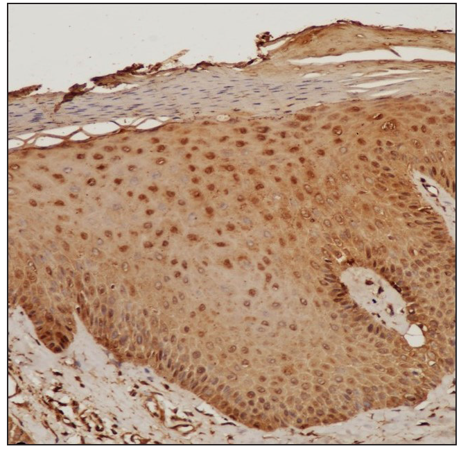
- IHC of the involved skin showing moderate nuclear positivity in the keratinocytes (STAT3, 100x).
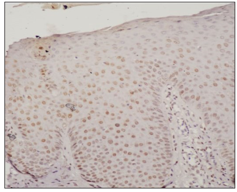
- IHC of the uninvolved skin showing weak nuclear positivity in the keratinocytes (STAT3, 400x).
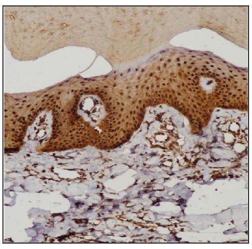
- IHC of involved skin showing strong nuclear positivity in the keratinocytes (STAT6, 400x).
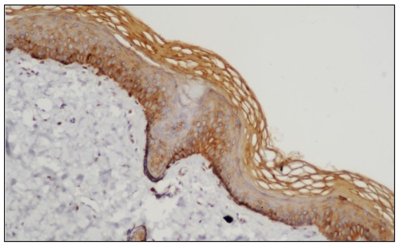
- IHC of the uninvolved skin showing absence of STAT6 staining in the nuclei of keratinocytes (STAT6, 400x).
| STAT | Positive | Negative | Chi-square test | P value* |
|---|---|---|---|---|
| STAT6 | 13 (8.5) [2.38] | 4 (8.5) [2.38] | 4.3714 | 0.036546 |
| STAT1 | 4 (8.5) [2.38] | 13 (8.5) [2.38] | ||
| STAT3 | 10 (7) [1.29] | 7 (10) [0.9] | 9.5294 | 0 .002022 |
| STAT1 | 4 (7) [1.29] | 13 (10) [0.9] |
Treatment with Systemic Tofacitinib
The mean treatment duration was 5.6 ± 2.2 months, with a 50% reduction in pruritus observed after 11.2 ± 6.44 days [Figure 2]. No significant correlation was found between disease duration and treatment response (p=0.16). The final mean PGSS was 4.1 ± 1.1, reflecting a statistically significant reduction of 66.1% [Table 3, Figure 3]. At the time of data analysis, 14 of the 16 patients were in remission on low-dose therapy, while 2 had completed treatment. One patient who relapsed after discontinuing treatment had therapy restarted [Figure 4].
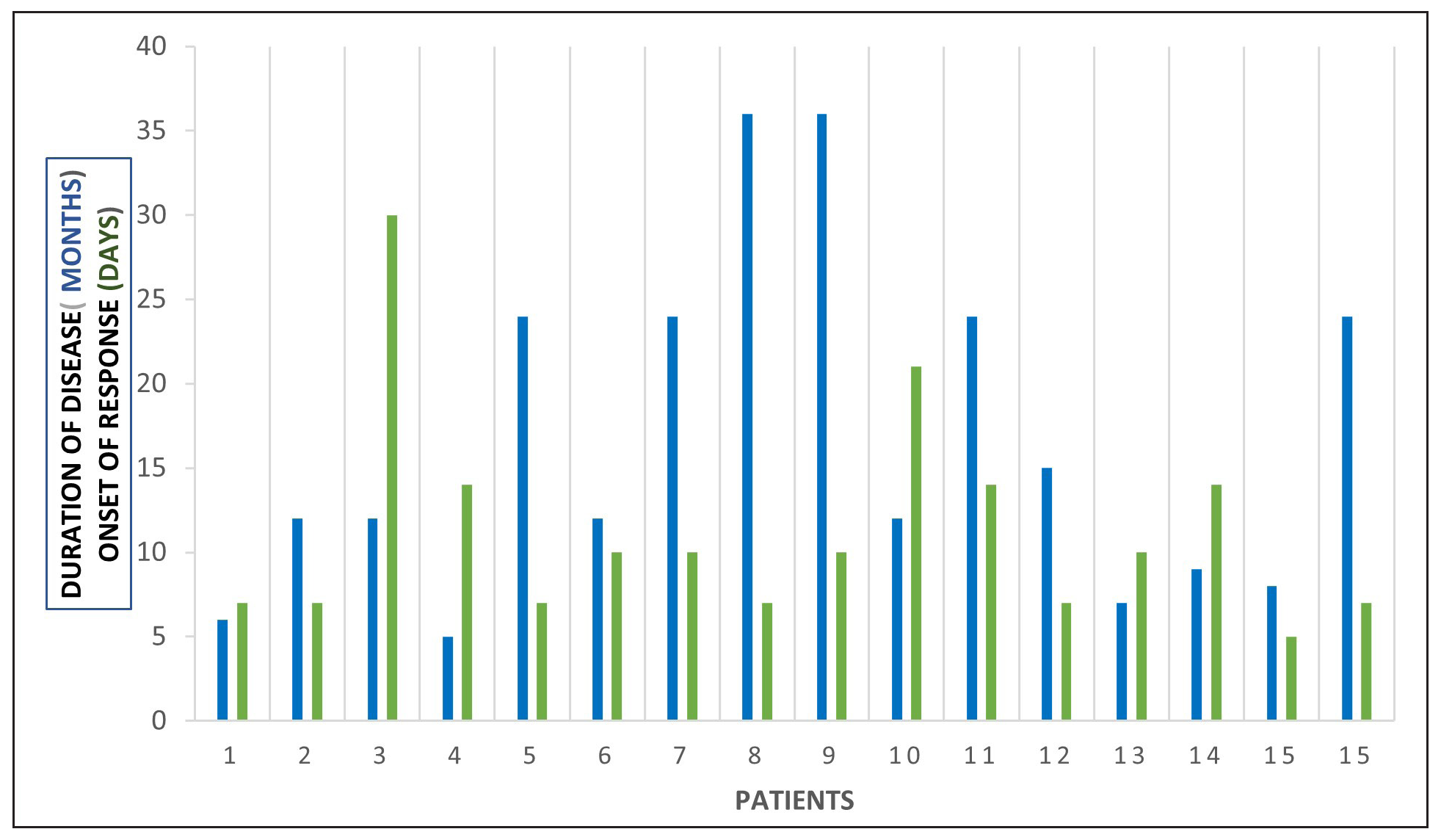
- A depiction of the duration of disease (months) and onset of response (days). The X axis represents individual patients.
| Mean duration of treatment | 5.6±2.2 months |
| Mean onset of action | 11.2±6.44 days |
| Mean final PGSS | 4.1±1.1 (P value 0.0004) |
| Number of PN cases as per PGSS (post-treatment) | |
| Mild | 14 |
| Moderate | 3 |
| Severe | 0 |
| Remission on low-dose therapy | 15 |
| Remission off therapy | 2 |
| Relapse | 1 |
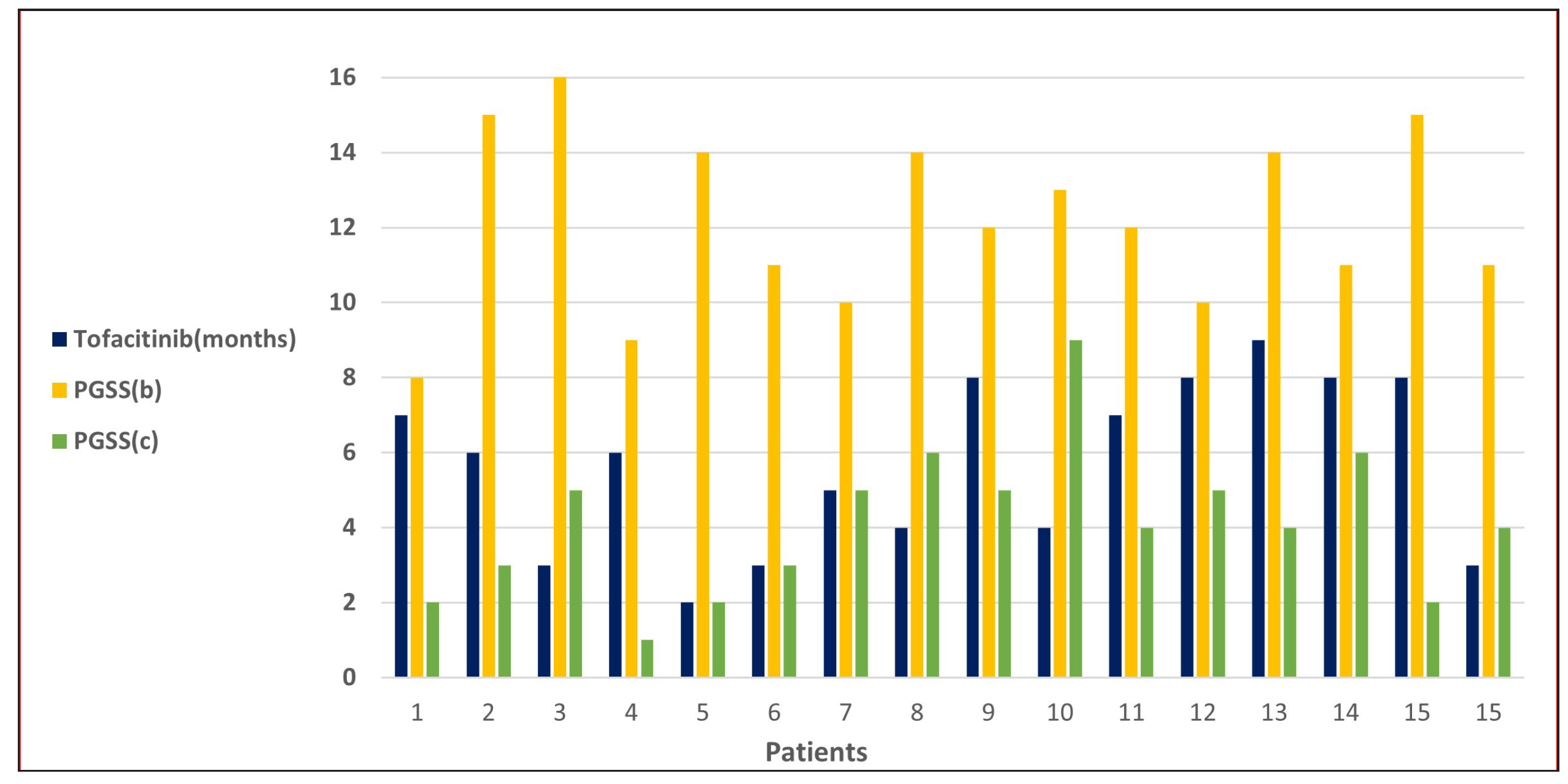
- A depiction of the duration of tofacitinib (months) and the baseline PGSS score (PGSSb) and current PGSS score (PGSSc).The X axis represents individual patients.
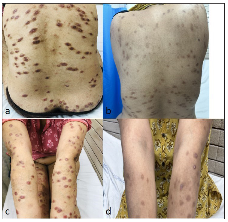
- (a) Multiple hyperpigmented to flesh-coloured nodules and plaques present over the back. (b) Resolution of lesions on the back with post-inflammatory hyperpigmentation post tofacitinib therapy. Tofacitinib (extended release tablet) 11 mg for mean duration of 3 months. (c) Multiple flesh-coloured nodules present over both upper limbs. (d) Resolution of lesions on upper limbs with post-inflammatory hyperpigmentation post-tofacitinib therapy. Tofacitinib (extended release tablet) 11 mg for mean duration of 3 months
Side effects observed included mild elevations in transaminases (SGOT or SGPT) in two patients and increased triglycerides in one patient and a reduction in lymphocyte/neutrophil counts in another patient. Acneiform eruptions were seen in two patients, and varicella occurred in a single patient. Except for the patient with varicella in whom treatment was temporarily paused for two weeks, no other patient had their treatment discontinued due to side effects.
Discussion
We observed increased tissue expression of STAT3 and STAT6 [Figure 1, Table 2]. STAT3 is activated by cytokines from the IL-6 and IL-10 families, as well as IL-21, IL-27, G-CSF, leptin, and IFN.8,9 STAT3 regulates the Th17 immune response, while STAT6 is primarily involved in the transduction of IL-4 and IL-13 signals.9,10 IL-4 is a key cytokine that mediates the differentiation of Th2 cells and immunoglobulin isotype switching.11,12 Additionally, STAT6 promotes the proliferation and maturation of B cells, enhances MHC-II and IgE expression, and plays a crucial role in mast cell activation. Our results explain the increased expression of mast cells and neural markers, suggesting STAT6 activation and are consistent with a previous study.1,6
Cytokines mediate their action through T helper cell subtypes, including Th1, Th2, Th17, and Th22, primarily via the JAK-STAT signaling pathway.9 These cytokines interact with cutaneous nerve fibers, keratinocytes, macrophages, mast cells, eosinophils and the JAK-STAT signalling pathway to shape the inflammatory response in PN. Interleukin (IL)-4 and IL-13 directly activate sensory neurons in the skin, driving itch sensation and enhancing the production of pro-inflammatory cytokines, IgE and fibroblasts through STAT6 signaling.11-13 IL-31, a Th2 cytokine, signals via the IL-31 receptor and oncostatin M receptor (OSMR) β to activate itch-sensing neurons, stimulate neuronal growth, and promote inflammatory cell activation.14,15 Additionally, IL-31 encourages fibroblast activity, drives T helper (Th) 2 polarisation and encourages further IL-31 production.16 While Th1, Th17, and Th22 cells play minor roles in inducing inflammation, IL-17 notably induces endothelin-1 (a histamine-independent pruritogen)17 while IL-22 facilitates epidermal differentiation, cutaneous inflammation, and keratinocyte proliferation.18
While a marked Th2 response is characteristic of PN, studies have shown that, unlike atopic dermatitis (AD), PN exhibits a strong fibrotic response in the stromal compartment, accompanied by abnormal activation of keratinocytes. This occurs alongside a relatively dampened type 2 inflammatory response compared to AD.19,20 Thus, the Th2 response in PN is less pronounced than in AD. Blocking IL-31Rα reduces both Th2/IL-13 and Th17/IL-17 responses, thereby stabilizing extracellular matrix remodeling.21
Our data suggest that JAK inhibitors can suppress the inflammatory activity of Th2/Th17 cells (STAT6/STAT3).22 Current treatment options for patients with PN are limited and many patients are dissatisfied with their therapy.21 The use of systemic tofacitinib has been largely speculative, with only a few reports addressing STAT expression.23 The cytokines involved, along with the STAT expression profile in PN, suggest that a treatment targeting multiple cytokines and immune cell pathways, such as JAK inhibitors, could offer a beneficial and potentially cost-effective approach to managing PN.24
Although most of our patients responded favorably to tofacitinib, the majority remained on low doses, suggesting that a maintenance dose may be necessary for patients with PN. A meta-analysis of 45 patients treated with dupilumab found that the mean time to first improvement was 10.15 ± 10.56 weeks, with final improvement achieved at 19.28 ± 13.71 weeks. Complete clearance of pruritus, when observed, occurred approximately four months after initiating dupilumab.23 However, our results suggest that tofacitinib is both more effective and acts more rapidly than dupilumab, with a 50% reduction in pruritus occurring in 11.2 ± 6.44 days and complete remission achieved on a low dose in 5.6 ± 2.2 months.
Published case reports have documented promising results with the use of tofacitinib (a JAK1/3 inhibitor), baricitinib (a JAK1/2 inhibitor), and upadacitinib (a JAK1 inhibitor) in the treatment of PN. Phase 2 clinical trials are currently investigating the efficacy of two other JAK1 inhibitors, abrocitinib (NCT05038982) and povorcitinib (NCT05061693), while a phase 3 trial is underway for ruxolitinib cream (a JAK1/JAK2 inhibitor) (NCT05755438, NCT05764161).25 Based on our findings, a pan-JAK inhibitor, such as tofacitinib, appears to be the most cost effective treatment option for PN.
Limitation and conclusions
Our study confirms the enhanced tissue expression of STAT3 and STAT6, which corresponds to the major cytokines involved in prurigo nodularis and in turn the therapeutic efficacy of a JAK inhibitor. The limitations of our work is that ideally tissue cytokine as well as JAK-STAT expression both prior to and following treatment with JAK inhibitors would be ideal, to validate the impact of tofacitinib on clinical outcomes and immune pathways. Nevertheless, the consistent reduction in itching, marked clinical improvement, and favorable safety profile suggest that tofacitinib holds significant potential for treating PN.
Ethical approval
The research/study was approved by the Institutional Review Board at ABVIMS & Dr. RML Hospital, number 1945, dated 24/10/2018.
Declaration of patient consent
The authors certify that they have obtained all appropriate patient consent.
Financial support and sponsorship
Nil.
Conflicts of interest
There are no conflicts of interest.
Use of artificial intelligence (AI)-assisted technology for manuscript preparation
The authors confirm that there was no use of artificial intelligence (AI)-assisted technology for assisting in the writing or editing of the manuscript and no images were manipulated using AI.
References
- A case-control study addressing the population of epidermal and dermal inflammatory infiltrate including neural milieu in primary prurigo nodularis using S-100 and toluidine blue stain and its therapeutic implications. Int J Dermatol. 2023;62:1352-8.
- [CrossRef] [PubMed] [Google Scholar]
- T cells in health and disease. Signal Transduct Target Ther. 2023;8:235.
- [CrossRef] [PubMed] [PubMed Central] [Google Scholar]
- Strategies to therapeutically modulate cytokine action. Nat Rev Drug Discov. 2023;22:827-54.
- [CrossRef] [PubMed] [PubMed Central] [Google Scholar]
- Molecular mechanisms of pruritus in prurigo nodularis. Front Immunol. 2023;14:1301817.
- [CrossRef] [PubMed] [PubMed Central] [Google Scholar]
- Nuclear localization of activated STAT6 and STAT3 in epidermis of prurigo nodularis. Br J Dermatol. 2011;165:990-6.
- [CrossRef] [PubMed] [Google Scholar]
- A prospective study examining the expression of STAT 1, 3, 6 in prurigo nodularis lesions with its immunopathogenic and therapeutic implications. J Cosmet Dermatol. 2022;21:4009-15.
- [CrossRef] [PubMed] [Google Scholar]
- Using pruritus grading system for measurement of pruritus in patients with diseases associated with itch. J Med J. 2012;46:39-44.
- [Google Scholar]
- Stat3: A STAT family member activated by tyrosine phosphorylation in response to epidermal growth factor and interleukin-6. Science. 1994;264:95-8.
- [CrossRef] [PubMed] [Google Scholar]
- The JAK/STAT signaling pathway: From bench to clinic. Signal Transduct Target Ther. 2021;6:402.
- [CrossRef] [PubMed] [PubMed Central] [Google Scholar]
- STAT6 as an asthma candidate gene: Polymorphism-screening, association and haplotype analysis in a Caucasian sib-pair study. Hum Mol Genet. 2002;11:613-21.
- [CrossRef] [PubMed] [Google Scholar]
- Lack of IL-4-induced Th2 response and IgE class switching in mice with disrupted Stat6 gene. Nature. 1996;380:630-3.
- [CrossRef] [PubMed] [Google Scholar]
- Stat6 is required for mediating responses to IL-4 and for development of Th2 cells. Immunity. 1996;4:313-9.
- [CrossRef] [PubMed] [Google Scholar]
- Antipruritic effects of janus kinase inhibitor tofacitinib in a mouse model of psoriasis. Acta Derm Venereol. 2019;99:298-303.
- [CrossRef] [PubMed] [Google Scholar]
- IL-31 inhibition as a therapeutic approach for the management of chronic pruritic dermatoses. Drugs. 2021;81:895-905.
- [CrossRef] [PubMed] [PubMed Central] [Google Scholar]
- IL-31: A new link between T cells and pruritus in atopic skin inflammation. J Allergy Clin Immunol. 2006;117:411-7.
- [CrossRef] [PubMed] [Google Scholar]
- Interleukin-31 promotes fibrosis and T helper 2 polarization in systemic sclerosis. Nat Commun. 2021;12:5947.
- [CrossRef] [PubMed] [PubMed Central] [Google Scholar]
- IL-17A Induces endothelin-1 expression through p38 pathway in prurigo nodularis. J Invest Dermatol. 2020;140:702-706.e2.
- [CrossRef] [PubMed] [Google Scholar]
- Prurigo nodularis is characterized by systemic and cutaneous t helper 22 immune polarization. J Invest Dermatol. 2021;141:2208-2218.e14.
- [CrossRef] [PubMed] [PubMed Central] [Google Scholar]
- Single-cell RNA sequencing defines disease-specific differences between chronic nodular prurigo and atopic dermatitis. J Allergy Clin Immunol. 2023;152:420-35.
- [CrossRef] [PubMed] [Google Scholar]
- Single-cell profiling of prurigo nodularis demonstrates immune-stromal crosstalk driving profibrotic responses and reversal with nemolizumab. J Allergy Clin Immunol. 2024;153:146-60.
- [CrossRef] [PubMed] [PubMed Central] [Google Scholar]
- Transcriptomic characterization of prurigo nodularis and the therapeutic response to nemolizumab. J Allergy Clin Immunol. 2022;149:1329-39.
- [CrossRef] [PubMed] [PubMed Central] [Google Scholar]
- Cytokine and cytokine receptors. In: Rich RR, Fleisher TA, Shearer WT, Schroeder HW Jr, Frew AJ, Weyand CM, eds. Clinical Immunology: Principles and Practice (5th ed). Amsterdam: Elsevier; 2019. p. :127-55.
- [Google Scholar]
- Treatment of recalcitrant paediatric prurigo nodularis with tofacitinib, an exquisite example of bench-to-bedside translation of JAK-STAT expression. Indian J Dermatol Venereol Leprol 2023:1-3.
- [CrossRef] [Google Scholar]
- Phase 3 trial of nemolizumab in patients with prurigo nodularis. N Engl J Med. 2023;389:1579-89.
- [CrossRef] [PubMed] [Google Scholar]
- Prurigo nodularis: New insights into pathogenesis and novel therapeutics. Br J Dermatol. 2024;190:798-810.
- [CrossRef] [PubMed] [PubMed Central] [Google Scholar]


