Translate this page into:
Trichosporon inkin and Trichosporon mucoides as unusual causes of white piedra of scalp hair
2 Department of Dermatology, Lokmanya Tilak Municipal Medical College and General Hospital, Sion, Mumbai, India
Correspondence Address:
Uma Tendolkar
Department of Microbiology, Lokmanya Tilak Municipal Medical College and General Hospital, Sion, Mumbai - 400 022
India
| How to cite this article: Tendolkar U, Shinde A, Baveja S, Dhurat R, Phiske M. Trichosporon inkin and Trichosporon mucoides as unusual causes of white piedra of scalp hair. Indian J Dermatol Venereol Leprol 2014;80:324-327 |
Abstract
White piedra of scalp hair is considered a rare entity. We report three cases of this disorder all of whom presented with nodules on the hair. Potassium hydroxide preparations of the hair revealed clustered arthrospores and mature, easily detachable nodules. Cultures grew Trichosporon inkin in 2 patients and Trichosporon mucoides in one patient. Both these fungi are unusual causes of white piedra.INTRODUCTION
Piedra, a superficial fungal infection of the hair shaft, often causes progressive weakness of the hair shaft leading to hair fall. The infection comprises of nodular concretions over the hair. Black piedra caused by Piedraia hortae is common in tropical climates of Central and south America and South East Asia whereas white piedra caused by Trichosporon species is more common in temperate and semitropical climates like Europe, Japan and parts of South USA. [1] The mode of infection in humans is not known. White piedra is considered a rare infection of scalp hair [2] and is commonly seen in pubic hair, axillary hair, beard, moustaches, eyebrows and eyelashes. Black piedra is considered to be more common in scalp hair. In Brazil, white piedra is reported to affect scalp hair most commonly. Black piedra occurs in monkeys and humans. [1] Both types of piedra ultimately can lead to hair breakage because the shaft is weakened by circular penetration by the fungus. T. ovoides is considered to be the common causative agent of white piedra, however an Indian report shows that T. inkin is also involved in white piedra. [2]
Three cases of white piedra are being reported, one caused by Trichosporon mucoides which is an unusual causative agent and two by Trichosporon inkin.
CASE REPORT
A 28-year-old man presented with history of hair fall for one month and of small lumps on the hair for three months. The second case was a seven year old Muslim girl who presented with similar nodules on the hair shaft which were noticed about two months back. The third case also was a Muslim female, thirty five years old, presenting with white nodules on the hair shaft for two months. She also had a history of intermittent exacerbations. Both the females had a history of tying wet hair. In all the three patients the nodules were distinct, compact and slightly creamy white in color [Figure - 1]. None of the patients had systemic complaints or a history suggestive of any disease or immunodeficiency. There was no history of similar complaints in the family. There was no history of nits or lice infesting the hair or regular use of any particular oil. Hair at other sites was normal. Their systemic examination was normal.
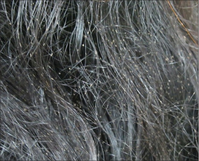 |
| Figure 1: Nodules of white piedra along the length of the hair |
Hair clippings were collected in a sterile petri dish for detailed mycological examination using a sterile scalpel blade. Small sections of the hair including the nodules were observed in 10% potassium hydroxide (KOH) mount. Some hair showed early clustering of the fungal arthrospores over the hair shaft [Figure - 2] and [Figure - 3] which represented a nodule in the making. The more mature nodules revealed complete coating of the hair shaft [Figure - 4], A gentle pressure on the cover slip revealed that the mature nodule was easily detachable and the underlying hair shaft appeared intact and normal [Figure - 5] and [Figure - 6].
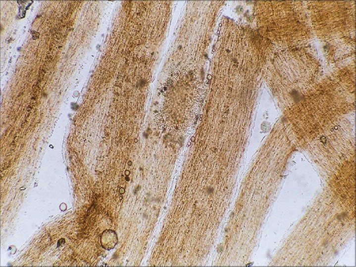 |
| Figure 2: Clustering of fungal arthrospores indicating beginning of nodule formation. KOH mount. ×100 |
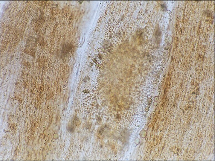 |
| Figure 3: Magnified view of the clustering of fungal arthrospores. KOH mount. ×200 |
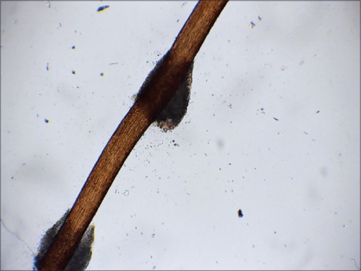 |
| Figure 4: Two mature nodules of white piedra on the hair shaft with normal hair intervening. KOH mount. ×100 |
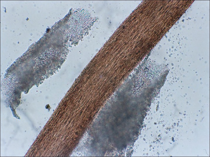 |
| Figure 5: Nodule on the hair shaft easily detachable with slight pressure. KOH mount. ×200 |
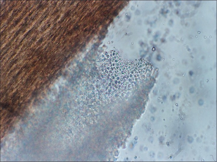 |
| Figure 6: Nodule on the hair shaft composed of closely aligned fungal arthrospores of Trichosporon mucoides. KOH mount. ×1000 |
For culture, the nodular concretions on the hair were gently pressed on Sabouraud′s dextrose agar (SDA) and the cultures were incubated at 26 °C in a cooling incubator. Within three days, rough, cream colored, cerebriform colonies were noted. Lactophenol cotton blue mount of the culture showed blastoconidia and arthrospores. For observing appressoria formation [Figure - 7], slide culture on potato dextrose agar was used. Growth at 37°C, assimilation tests and cycloheximide tolerance tests were used for species identification. [3] It is difficult to identify the species of Trichosporon from colony characteristics [Figure - 8]. The species of Trichosporon, T. mucoides was identified by a positive growth at 37°C and sorbitol assimilation test and T. inkin was identified by a positive growth at 37°C, inositol assimilation and appressoria formation and a negative sorbitol assimilation test. Both of these species are tolerant to 0.1% cycloheximide. The isolates from the adult man and the young girl were identified as Trichosporon inkin. The hair of the adult female on the other hand yielded T. mucoides. The patients
were advised cutting of hair and 2% selenium sulphide shampoo wash on alternate days. Oral itraconazole 100 mg. twice a day for 15 days was advised.
Since the patients did not return for follow up, the efficacy of the treatment cannot be commented upon.
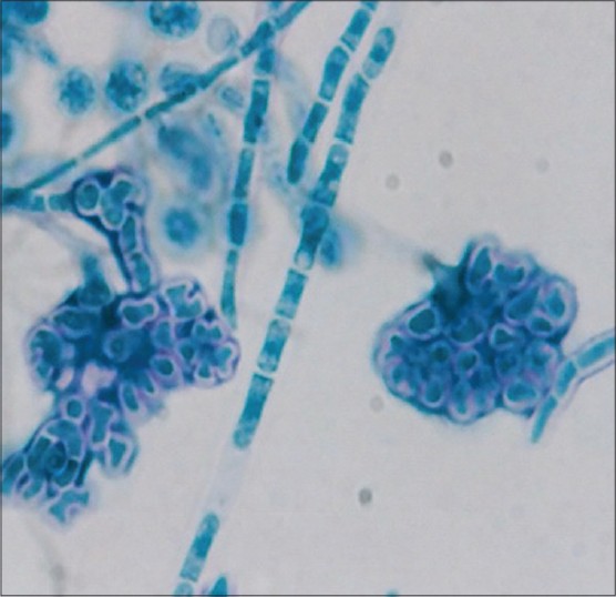 |
| Figure 7: Appressoria formation by Trichosporon inkin on potato dextrose agar. Lacto Phenol Cotton Blue Mount. ×200 |
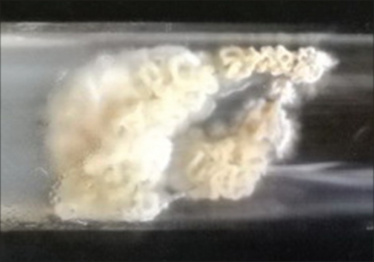 |
| Figure 8: Convoluted white colony of Trichosporon mucoides on Sabouraud's dextrose agar |
DISCUSSION
Six species of Trichosporon belonging to two clades identified by molecular studies appear to be of clinical importance, namely, Trichosporon ovoides, T. asahii, T. asteroids and T. inkin, belonging to the Ovoides clade and T. cutaneum and T. mucoides, belonging to the Cutaneum clade. [4] T. mucoides generally causes pubic white piedra, [5] onychomycosis and rarely, systemic infection. [6] Besides white piedra on pubic hair, T. inkin can occasionally cause endocarditis and pericarditis.
Patients with piedra of scalp generally seek medical advice because of cosmetic issues. In India both black and white piedra are found. Pasricha et al., [7] in their report mention that in India, black piedra cases have been reported from Varanasi, Mumbai and Delhi and cities from Kerala and white piedra cases have been reported from various cities like Mumbai, Chennai and Kolkata.
An increased occurrence in females has been reported, probable predisposing factors being the practice of tying up long hair when still wet, a feature seen in both of our female patients. Spread of the infection to family members has been noted. [8] Sharing of combs and application of oils and conditioners have also been proposed as predisposing factors but there is insufficient evidence. Piedra appears to be more common in Muslim females which could be related to the custom of wearing head covering veils adding to the warmth and humidity predisposing to yeast infections. [2] White piedra is common in young adults but is not uncommon in pediatric patients.
Other species, Trichosporon asahii and T. mucoides are more commonly associated with systemic infections and T. asteroids and T. cutaneum, with superficial skin lesions. [4]
Recent reports from India have implicated T. ovoides[9] and T. inkin in white piedra of the scalp hair. [2] One case due to T. ovoides reported by Shivprakash et al., was in a Sikh male in whom tying up of wet hair and increased humidity due to regular use of a turban may have been contributing factors. [10] With our report of T. mucoides, it appears that diverse species of Trichosporon may be implicated in causing white piedra. Antifungal drug susceptibilities may vary with species. Itraconazole and voriconazole appeared to be more active than amphotericin B and fluconazole in an in vitro study and T. mucoides and T.inkin appeared more resistant to triazoles than
T. asahii. [11]
| 1. |
Schwartz A, Altman R. Piedra EMedicine, 2009. Available from: http://emedicine.medscape.com/article/1092330-overview [Last accessed on 2013 Jul 1].
[Google Scholar]
|
| 2. |
Viswanath V, Kriplani D, Miskeen A, Patel B, Torsekar RG. White piedra of the Scalp hair by Trichosporon inkin. Indian J Dermatol Venereol Leprol 2011;77:591-3.
[Google Scholar]
|
| 3. |
de Hoog GS, Guarro J, Gene J, Figueras MJ, editors. Atlas of Clinical Fungi. Utrecht, The Netherlands: CBS; 2000.
[Google Scholar]
|
| 4. |
Reiss E, Shadomy HJ, Mars hall Lyon 3 rd G, editors. Fundamental Medical Mycology. New Jersey, USA: Wiley-Blackwell; 2012.
[Google Scholar]
|
| 5. |
Therizol-Ferly M, Kombila M, Gomez de diaz M, Douchet C, Salaum Y, Barrabes A, et al. White piedra and Trichosporon species in equatorial Africa II. Clinical and mycological associations: An analysis of 449 superficial inguinal specimens. Mycoses 1994;37:255-60.
[Google Scholar]
|
| 6. |
Kendirli T, Ciftci E, Ince E, Oncel S, Dalgic N, Guriz H, et al. Successful treatment of Trichosporon mucoides disseminated infection with lipid complex amphotericin B and 5-urocytosine. Mycoses 2006;49:251-3.
[Google Scholar]
|
| 7. |
Pasricha JS, Nigam PK, Banerjee U. White piedra in Delhi. Indian J Dermatol Venereol Leprol 1990;56:56-7.
[Google Scholar]
|
| 8. |
Roshan AS, Janaki C, Parveen B. White piedra in mother and daughter. Int J Trichol 2009;1:140-1.
[Google Scholar]
|
| 9. |
Tambe SA, Dhurat RS, Kumar CA, Thakare P, Lade N, Jerajani H, et al. Two cases of scalp white piedra caused by Trichosporon ovoides. Indian J Dermatol Venereol Leprol 2009;75:293-5.
[Google Scholar]
|
| 10. |
Shivprakash MR, Singh G, Gupta P, Dhaliwal M, Kanwar AJ, Chakrabarti A. Extensive white piedra of the scalp caused by Trichosporon inkin: A case report and review of literature. Mycopathologia 2011;172:481-6.
[Google Scholar]
|
| 11. |
Pahitou N, Otrosky-Zeichner L, Paetznick VL, Rodriguez JR, Chen E, Rex JH. In Vito Antifungal Susceptibilities of Trichosporon species. Antimicrob Agents Chemother 2002;46:1144-6.
[Google Scholar]
|
Fulltext Views
11,980
PDF downloads
2,244


