Translate this page into:
Ultrasound biomicroscopy of the skin to detect a subclinical neuroma of the proximal nail-fold
2 Consultant Dermatologist, Bandra, Mumbai, India
3 Department of Dermatology, LTMC, Sion, Mumbai, India
Correspondence Address:
Kalpana D Bhatt
UBM Institute, Ganesh Baug, Behind Ruia College, Dadar, Mumbai 400019, Maharashtra
India
| How to cite this article: Bhatt KD, Fernandes R, Dhurat R. Ultrasound biomicroscopy of the skin to detect a subclinical neuroma of the proximal nail-fold. Indian J Dermatol Venereol Leprol 2006;72:60-62 |
 |
 |
 |
 |
 |
 |
Sir,
Ultrasound is a very useful, noninvasive modality to visualize normal and abnormal tissue characteristics in situ . Recent advances in transducer technology have led to its application in dermatology.[1],[2] Here, ultrasound is used to measure skin thickness. It gives information on the axial and lateral extension of tumors[3] and inflammatory processes. It can also be used to monitor various inflammatory conditions such as psoriasis, scleroderma, etc, and to study the therapeutic effects of drugs on skin.
Dermatological ultrasound is performed using a 20-MHz probe, giving a resolution of 80 µm and depth penetration of 8-10 mm. Ultrasound biomicroscopy (UBM) is high-frequency ultrasonography performed using a 50-MHz transducer[4] or probe (the resolution is 40 mm and depth penetration is only 4 mm). In dermatology, UBM gives a better resolution of the dermis and epidermis compared with the lower-frequency probes. We present a case in which a subclinical tumor (neuroma) in the dermis and epidermis of the skin of the proximal nail-fold of the left thumb was detected by performing UBM.
A 35-year-old female presented with gradually increasing excruciating intermittent pain and tingling numbness at the tip of the left thumb since 5 years. There were no aggravating factors. There was no history of trauma or neck pain. Cutaneous examination revealed normal skin on the left thumb [Figure - 1] and no mass or any other abnormality could be palpated. The only positive finding was local tenderness in an area of 2 mm in the region of the proximal nail-fold of the left thumb.
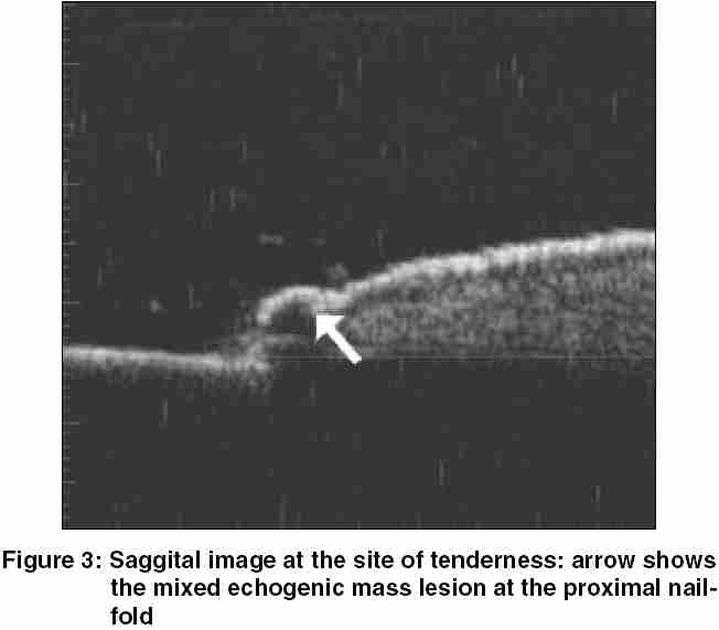
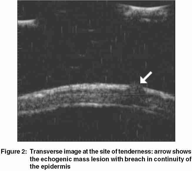
An X-ray of the cervical spine was normal and showed no evidence of spondylosis. A 20-MHz ultrasound of the affected area was indeterminate. Subsequently, UBM was performed. It showed a well-defined hypoechoic mass measuring 350 x 400 µm in the dermis extending into and causing a breach in the continuity of the overlying epidermis [Figure - 2][Figure - 3]. UBM of the opposite thumb was normal, showing a normal hyperechoic entry echo of the epidermis and a homogenous isoechoic dermis. A diagnosis of glomus tumor of the proximal nail fold was made. Excisional biopsy of the lesion was suggestive of neuroma. The patient is asymptomatic postoperatively.
Neuromas are solitary skin colored or pink firm papules or nodules at the site of trauma or a scar. In mature neuromas, a lancinating pain may be elicited in response to local pressure. Initially, as a routine, a 20-MHz ultrasound was performed. [5],[6],[7],[8] Because the findings were indeterminate, a UBM was performed and showed us a well-defined subclinical tumor.
A non-invasive investigation, UBM, is best suited for epidermal and dermal lesions as its resolution is far superior to that of a 20-MHz ultrasound probe. It exhibits very small anatomical details in the epidermis and dermis because its penetration depth is only 4 mm. Its resolution is comparable with that of a low-power microscope.
Since it is a non-invasive, investigative modality, it can be repeatedly performed with good patient compliance. However, UBM has its limitations in that it does not give tissue diagnosis, which rests on biopsy.
ACKNOWLEDGMENT
We thank Dr. H. R. Jerajani, Professor and Head, Department of Dermatology, LTMC, Sion, Mumbai, for her guidance.
| 1. |
Serup J. Ten years experience with high frequency ultrasound examination of the skin. In: Hoffman K, editor. Ultrasound in Dermatology. 1st ed. Germany: Springer-Verlag; 1992. p. 41-54.
[Google Scholar]
|
| 2. |
Thiboutot D. Dermatological application of high-frequency ultrasound. SPIE Conference Proceedings. 1999. Abstract No. 3664. Available at http://www.bioe.psu.edu/labs/NIH/derm_app. pdf.
[Google Scholar]
|
| 3. |
Breitbart EW, Mohr P. Sonographic structures of benign skin tumors. In: Hoffman K, editor. Ultrasound in Dermatology. 1st ed. Germany: Spinger-Verlag; 1992. p. 171-80.
[Google Scholar]
|
| 4. |
El-Gammal S, Hoffman K, Auer T, Korten M, Atlmeyer P, Hoss A, et al. A 50 MHz high-resolution ultrasound imaging system for dermatology. In : Hoffman K, editor. Ultrasound in Dermatology. 1st ed. Germany: Spinger-Verlag; 1992. p. 297-321.
[Google Scholar]
|
| 5. |
Jovanovic DL, Katic V, Jovanovic B. Value of preoperative determination of skin tumor thickness with 20-MHz ultrasound. Arch Dermatol 2005;141:269-70.
[Google Scholar]
|
| 6. |
Chen SH, Chen YL, Cheng MH, Yeow KM, Chen HC, Wei FC. The use of ultrasonography in preoperative localization of digital glomus tumors. Plast Reconstr Surg 2003;112:115-9.
[Google Scholar]
|
| 7. |
Hirai T, Fumiiri M. Ultrasonic observation of the nail matrix. Dermatol Surg 1995;21:158-61.
[Google Scholar]
|
| 8. |
Ogino T, Ohnishi N. Ultrasonography of a subungual glomus tumour. J Hand Surg 1993;18:746-7.
[Google Scholar]
|
Fulltext Views
3,279
PDF downloads
2,420


