
Translate this page into:
Ultraviolet dermoscopy of cutaneous larva migrans
Corresponding author: Dr. Sheetanshu Kumar, Department of Dermatology, Jawaharlal Institute of Postgraduate Medical Education and Research, Puducherry, India. kumar.sheetanshu@gmail.com
-
Received: ,
Accepted: ,
How to cite this article: Sivakumar A, Arora K, Kumar S. Ultraviolet dermoscopy of cutaneous larva migrans. Indian J Dermatol Venereol Leprol. doi: 10.25259/IJDVL_1835_2024
A middle-aged woman presented with itchy, red, raised lesions on her waist for 10 days. Examination revealed a well-defined serpiginous erythematous tract on the left flank [Figure 1]. Polarised dermoscopy (Dermlite DL5, 10x magnification) showed fine white scaling arranged in a serpiginous fashion with brownish-black dots and clods along the length [Figure 2a]. These brownish-black dots indicate spongiotic dermatitis, accompanied by an inflammatory infiltrate, vasodilation, red blood cell extravasation, and hemosiderin deposition. The parallelly arranged brown clods correspond to the helminth’s body. Ultraviolet fluorescence dermoscopy revealed bluish fluorescence along the tracts, and enhanced visibility of hidden larval tracts [Figure 2b]. This technique aids in identifying the advancing larvae, guiding targeted biopsies, and facilitating appropriate treatment.
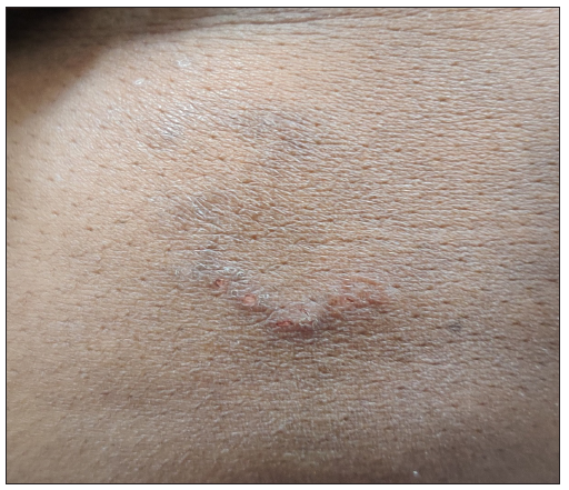
- A well-defined serpiginous tract noted over the left flank.
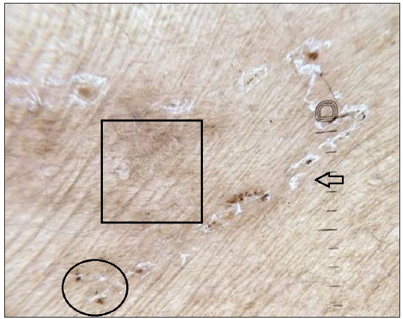
- Dermoscopy of the lesion (Dermlite DL5, polarised mode,10x magnification) revealing the presence of serpiginous scaling (arrow), with brownish-black dots and clods (circle) and parallel brown clods (square).
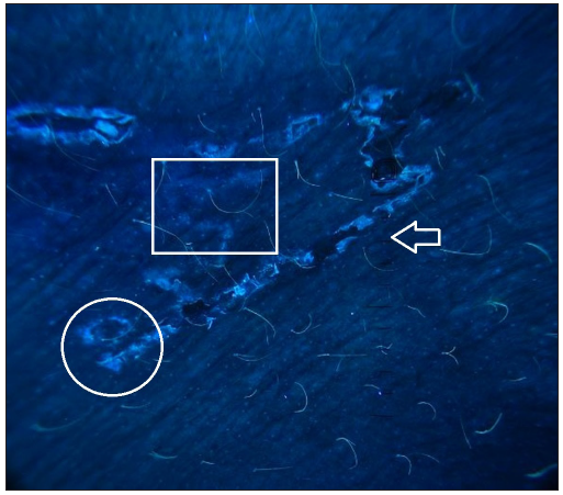
- Dermoscopy of the lesion (Dermlite DL5, ultraviolet mode, 10x magnification) shows the presence of bluish fluorescence overlying the serpiginous tracts (arrow) along with visualisation of the hidden larval tract (rectangle) originating from the previously thought area of termination (circle).
Declaration of patient consent
The authors certify that they have obtained all appropriate patient consent.
Financial support and sponsorship
Nil.
Conflicts of interest
There are no conflicts of interest.
Use of artificial intelligence (AI)-assisted technology for manuscript preparation
The authors confirm that there was no use of artificial intelligence (AI)-assisted technology for assisting in the writing or editing of the manuscript and no images were manipulated using AI.


