Translate this page into:
Umbilical metastasis: An unusual presentation of pancreatic adenocarcinoma
2 Department of Pathology, Tata Memorial Hospital, Parel, Mumbai, India
Correspondence Address:
K Prabhash
Room no-22, Medical Oncology Department of Tata Memorial Hospital, Parel - 400012. Mumbai
India
| How to cite this article: Pranjali K, Prabhash K, Karanth V N, Kane S, Nair R, Parikh P M. Umbilical metastasis: An unusual presentation of pancreatic adenocarcinoma. Indian J Dermatol Venereol Leprol 2007;73:199-200 |
 |
| Figure 2: FNAC: Clusters of adenocarcinoma cells (Papanicolaou, x200) |
 |
| Figure 2: FNAC: Clusters of adenocarcinoma cells (Papanicolaou, x200) |
 |
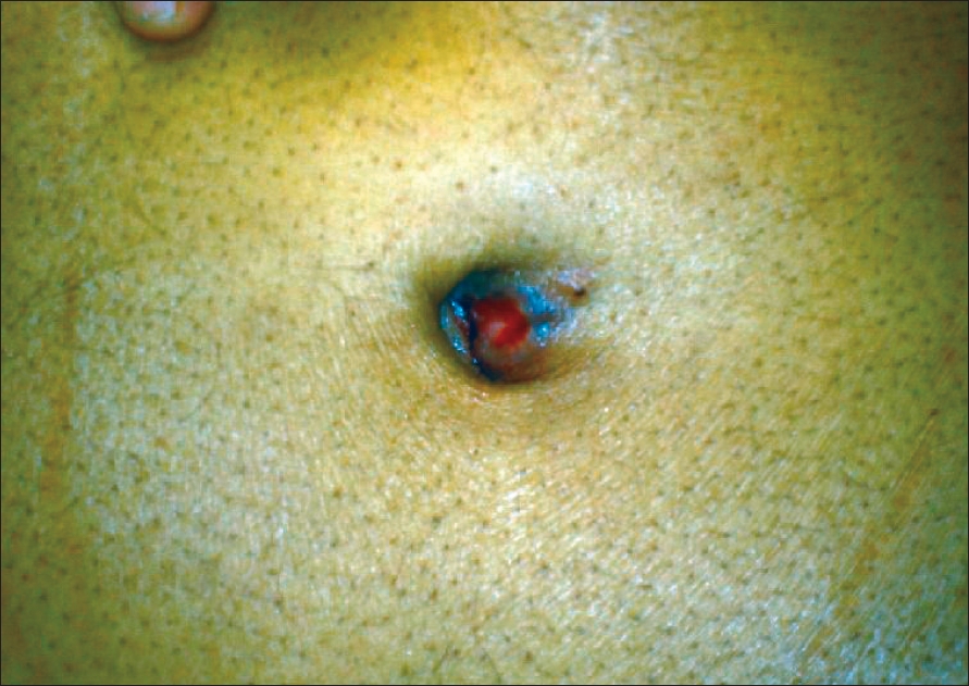
| Figure 1: Reddish smooth surfaced umbilical nodule |
 |
| Figure 1: Reddish smooth surfaced umbilical nodule |
Sir,
Metastasis to the umbilicus is rare. It may be a presenting feature of visceral malignancy or may occur after the diagnosis of the primary site is made. Sister Mary Joseph was the first to note the link between umbilical nodule and intra-abdominal malignancy. Here we present a case of carcinoma of pancreas that presented with an umbilical nodule. To our knowledge, only 12 cases of carcinoma of pancreas with umbilical metastasis have been reported. [1]
A 54-year-old man presented with painless swelling in the umbilical area of two weeks duation. There was no discharge from the lesion. On examination, there was a small nodule of 2x2 cm size, oval in shape, confined to the skin and sparing the underlying muscle. Its surface was smooth with a reddish hue and on palpation it was firm to hard in consistency, nontender, nonpulsatile and nonfluctuant [Figure - 1]. The surrounding skin was normal. Systemic examination was normal. Fine needle aspiration from the lesion revealed adenocarcinoma [Figure - 2]. The patient was investigated further to detect the site of primary malignancy. CT scan of abdomen and pelvis revealed a mass lesion in the head of the pancreas and involving the surrounding structures. The pancreatic lesion was nonresectable. The patient was planned for palliative chemotherapy with gemcitabine 1200 mg/m 2 on day 1 and 8 and every 3 weeks with cisplatin 75 mg/m 2 on day one and every three weeks. He received three cycles of chemotherapy. Re-evaluation suggested that there was decrease in the size of the tumor. Subsequently the patient completed six cycles of chemotherapy. However, the disease progressed two months after completion of chemotherapy and the patient is now only on symptomatic care.
Cutaneous metastasis occurs in 1-9% of patients with carcinomas. Umbilical metastasis represents only 10% of these lesions. [2] Site of primary of metastatic umbilical nodule is commonly the gastrointestinal tract (35-65%) and the genitourinary tract (12-35%) while in 15-30% of the patients the primary site is unknown. [3] The umbilical nodule is usually a painful lump with ulcerated surface and irregular margins unlike our case and is hard in consistency. [4]
The exact mechanism for spread of umbilical metastasis is not known. It has been postulated that it may occur either due to direct spread or through arterial, venous or lymphatic vessels or via the umbilical ligament. The most common form of umbilical involvement is the direct invasion of peritoneal metastasis. [1],[3] The so-called Sister Mary Joseph′s nodule is formed by localization of metastatic tumors to the umbilicus. [5] FNAC is a useful diagnostic intervention with 92.8% sensitivity and 100% positive predictive value. Once metastatic lesion is confirmed the primary lesion should be looked for with the help of clinical examination and further investigations.
Being a metastatic disease, the treatment remains palliative. Patients may be treated with chemotherapy, radiotherapy or both. In some patients if the primary lesion is resectable and umbilical metastasis is the only lesion then resection of primary with metastatic lesion can be done followed by adjuvant chemotherapy. In spite of the best of treatment, outcome of carcinoma pancreas with skin metastasis is poor as seen in our patient.
| 1. |
Crescentini F, Deutsch F, Sobrado CW, Araujo S. Umbilical mass as the sole presenting symptom of pancreatic cancer: A case report. Rev Hosp Clin Fac Med S Paulo 2004;59:198-202.
[Google Scholar]
|
| 2. |
Lookingbill D, Spangler N, Sexton FM. Skin involvement as the presenting sign of internal carcinoma. A retrospective study of 7316 cancer patients. J Am Acad Dermatol 1990;22:19-26.
[Google Scholar]
|
| 3. |
Gabriele R, Conte M, Egidi F, Borghese M. Umbilical metastasis: Current viewpoint. World J Surg Oncol 2005;3:13.
[Google Scholar]
|
| 4. |
Barrow MV. Metastatic tumors of the umbilicus. Chron Dis 1966;19:1113-7.
[Google Scholar]
|
| 5. |
Inamdar CA, Mavarkar L. Sister Mary Joseph's nodule as presenting sign of ovarian carcinoma. Indian J Dermatol Venereol Leprol 1995;61:383.
[Google Scholar]
|
Fulltext Views
2,210
PDF downloads
1,295


