Generic selectors
Exact matches only
Search in title
Search in content
Post Type Selectors
Search in posts
Search in pages
Filter by Categories
15th National Conference of the IAOMFP, Chennai, 2006
Abstract
Abstracts from current literature
Acne in India: Guidelines for management - IAA Consensus Document
Addendum
Announcement
Art & Psychiatry
Article
Articles
Association Activities
Association Notes
Award Article
Book Review
Brief Report
Case Analysis
Case Letter
Case Letters
Case Notes
Case Report
Case Reports
Clinical and Laboratory Investigations
Clinical Article
Clinical Studies
Clinical Study
Commentary
Conference Oration
Conference Summary
Continuing Medical Education
Correspondence
Corrigendum
Cosmetic Dermatology
Cosmetology
Current Best Evidence
Current Issue
Current View
Derma Quest
Dermato Surgery
Dermatopathology
Dermatosurgery Specials
Dispensing Pearl
Do you know?
Drug Dialogues
e-IJDVL
Editor Speaks
Editorial
Editorial Remarks
Editorial Report
Editorial Report - 2007
Editorial report for 2004-2005
Errata
Erratum
Focus
Fourth All India Conference Programme
From Our Book Shelf
From the Desk of Chief Editor
General
Get Set for Net
Get set for the net
Guest Article
Guest Editorial
History
How I Manage?
IADVL Announcement
IADVL Announcements
IJDVL Awards
IJDVL AWARDS 2015
IJDVL Awards 2018
IJDVL Awards 2019
IJDVL Awards 2020
IJDVL International Awards 2018
Images in Clinical Practice
Images in Dermatology
In Memorium
Inaugural Address
Index
Knowledge From World Contemporaries
Leprosy Section
Letter in Response to Previous Publication
Letter to Editor
Letter to the Editor
Letter to the Editor - Case Letter
Letter to the Editor - Letter in Response to Published Article
LETTER TO THE EDITOR - LETTERS IN RESPONSE TO PUBLISHED ARTICLES
Letter to the Editor - Observation Letter
Letter to the Editor - Study Letter
Letter to the Editor - Therapy Letter
Letter to the Editor: Articles in Response to Previously Published Articles
Letters in Response to Previous Publication
Letters to the Editor
Letters to the Editor - Letter in Response to Previously Published Articles
Letters to the Editor: Case Letters
Letters to the Editor: Letters in Response to Previously Published Articles
Media and news
Medicolegal Window
Messages
Miscellaneous Letter
Musings
Net Case
Net case report
Net Image
Net Images
Net Letter
Net Quiz
Net Study
New Preparations
News
News & Views
Obituary
Observation Letter
Observation Letters
Oration
Original Article
ORIGINAL CONTRIBUTION
Original Contributions
Pattern of Skin Diseases
Pearls
Pediatric Dermatology
Pediatric Rounds
Perspective
Presedential Address
Presidential Address
Presidents Remarks
Quiz
Recommendations
Regret
Report
Report of chief editor
Report of Hon : Treasurer IADVL
Report of Hon. General Secretary IADVL
Research Methdology
Research Methodology
Resident page
Resident's Page
Resident’s Page
Residents' Corner
Residents' Corner
Residents' Page
Retraction
Review
Review Article
Review Articles
Reviewers 2022
Revision Corner
Self Assessment Programme
SEMINAR
Seminar: Chronic Arsenicosis in India
Seminar: HIV Infection
Short Communication
Short Communications
Short Report
Snippets
Special Article
Specialty Interface
Studies
Study Letter
Study Letters
Supplement-Photoprotection
Supplement-Psoriasis
Symposium - Contact Dermatitis
Symposium - Lasers
Symposium - Pediatric Dermatoses
Symposium - Psoriasis
Symposium - Vesicobullous Disorders
SYMPOSIUM - VITILIGO
Symposium Aesthetic Surgery
Symposium Dermatopathology
Symposium-Hair Disorders
Symposium-Nails Part I
Symposium-Nails-Part II
Systematic Review and Meta-Analysis
Systematic Reviews and Meta-analyses
Systematic Reviews and Meta-analysis
Tables
Technology
Therapeutic Guideline-IADVL
Therapeutic Guidelines
Therapeutic Guidelines - IADVL
Therapeutics
Therapy
Therapy Letter
Therapy Letters
View Point
Viewpoint
What’s new in Dermatology
View/Download PDF

Translate this page into:

Observation Letter
87 (
2
); 265-267
doi:
10.25259/IJDVL_55_20

Unexpected high fluorine-18 fluorodeoxyglucose uptake on positron emission tomography/computed tomography of a benign cellular blue nevus

Department of Dermatology, School of Medicine, Kyungpook National University, Kyungpook National University Hospital, Daegu, South Korea

Corresponding author: Dr. Jun Young Kim, Department of Dermatology, Kyungpook National University Hospital, 130, Dongdeok-ro, Jung-gu, Daegu 41944, South Korea. 198kjy@hanmail.net

Licence
This is an open-access article distributed under the terms of the Creative Commons Attribution-Non Commercial-Share Alike 4.0 License, which allows others to remix, tweak, and build upon the work non-commercially, as long as the author is credited and the new creations are licensed under the identical terms.

How to cite this article: Lee HJ, Kim JY. Unexpected high fluorine-18 fluorodeoxyglucose uptake on positron emission tomography/computed tomography of a benign cellular blue nevus. Indian J Dermatol Venereol Leprol 2021;87:265-7.

Sir,

Positron emission tomography/computed tomography (PET/ CT) is useful for investigating suspected malignancies. Fluorine-18 fluorodeoxyglucose (18F-FDG) is a tracer used with such studies to detect hypermetabolic lesions and high uptake of 18F-FDG usually indicates the presence of malignancy.1

A 31-year-old female visited the dermatology department, after consulting a breast surgeon, because of an incidental finding of high uptake (standardized uptake value max 6.1) of 18F-FDG by the skin of her left buttock on PET/ CT [Figure 1a] conducted for the staging of her breast cancer. Cutaneous physical examination showed a blue-to-gray, non-fixed, hard mass, measuring 5.5 cm × 4.1 cm on the left buttock [Figure 1b]. She stated that the mass had been present since birth and had persisted without any noticeable change. An incisional biopsy was performed, revealing the mass as a benign blue nevus. Following consultation with a nuclear medicine physician it was understood that the hypermetabolic uptake on PET/CT imaging was localized to a soft tissue lesion of her left buttock rather than a peripheral lesion; therefore, this was considered a true positive finding. However, he could not explain the exact cause of the high uptake. Considering the possibility of malignant change of cellular blue nevus or metastasis of breast cancer, subsequent total excision was performed. We carefully and closely repeated the pathological examination and it showed dense pigmented melanocytic infiltration throughout whole dermis to subcutaneous tissue [Figure 2a]. Even after bleaching of the dense pigmentation there were nearly absent necrosis, mitotic rate more than 1-2/mm2, cytologic atypia, asymmetry and large expansile tumor nests, all featuring benign blue nevus again [Figure 2b]. Various immunohistochemical (IHC) stains including the markers of proliferation and mitosis (focal positive of HMB45, S-100, Melan-A and negative of Ki-67, PHH-3) were used [Figure 2c and d], but we could not find any evidence to suspect melanoma or other malignancies. As cellular blue nevus, atypical cellular blue nevus and blue nevus like melanomas have recently been reported as within the same spectrum, differential diagnosis of them is still subjective.2 By the histologic criteria shown by Hung et al., this case demonstrates two of the findings of atypical cellular blue nevus; size >1cm and cellular crowding, but without cellular atypia.2 It was difficult to define this case obviously as atypical cellular blue nevus. Therefore, we finally diagnosed the lesion as a benign cellular blue nevus showing high uptake of 18F-FDG. We did not perform sentinel lymph node biopsy because of the benign features of the primary lesion. The patient was followed up for 11 months postoperatively without recurrence or metastasis in primary lesion and chest/ abdominal CT imaging studies.
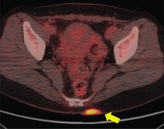
Positron emission tomography/computed tomography showed high fluorine-18 fluorodeoxyglucose uptake in left buttock (yellow arrow) of breast cancer patient
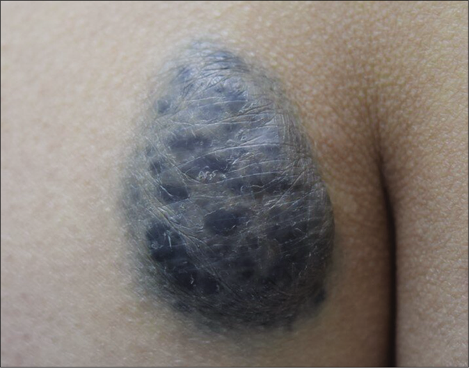
Figure 1a:
Positron emission tomography/computed tomography showed high fluorine-18 fluorodeoxyglucose uptake in left buttock (yellow arrow) of breast cancer patient
A blue-to-gray, non-fixed, hard mass, measuring 5.5 cm × 4.1 cm on left buttock
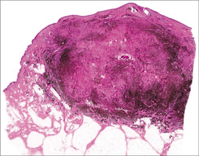
Figure 1b:
A blue-to-gray, non-fixed, hard mass, measuring 5.5 cm × 4.1 cm on left buttock
Dense pigmented melanocytic infiltration throughout whole dermis to subcutaneous tissue (hematoxylin and eosin, ×40)
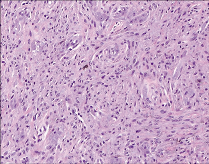
Figure 2a:
Dense pigmented melanocytic infiltration throughout whole dermis to subcutaneous tissue (hematoxylin and eosin, ×40)
Even after bleaching the pigment, it showed rare cellular atypia and mitosis (hematoxylin and eosin, ×200)
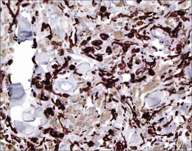
Figure 2b:
Even after bleaching the pigment, it showed rare cellular atypia and mitosis (hematoxylin and eosin, ×200)
Melan A focal positivity (Immunohistochemistry with 3-Amino-9-ethylcarbozole chromogen, ×200)
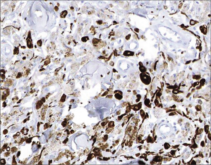
Figure 2c:
Melan A focal positivity (Immunohistochemistry with 3-Amino-9-ethylcarbozole chromogen, ×200)
Absence of Ki-67 (Immunohistochemistry with 3-Amino-9-ethylcarbozole chromogen, ×200)
Figure 2d:
Absence of Ki-67 (Immunohistochemistry with 3-Amino-9-ethylcarbozole chromogen, ×200)

In PET/CT imaging, FDG, an analog of glucose, is taken up, prevented from being further catabolized and becomes trapped in metabolically active cells.3 As a result, a variety of benign lesions with increased metabolic activity and glucose metabolism, such as inflammatory or infectious cutaneous conditions, are observed as false positives.3 Benign tumors that result in false-positive results include fibrous mesothelioma, schwannoma, aggressive neurofibromas and enchondromas.3 Recently Robinson et al. reported a rare case of benign intradermal nevus yielding a false-positive finding on PET/ CT.4 However, we were unable to find any previous reports of false-positive high uptake of 18F-FDG in a benign blue nevus.

The mechanism by which this case of benign blue nevus resulted in a false-positive 18F-FDG uptake result is unclear. Infiltration of inflammatory cells (neutrophils and activated macrophages) or proliferation of pigmented spindle cells and hypercellularity rather than malignant change or metastasis might have caused alterations in glucose metabolism and accumulation of FDG. Previous research has suggested that idiopathic chromosomal aberrations or genetic mutations associated with melanoma might be one of the possibilities.5 But we could not perform them in this case. The patient strongly refused molecular tests like fluorescence in-situ hybridization or chromosomal aberrations because of the cost and her good prognosis during follow-ups.

A sound understanding of the fundamentals regarding molecular and genetic evaluation for false-positive 18F-FDG uptake of benign skin tumors, including blue nevi, is necessary to avoid misdiagnoses. We may also consider benign blue nevus as possibly one of the skin diseases that can show false positive findings on PET/CT.

Declaration of patient consent

The authors certify that they have obtained all appropriate patient consent.

Financial support and sponsorship

This work was supported by a National Research Foundation of Korea (NRF) grant funded by the Korean government (MSIT) (No. 2018R1C1B5085905).

Conflicts of interest

There are no conflicts of interest.

References

  1. , , , , . The role of imaging in the management of patients with nonmelanoma skin cancer: Diagnostic modalities and applications. J Am Acad Dermatol. 2017;76:579-88.
    [CrossRef] [PubMed] [Google Scholar]
  2. , , , , , , et al. Cellular blue nevomelanocytic lesions: Analysis of clinical, histological, and outcome data in 37 cases. Am J Dermatopathol. 2016;38:499-503.
    [CrossRef] [PubMed] [Google Scholar]
  3. , . Common causes of false positive F18 FDG PET/CT scans in oncology. Braz Arch Biol Technol. 2007;50:29-35.
    [CrossRef] [Google Scholar]
  4. , , . Unexpected positron emission tomography/computed tomography uptake in benign dermal naevus. Australas J Dermatol. 2019;60:e58-60.
    [CrossRef] [PubMed] [Google Scholar]
  5. , , , , , , et al. Proliferative activity, chromosomal aberrations, and tumor specific mutations in the differential diagnosis between blue nevi and melanoma. Am J Pathol. 2013;182:640-5.
    [CrossRef] [PubMed] [Google Scholar]

Fulltext Views
3,852

PDF downloads
3,273
View/Download PDF
Download Citations
BibTeX
RIS
Show Sections