Translate this page into:
Unusual presentation of cystic fibrosis as diffuse dermatitis
2 Post-Graduation Program in Interactive Processes of Systems and Organs, Institute of Health Sciences, Federal University of Bahia, Bahia, Brazil
3 State University of Bahia, Bahia, Brazil
4 Climério de Oliveira Maternity Hospital, Federal University of Bahia, Bahia, Brazil
5 Department of Biology, Institute of Biology, Federal University of Bahia, Bahia, Brazil
6 Department of Pediatrics, School of Medicine of Bahia and Teaching Hospital Professor Edgard Santos, Federal University of Bahia, Bahia, Brazil
Correspondence Address:
Edna Lúcia Souza
Santa Luzia Avenue, 379/902, Horto Florestal, Salvador, Bahia
Brazil
| How to cite this article: Bonfim BS, Mota LR, Almeida Cd, de Brito Aguiar AP, Ferreira de Lima RL, de Mattos &P, Souza EL. Unusual presentation of cystic fibrosis as diffuse dermatitis. Indian J Dermatol Venereol Leprol 2018;84:461-463 |
Sir,
Cystic fibrosis is one of the most common autosomal recessive disorders among Caucasians. Most of the presentations are related to lung disease and exocrine pancreatic deficiency. Severe dermatitis is a rare initial presentation of the disease.[1]
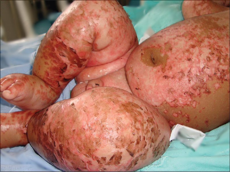
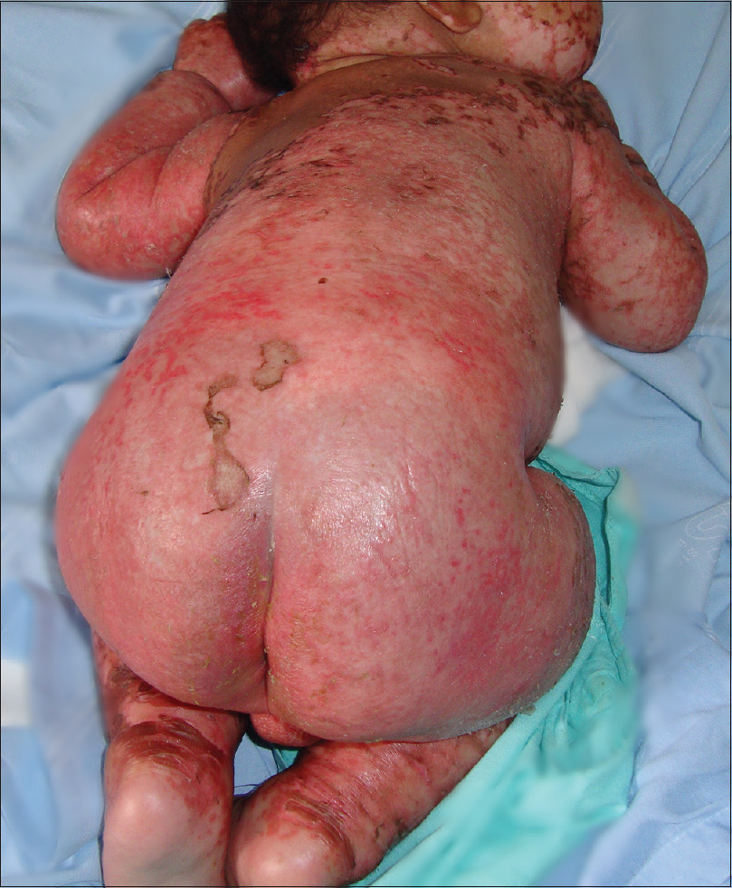
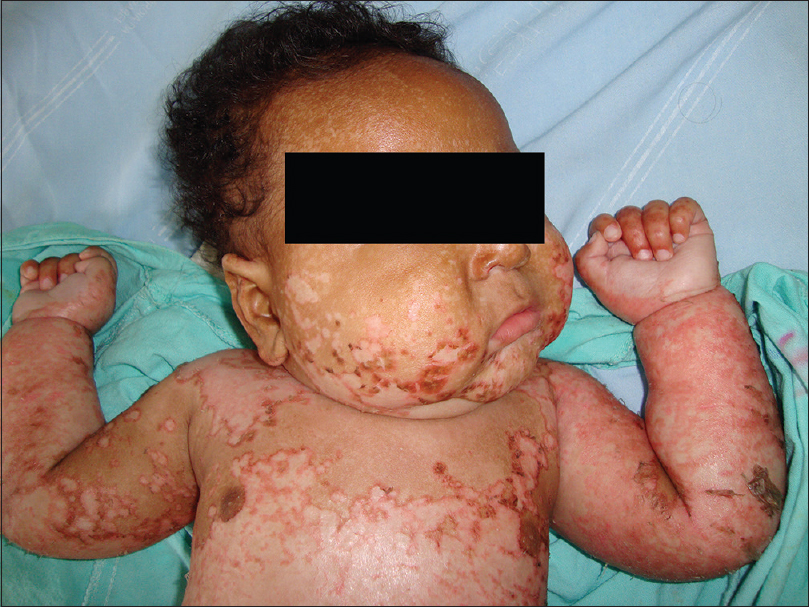
A non-Caucasian 5-month-old boy born at term to unrelated parents (weight 3.900 g) was healthy and exclusively breastfed until 4 months of age when he developed an erythematous eruption on the chest. The child was evaluated and diverse diagnoses were proposed: atopic dermatitis, contact dermatitis and scabies. The lesions were treated with several topical formulations including corticosteroids, antibiotics and antifungals, with no resolution. Following this, the rash became generalized and the patient further developed cough and wheeze; therefore, oral antibiotics were prescribed. However, as his general condition worsened, and he also developed edema on his lower limbs, he was transferred to Teaching Hospital Edgard Santos, Salvador, Bahia, Brazil. Upon admission, the patient was very irritable with generalized edema, wheezing and diffuse erythematous papules with overlying scaling or peeling of skin involving face, chest, limbs and genital area [Figure - 1] and [Figure - 2]. Body weight and length recorded were 7.090 kg and 62.2 cm, and his nutritional evaluation showed a weight/age (W/A) Z-score = +0.99; height/age (H/A) Z-score = −1.75 and weight/height (W/H) Z-score = −0.52. Mucosal examination was normal and no abnormalities were found. Abnormal laboratory values were as follows: hemoglobin 9.7 g/dL, albumin 1.8 g/dL, aspartate aminotransferase 116 IU/L, alanine transaminase 60 IU/L, Na + 133 mEq/L. Blood, urine and faecal cultures were negative. Initial diagnosis was drug eruption with secondary hypoalbuminemia. However, cystic fibrosis was investigated for and he presented two positive sweat tests (chloride 99/98 and 86/85 mEq/L). The steatocrit value was 22% and oropharyngeal cultures isolated Staphylococcus aureus, Klebsiella pneumoniae and Pseudomonas aeruginosa. Agenetic test was performed and the mutations F508del and 3120+1G>A were identified. Fecal elastase-1 level was lower than 15 μE1/g. Pancreatic enzyme replacement was instituted, as well as vitamins, antibiotics and nutritional therapy. The patient exhibited improvement in skin lesions, edema and nutritional status. He was discharged in good clinical condition after 37 days of hospitalization. Currently, he is 7 years old and is being followed-up as an outpatient at the same multidisciplinary cystic fibrosis clinic with good nutritional status: W/A Z-score = 0.6; H/A Z-score = −0.1; body mass index Z-score = +1.38, and stable clinical parameters [Figure - 3] and [Figure - 4].
 |
| Figure 1: Five-month-old patient presenting with generalized edema, wheezing and erythematous papules with overlying diffuse desquamation |
 |
| Figure 2: Five-month-old boy presenting with generalized edema, wheezing and erythematous papules with overlying desquamation involving face, back and limbs |
 |
| Figure 3: Patient's recovery after a few weeks of treatment showing initial regression of edema and skin lesions |
 |
| Figure 4: Patient after a few years of cystic fibrosis treatment with adequate growth and nutritional status |
Severe and generalized dermatitis as an initial presentation is rare and approximately 30 cases have been described.[1],[2] Edema, anemia and malnutrition are serious clinical manifestations of cystic fibrosis in infants. It usually occurs in infants with ages varying from 2 weeks to 15 months old.[1] The rash typically appears as erythematous papules which evolve in weeks to months into extensive plaques with desquamation. The lesions are first noted in perioral or diaper areas and extremities; subsequently, the dermatitis can become generalized with no response to topical formulations, including corticosteroids, antibiotics, antifungals or oral zinc supplementation.[1]
The skin lesions could be mistaken for other diseases, delaying the diagnosis and appropriate treatment. Differential diagnosis of this kind of rash without associated systemic manifestations includes atopic dermatitis, psoriasis, seborrheic dermatitis, Langerhans cell histiocytosis, immunodeficiency syndromes and acrodermatitis enteropathica. Kwashiorkor and essential fatty acids deficiency must be included in the differential diagnosis, especially if other clinical features are present.[1],[2],[3] This patient was previously diagnosed with atopic dermatitis, contact dermatitis and scabies.
Edema, gastrointestinal and pulmonary symptoms usually occur 1–2 months after the beginning of the rash; these symptoms were also noted in the case described. Laboratory abnormalities include hypoproteinemia, hypoalbuminemia, anemia, low cholesterol, zinc deficiency, undetectable liposoluble vitamins, steatorrhea, elevated transaminases and alkaline phosphatase.[1],[2],[4] Most of these abnormalities were observed in our patient.
The etiopathogenesis of the skin changes in cystic fibrosis still remain elusive, but it is apparently related to concomitant deficiencies of protein, zinc, essential fatty acids and possibly copper.[3],[4],[5] The essential fatty acids deficiency occurs even in patients who have pancreatic sufficiency.[4] Furthermore, children are more susceptible to essential fatty acids deficiency due to high metabolic demands.[2]
This atypical clinical presentation of cystic fibrosis may be mistaken for dermatitis of different etiologies, and the presence of edema and protein deficiency, which can cause false negativity in the sweat test, can contribute to diagnostic delay.[2],[3] Moreover, there are no pathognomonic histopathologic findings of this rash, and the microscopic aspect of the lesion is common to various diseases such as eczematous dermatitis, seborrheic dermatitis and drug reactions.[1]
Early diagnosis and proper cystic fibrosis treatment are crucial for this clinical presentation since it is associated with poor prognosis and the lesions may evolve with secondary infection and septicemia, a life-threatening condition whose sequelae can be severe and progress to death.[1],[3] Nevertheless, the rash usually improves after 2 weeks of nutritional therapy and pancreatic enzyme replacement, as in this case.[1]
Declaration of patient consent
The authors certify that they have obtained all appropriate patient consent forms. In the form, the legal guardian has given his consent for images and other clinical information to be reported in the journal. The guardian understands that names and initials will not be published and due efforts will be made to conceal patient identity, but anonymity cannot be guaranteed.
Financial support and sponsorship
This work was partially supported by the Research Support Foundation of the State of Bahia (FAPESB) PPSUS 020/2013.
Conflicts of interest
There are no conflicts of interest.
| 1. |
Lovett A, Kokta V, Maari C. Diffuse dermatitis: An unexpected initial presentation of cystic fibrosis. J Am Acad Dermatol 2008;58:S1-4.
[Google Scholar]
|
| 2. |
O'Regan GM, Canny G, Irvine AD. 'Peeling paint' dermatitis as a presenting sign of cystic fibrosis. J Cyst Fibros 2006;5:257-9.
[Google Scholar]
|
| 3. |
Bernstein ML, McCusker MM, Grant-Kels JM. Cutaneous manifestations of cystic fibrosis. Pediatr Dermatol 2008;25:150-7.
[Google Scholar]
|
| 4. |
Darmstadt GL, McGuire J, Ziboh VA. Malnutrition-associated rash of cystic fibrosis. Pediatr Dermatol 2000;17:337-47.
[Google Scholar]
|
| 5. |
Muñiz AE, Bartle S, Foster R. Edema, anemia, hypoproteinemia, and acrodermatitis enteropathica: An uncommon initial presentation of cystic fibrosis. Pediatr Emerg Care 2004;20:112-4.
[Google Scholar]
|
Fulltext Views
3,872
PDF downloads
2,078


