Translate this page into:
When in doubt look up to the skies: A skywatchers perspective in dermatology
Corresponding author: Dr. Aravind Sivakumar, Department of Dermatology and STD, Jawaharlal Institute of Postgraduate Medical Education & Research, Puducherry, India. aravinddermat@gmail.com
-
Received: ,
Accepted: ,
How to cite this article: Sivakumar A, Kumari R. When in doubt look up to the skies: A skywatchers perspective in dermatology. Indian J Dermatol Venereol Leprol 2023;89:482-6.
Introduction
Dermatologists in the past have been on the lookout for visual cues and similarities for the nomenclature of clinical conditions and signs and have been comparing them to various foods, animals, birds, and objects that have a common occurrence in everyday life. This has led to the easier memory and retention of complex dermatological nosology that would have been long forgotten otherwise. This article highlights the aerial perspective on such terminology.
Asteroid bodies: These are stellate acidophilic inclusion bodies with 20-30 radial projections seen in the cytoplasm and staining positively with PTAH, aldehyde fuchsin, and Masson trichrome stain. They are composed of microfilaments made up of tyrosine-containing protein. These are not specific for any condition and can be seen in any granulomatous reaction pattern, being most commonly observed in sarcoidosis and sporotrichosis. Other rare conditions with asteroid bodies include mycobacterial infections, other deep fungal infections, foreign body granulomas, necrobiotic xanthogranuloma, annular elastolytic granuloma, schistosomiasis and soft tissue tumours like fibroxanthosarcoma.1
Aurora borealis: It is a distinctive pattern seen in distal subungual onychomycosis due to the irregular matte pigmentation of different colours in parallel patterns thus giving similarity to the northern lights. It arises due to the presence of underlying fungal colonies and accumulated scales and subungual debris beneath the onycholytic nail plate.2
Chicago sky blue stain: Named due to its bright blue colour that it imparts, it is a fungal stain mainly used as an adjunct to potassium hydroxide mount mainly for diagnosing dermatophyte infections and pityriasis versicolor. It increases the colour contrast and eases identification of varied fungal morphology by imparting a bluish colour. It can be used with a light microscope unlike calcofluor white stain and also is better than Parker blue ink in the identification of dermatophytes, hence serves as a useful alternative for Swartz Lamkins stain for dermatophyte.3,4
Cloudy sky pattern: Described as a dermoscopic pattern in idiopathic guttate hypomelanosis characterized by ill-defined whitish homogeneous round macules coalescing to form irregular, polycyclic macules with surrounding hyperpigmentation [Figure 1]. It has also been described as a clue for the diagnosis of periorbital syringomas.5,6
Comet tail sign: This finds its place in more than one context in dermatology. Firstly in vitiligo where comet tail pattern observed dermoscopically can be an indicator of disease activity.7 Comet sign has also been described in infestation with the mite Pyemotes ventricosus which presents as erythematous macules tapering distally with a linear tract resembling a comet, which might herald the onset of lymphangitis.8 Comet tail lesions are also seen in the fundus of patients with pseudoxanthoma elasticum and are pathognomonic for this condition. These arise earlier than angioid streaks and peau d’ orange appearance which are nonspecific.9
Craters of the moon: It refers to the specific trichoscopic pattern seen in alopecia areata characterized by extensive depressed yellow dots and signifies poor prognosis. It also helps to differentiate from the yellow dots of the DLE which lack these depressions on trichoscopy.10
Delta wing sign: Refers to the dark triangular V-shaped proximal portion of the scabies mite when examined dermoscopically while the rest of the body appears translucent resembling a delta wing jet. Also referred to as the triangle sign /hang glider sign or spermatozoid appearance.11
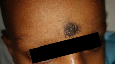
Eclipse nevus: This is a particular type of scalp nevus most seen in children. Characterized by the presence of tan centre which may be elevated surrounded by an irregular brownish rim with a stellate appearance resembling a solar eclipse. It may bear resemblance to a cockade nevus which has the reverse of this pigment pattern. Clinically it is a benign lesion that shouldn’t raise any alarming signs.12
Galaxy sign: Although not a cutaneous sign it is seen in sarcoidosis. It might be an important imaging clue for diagnosis, where there is a single pulmonary nodule with surrounding smaller nodules in high resolution computed tomography (HRCT) of the chest which is due to the less dense arrangement of smaller satellite nodules around the main nodule hence resembling a galaxy.13
Haloes: Halo nevus or Sutton’s nevus refers to the appearance of rim of depigmentation surrounding benign melanocytic nevi. Seen most commonly in children, multiple halo nevi may be associated with atopic dermatitis or autoimmune disorders like vitiligo or Hashimoto thyroiditis.14 Halo phenomenon or Meyerson phenomenon refers to the symmetrical area of erythema and scaling surrounding a lesion such as melanocytic nevus due to eczematisation. This can also be observed in molluscum contagiosum, seborrheic keratosis and capillary malformation.15
Jet with contrail appearance: Seen in scabies dermoscopy where the burrow with the mite at its end resembles the trail of a jet plane.16
Lichtenberg figures: Also referred to as arborescent burns or filigree burns, these are skin manifestations which are pathognomonic of lightning injury either directly or as a side flash from surrounding objects like trees causing a superficial burn in a fern or feather-like pattern. It is a transient phenomenon lasting from 30 minutes up to 2 days and disappears without pigmentation.17 Sometimes similar appearance can be observed in pressure-induced vasodilatation causing arborescent appearance which can be differentiated with diascopy.18
Lone star ticks: Refers to Ambylomma americanum due to the prominent white dot on the back of the adult female. It serves as a vector for various rickettsial diseases such as Rocky Mountain spotted fever, Q fever, tularaemia, ehrlichiosis, and southern tick-associated rash illness (STARI).19
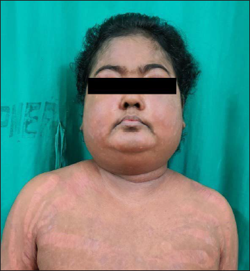
Moon facies: Coined by Harvey Cushing, an American neurosurgeon who first described it in people with Cushing’s syndrome who had round faces. It occurs due to the excessive fat deposition on the face that accompanies primary or exogenous Cushing’s syndrome [Figure 2].20
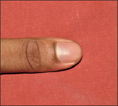
Half-moon appearance of lunula: Lunula is the visible portion of the distal nail matrix that appears whitish with distal convex and proximal straight borders [Figure 3]. It is most prominent on the thumb, index finger, and great toe and guides the shape of the nail plate distally. Abnormalities of the lunula i.e., micro lunula, macrolunula, erythema, lunular dyschromia all point to underlying systemic disorders.21
Nebuloid appearance: Seen in the dermoscopy of idiopathic guttate hypomelanosis where there are whitish homogeneous areas with indistinct borders. Nebula refers to clouds of celestial dust and gases usually describing distant galaxies which are indistinct.6
Papular epidermal nevus with ‘skyline’ basal layer (PENS): Presents as single or multiple skin-coloured keratotic skin lesions that do not follow the lines of Blaschko, usually present since birth. Histologically it has columnar cells with uniform small nuclei in a palisaded pattern in the basal layer like a ‘skyline’ or ‘eyeliner’ appearance.22
Pink rim sign: In the dermoscopy of melanoma there are varied shades of pink and the location in the periphery suggests the presence of underlying invasive melanoma. This resembles the pink rim of the sun at sunset or the lunar eclipse.23
Raindrops: Raindrops are described in a myriad of conditions, most seen in miliaria crystallina [Figure 4] where the flaccid vesicles are seen on a non-erythematous base. Raindrop pigmentation has been described in arsenic toxicity which is characterized by truncal hyperpigmentation with scattered depigmented macules present over it. Sometimes the distribution of seborrheic keratosis over the back resembles that of raindrops and stream appearance.24
Rainbow appearance: It is a specific appearance seen in the polarized dermoscopy where there is the presence of bluish-red discolouration along with varied colours of the rainbow. It occurs due to diffraction of the incident light over the skin due to the difference in refractive indices of the layers of skin termed optical dichroism. Classically seen in Kaposi sarcoma it can also be seen in atypical fibroxanthoma, basal cell carcinoma, aneurysmal dermatofibroma, Merkel cell carcinoma, and acral pseudolymphomatous angiokeratoma.25
Red half-moon nail sign: Seen particularly in COVID-19 infection where there are reddish half-moon-shaped convex bands over the margin of the lunula due to the localized microvascular injury to the distal subungual arterial arcade.26
Subungual red comets: This is a specific nail finding seen on dermoscopy in tuberous sclerosis complex. Characterized by the presence of tortuous capillaries with surrounding whitish halo and can be associated with periungual or subungual fibromas. It can be differentiated from splinter haemorrhage by the absence of whitish halo, shorter length, and straight linear appearance reaching the distal free end of the nail plate.27
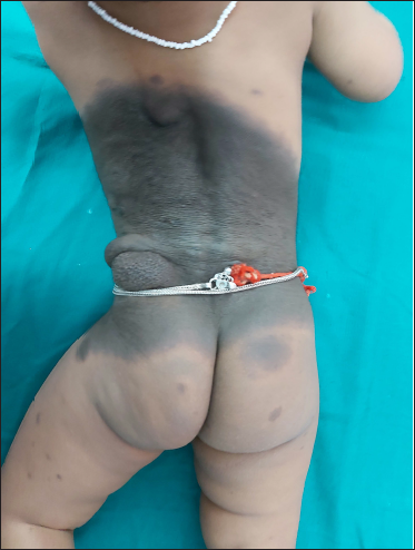
Satellite lesions: This is common terminology for the arrangement of multiple smaller lesions around larger skin lesions situated nearby. It is usually seen in conditions such as borderline tuberculoid Hansen’s, congenital melanocytic nevus [Figure 5], and flexural candidiasis.28
Setting sun sign: Describes the dermoscopic picture characterized by yellow-orange background surrounded by an erythematous border seen in juvenile xanthogranuloma. Also, it can be seen in other conditions such as reticulohistiocytoma, mastocytoma, xanthomatous dermatofibroma, and histiocytic sarcoma.29
Sky full of stars appearance: Is a specific dermoscopic finding in desmoplastic giant congenital melanocytic nevus where there is the presence of reticular pigmentation with radially arranged accentuation around the follicular ostia.30
Starburst appearance: Classically described as a dermoscopic finding in Spitz nevus where there is a radial arrangement of sharply focused streaks in a regular fashion around the lesion. Can also be observed in pigmented Bowens disease and malignant melanoma.31 The starburst giant cell refers to the multinucleated melanocytes with prominent dendritic processes seen in lentigo maligna melanoma.32 Starburst hair follicles refer to the radially oriented, horizontally extending hair follicles seen in aplasia cutis congenita. It occurs due to the abnormal shearing forces during neuroectodermal development and can be useful when the hair collar sign is not evident.33 Lastly starburst appearance in vitiligo also denotes disease activity and progressive vitiligo.34
Starry sky: Histologically it is described in cases of Burkitt’s lymphoma where the presence of tingible body macrophages (containing cellular apoptotic debris) is seen scattered among the background of tumour cells.35 Dermoscopic appearance in sweat dermatitis seen as yellowish-white lacunar areas interspersed with areas of necrosis distributed over diffuse background erythema due to obstructed sweat duct openings is referred to as starry skin appearance.36
Starry night sky appearance: Seen particularly with ultraviolet light enhanced trichoscopy where the hair follicles are illuminated due to the presence of follicular Propionibacterium acnes which has porphyrins and hence has a reddish-orange fluorescence. This technique can be used to check hair follicle viability (positive ultraviolet enhanced trichoscopy) and assess prognosis in case of scarring alopecia especially frontal fibrosing alopecia.37
Stars in heaven: Seen in cases of heavy metal poisoning, especially argyria where there is presence of silver deposits seen around the eccrine glands and dispersed in the dermis which in darkfield microscopy appears as brilliantly refractile white particles against a black background.38
Stellate appearance: Stellate pseudoscars are whitish irregular or star-shaped pseudoscars mostly seen in elderly individuals over the photo exposed sites, but can also arise due to topical steroid abuse.39
Uremic frost: It is a sign of severe azotaemia seen in patients with end-stage renal disease characterized by the presence of whitish tiny crystalline deposits on the skin resembling frosting that occurs during the time of the year when it snows from the skies. It occurs due to the crystallization of urea and other nitrogenous waste products onto the surface of the skin also referred to as ‘uridrosis’ or ‘urinous sweat’.40
Weather-beaten appearance: Resembles skin that is damaged due to chronic sun damage during the summer season, clinically presents as shallow puckered linear or elliptical scars accompanied by waxy thickening and sclerodermoid changes. It is described classically in children with erythropoietic protoporphyria and lipoid proteinosis.41

- Cloudy skin pattern observed on dermoscopy in idiopathic guttate hypomelanosis

- Moon facies seen in exogenous Cushing syndrome due to steroid intake

- Half-moon appearance of lunula

- Raindrop-like appearance in miliaria crystallina

- Multiple satellite lesions seen in congenital melanocytic nevus
Declaration of patient consent
Informed consent for publication of images has been taken from the patients by the authors.
Financial support and sponsorship
Nil.
Conflicts of interest
There are no conflicts of interest.
References
- Asteroid bodies and other cytoplasmic inclusions in necrobiotic xanthogranuloma with paraproteinemia. J Am Acad Dermatol. 1998;38:967-70.
- [CrossRef] [PubMed] [Google Scholar]
- Onychoscopy: Dermoscopy of the nails. Dermatol Clin. 2018;36:431-8.
- [CrossRef] [PubMed] [Google Scholar]
- Practical tip: Chicago sky blue (CSB) stain can be added to the routine potassium hydroxide (koh) wet-mount to provide a color contrast and facilitate the diagnosis of dermatomycoses. Dermatol Online J. 2011;17:11.
- [CrossRef] [PubMed] [Google Scholar]
- Comparison of modified Chicago sky blue stain and potassium hydroxide mount for the diagnosis of dermatomycoses and onychomycoses. J Microbiol Methods. 2015;112:21-3.
- [CrossRef] [PubMed] [Google Scholar]
- Dermoscopy of idiopathic guttate hypomelanosis. J Dermatol. 2015;42
- [CrossRef] [PubMed] [Google Scholar]
- Dermoscopic evaluation of idiopathic guttate hypomelanosis: A preliminary observation. Indian Dermatol Online J. 2015;6:164.
- [CrossRef] [PubMed] [Google Scholar]
- Pyemotesventricosus dermatitis, southeastern France. Emerg Infect Dis. 2008;14:1759-61.
- [CrossRef] [PubMed] [PubMed Central] [Google Scholar]
- “Comet-tail” lesions of pseudoxanthoma elasticum. Indian J Ophthalmol. 2018;66:300.
- [CrossRef] [PubMed] [Google Scholar]
- Craters of the moon: A marker for disease severity in alopecia areata? Indian J Paediatr Dermatol. 2021;18:341-16.
- [CrossRef] [Google Scholar]
- Diagnosis of scabies by dermoscopy. BMJ Case Rep. 2009;2009:bcr0620080279.
- [CrossRef] [PubMed] [Google Scholar]
- No biopsy needed for eclipse and cockade nevi found on the scalps of children. Arch Dermatol. 2009;145:1334-6.
- [CrossRef] [PubMed] [Google Scholar]
- Pulmonary sarcoidosis: Calcification within the galaxy sign. Case Rep. 2016;2016:bcr2016218187.
- [CrossRef] [PubMed] [Google Scholar]
- Dermoscopy patterns of halo nevi. Arch Dermatol. 2006;142:1627-32.
- [CrossRef] [PubMed] [Google Scholar]
- Lichtenberg figures and lightning: Case reports and review of the literature. Cutis. 2007;80:141-3.
- [PubMed] [Google Scholar]
- Arborescent vascular dilatation mimicking lichtenberg figures from lightning. Pediatr Dermatol. 2014;31:522-3.
- [CrossRef] [PubMed] [Google Scholar]
- Multiple pruritic papules from lone star tick larvae bites. Arch Dermatol. 2006;142:491-4.
- [CrossRef] [PubMed] [Google Scholar]
- The “moon face” of cushing syndrome. JAMA Dermatol. 2018;154:329.
- [CrossRef] [PubMed] [Google Scholar]
- Papular epidermal nevus with “skyline” basal cell layer (PENS) J Am Acad Dermatol. 2011;64:888-92.
- [CrossRef] [PubMed] [Google Scholar]
- The pink rim sign: Location of pink as an indicator of melanoma in dermoscopic images. J Skin Cancer. 2014;2014:719740.
- [CrossRef] [PubMed] [Google Scholar]
- Seborrheic keratoses in five elderly patients: An appearance of raindrops and streams. Indian J Dermatol. 2011;56:432.
- [CrossRef] [PubMed] [Google Scholar]
- The dermoscopic rainbow pattern - A review of the literature. Acta Dermato venerol Croat. 2019;27:111-5.
- [PubMed] [Google Scholar]
- The red half-moon nail sign: A novel manifestation of coronavirus infection. J Eur Acad Dermatol Venereol. 2020;34:e663-5.
- [CrossRef] [PubMed] [Google Scholar]
- Dermoscopy of subungual red comets associated with tuberous sclerosis complex. Pediatr Dermatol. 2019;36:408-10.
- [CrossRef] [PubMed] [Google Scholar]
- Satellite lesions in congenital melanocytic nevi-time for a change of name. Pediatr Dermatol. 2011;28:212-3.
- [CrossRef] [PubMed] [Google Scholar]
- Is the setting sun dermoscopic pattern specific to juvenile xanthogranuloma? J Am Acad Dermatol. 2018;78:e49.
- [CrossRef] [PubMed] [Google Scholar]
- “Sky full of stars” pattern: Dermoscopic findings in a desmoplastic giant congenital melanocytic nevus. Pediatr Dermatol. 2017;34:e142-3.
- [CrossRef] [PubMed] [Google Scholar]
- Pigmented Bowen’s disease presenting with a “starburst” pattern. Dermatol Pract Concept. 2016;6:47-9.
- [CrossRef] [PubMed] [PubMed Central] [Google Scholar]
- The starburst giant cell is useful for distinguishing lentigomaligna from photodamaged skin. J Am Acad Dermatol. 1996;35:962-8.
- [CrossRef] [PubMed] [Google Scholar]
- Starburst hair follicles: A dermoscopic clue for aplasia cutis congenita. J Am Acad Dermatol. 2016;75:e141-2.
- [CrossRef] [PubMed] [Google Scholar]
- Dermoscopy in vitiligo: Diagnosis and beyond. Int J Dermatol. 2018;57:50-4.
- [CrossRef] [PubMed] [Google Scholar]
- Burkitt lymphoma with cutaneous involvement. Dermatol Online J. 2008;14:14.
- [CrossRef] [PubMed] [Google Scholar]
- A typical presentation of sweat dermatitis with review of literature. Indian Dermatol Online J. 2019;10:698-703.
- [CrossRef] [PubMed] [Google Scholar]
- The “Starry night sky sign” Using ultraviolet-light-enhanced trichoscopy: A new sign that may predict efficacy of treatment in frontal fibrosing alopecia. Int J Trichology. 2018;10:241-3.
- [CrossRef] [PubMed] [Google Scholar]
- Localized argyria with pseudo-ochronosis. J Am Acad Dermatol. 2002;46:222-7.
- [CrossRef] [PubMed] [Google Scholar]
- “Pseudo” nomenclature in dermatology: What’s in a name? Indian J Dermatol. 2013;58:369-76.
- [CrossRef] [PubMed] [Google Scholar]
- Uremic frost: A harbinger of impending renal failure. Int J Dermatol. 2016;55:17-20.
- [CrossRef] [PubMed] [Google Scholar]
- A young girl with weather-beaten, waxy knuckles. J Pediatr. 2010;156:671-4.
- [CrossRef] [PubMed] [Google Scholar]


