Translate this page into:
Zosteriform lymphangitis carcinomatosis: A dermatologist’s enigma
Corresponding author: Dr. Manoj Kumar Nayak, Department of Dermatology, Venereology and Leprosy, Institute of Medical Sciences, Bhubaneswar, Odisha, India. trriger2010@gmail.com
-
Received: ,
Accepted: ,
How to cite this article: Singh BSTP, Nayak MK, Biswal R, Singh S, Biswal A. Zosteriform lymphangitis carcinomatosis: A dermatologist’s enigma. Indian J Dermatol Venereol Leprol. 2025;91:374-6. doi: 10.25259/IJDVL_417_2023
Dear Editor,
A 46-year-old man presented with tender well-defined reddish plaques over the neck and upper back for the past 3-months. His medical history was unremarkable, except for a history of mucoepidermoid carcinoma of the left parotid gland. He had undergone left radical parotidectomy with pectoralis major myo-cutaneous flap reconstruction of the neck along with chemotherapy and radiotherapy 3 years back. The patient was declared free of malignancy after the treatment.
On examination, there was a well-demarcated, indurated, erythematous plaque extending from the posterior to the front of the neck on the left side and the upper back [Figure 1a]. It also involved the left side of the cheek and ear with overlying haemorrhagic and translucent papulovesicular lesions and erosions in the root of the neck [Figure 1b]. Tzanck smear did not show any multinucleated giant cells. Histopathology of the lesion showed keratinised stratified squamous epithelium. The dermis showed tumour cells arranged in an acinar pattern, cord, nest and comedo pattern [Figure 2a]. The tumour cells were round to oval with moderate cytoplasm and hyperchromatic nuclei [Figure 2b]. This suggested metastatic invasion from the salivary gland as the primary site. On immunohistochemical staining, tumour cells were strongly and diffusely positive for Androgen receptor (AR) [Figure 3a] and epithelial membrane antigen (EMA) [Figure 3b] and negative for estrogen receptor (ER) [Figure 3c]. On further investigation, a positron emission tomography (PET) scan revealed recurrence of malignancy in the parotid gland. A diagnosis of cutaneous zosteriform lymphangitis carcinomatosis was made based on the clinical, histopathological and imaging features.
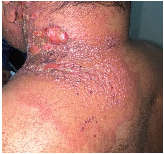
- Shows well-demarcated, indurated erythematous plaque with papulovesicles and few erosions over the neck and upper back.
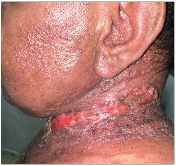
- Shows well-demarcated,indurated erythematous plaque with papulovesicles and few erosions over neck, cheek and ear.
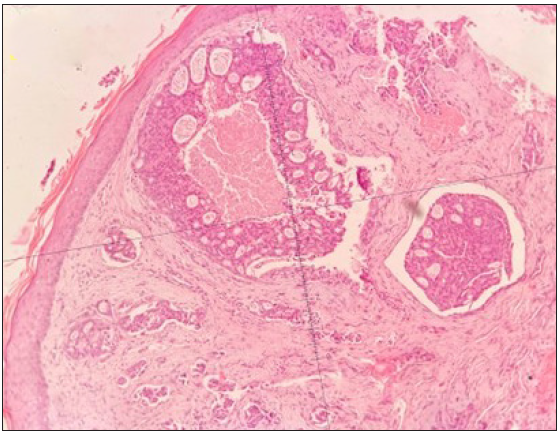
- Histopathology showing the source of tumour cells was discovered in later nests inside lymphatic channels. (Haematoxylin and eosin, 100x).
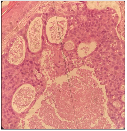
- Histopathology showing tumour cell nests with hyperchromatic nuclei with a high N:C ratio inside lymphatic channels (Haematoxylin and eosin, 400x).
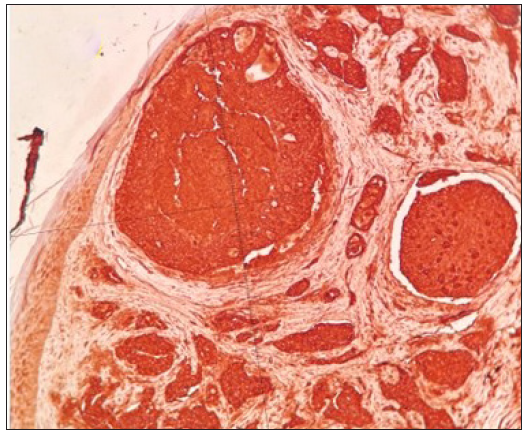
- Show immunohistochemistry results for androgen receptor (AR).
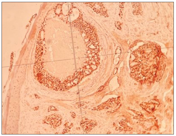
- Show immunohistochemistry results for epithelial membrane antigen (EMA).
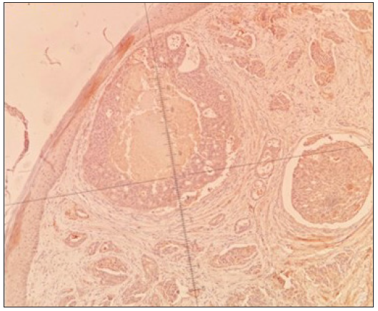
- Show immunohistochemistry results for estrogen receptor (ER).
Cutaneous metastases occur only in 0.7–0.9% of cancer patients.1 Cutaneous lymphangitis carcinomatosis is a rare presentation of the metastatic spread of cancer to the skin. It is caused by the occlusion of lymphatic channels of the dermis by neoplastic cells.2 Clinically, it presents as erythematous tender plaques closely resembling lymphangioma circumscriptum, erysipelas, cellulitis, herpes zoster, irritant contact dermatitis or post-irradiation lymphangiectasia. The clinical features may be diagnostic but may mimic several infectious and benign dermatoses. The most common clinical mimicker of this condition would be radiotherapy-induced acquired lymphangiectasia which is a vesicular dilation of the lymphatic channels. The vesicles appear on the normal skin in acquired lymphangiectasia, in contrast to lymphangitic carcinomatosis, where papulovesicles appear on infiltrated, erythematous skin. There was no temporal association of the eruption with the institution of chemotherapy, thereby ruling out the radiation recall phenomenon.
Lymphangitic carcinomatosis has been previously reported with mostly the lung and the breast as the site of primary malignancy.1 A few reported cases of carcinomatosis lymphangitis presenting over the neck have been tabulated in Table 1. These cases presented with metastases from the palate, lungs, pyriform fossa, larynx and the parotid; the primary was unknown in one case.1–5 Bishnoi et al. proposed the term ‘carcinoma en gorget’ to describe the malignant infiltration of the skin over the neck arising from different malignancies similar to ‘carcinoma en cuirasse’ for infiltration of the chest wall.5
| Author | Presentation | Type of malignancy | Immunohistochemistry | Primary |
|---|---|---|---|---|
| Hari Kishan et al. 20131 | Confluent papules, nodules, and on the left side of the neck in a zosteriform distribution | SCC | Not Done | Palate |
| Lola Prat et al. 20132 | Erythematous, eczematous, itchy rash localised on the right anterior chest and left shoulder | Adenocarcinoma | Positive for cytokeratin 7 (CK7+) and thyroid transcription factor-1 (TTF-1+) and negative for cytokeratin 20 (CK20-). | Lung |
| Echeverria-Garcia et al. 20133 | Erythematous indurated plaque on the right side of neck and jaw, mimicking erysipelas | Lymphoepithelial carcinoma of the parotid | Positive for cytokeratin, lymphocytic expression of CD45 | Parotid |
| Shamsadini et al. 20034 | Nodules, ulcers, vesicles, inflammatory areas, sclerotic areas, lesions on the left side of the neck | SCC | Not Done | Larynx |
| Schmitt et al.6 2011 | Violaceous plaque in a collar like distribution over the right side of the neck and trunk | Adenocarcinoma | Positive for TTF-1 and CK7 | No primary identified even with exhaustive imaging |
| Bishnoi A et al. 20195 | Ill-defined, indurated, erythematous plaque encircling the right side of the patient’s neck. | Adenocarcinoma | Positive for CK7 and negative for p63, CK20, PSA, GAT A3 and TTF-1 | Pyriform fossa |
SCC: Squamous cell carcinoma
This case is presented to highlight the occurrence of rare zosteriform lymphangitis carcinomatosis metastasizing from the parotid gland. Also, lymphangitis carcinomatosis may be the first clinical marker of recurrence of malignancy after surgical resection as was seen in this case.
Declaration of patient consent
The authors certify that they have obtained all appropriate patient consent.
Financial support and sponsorship
Nil.
Conflicts of interest
There are no conflicts of interest.
Use of artificial intelligence (AI)-assisted technology for manuscript preparation
The authors confirm that there was no use of artificial intelligence (AI)-assisted technology for assisting in the writing or editing of the manuscript and no images were manipulated using AI.
References
- A rare case of zosteriform cutaneous metastases from squamous cell carcinoma of hard palate. Ann Med Health Sci Res. 2013;3:127-30.
- [CrossRef] [PubMed] [Google Scholar]
- Cutaneous lymphangitis carcinomatosa in a patient with lung adenocarcinoma: Case report and literature review. Lung Cancer. 2013;79:91-3.
- [CrossRef] [PubMed] [Google Scholar]
- Carcinomatous lymphangitis in lymphoepithelialcarcinoma of the parotid. J Am Acad Dermatol. 2013;68:e194-5.
- [CrossRef] [PubMed] [Google Scholar]
- Grouped skin metastases from laryngeal squamous cell carcinoma and overview of similar cases. Dermatol Online J. 2003;9:27.
- [PubMed] [Google Scholar]
- Zosteriform lymphangitis carcinomatosis in the cervical area arising from pyriform fossa adenocarcinoma. Clin Exp Dermatol. 2019;44:708-11.
- [CrossRef] [PubMed] [Google Scholar]
- Violaceous plaques in a collarlike distribution--quiz case. Carcinoma erysipelatoides from carcinoma of unknown primary. Arch Dermatol. 2011;147:345-50.
- [CrossRef] [PubMed] [Google Scholar]


