Translate this page into:
Multiple skin nodules over the scalp and trunk
2 Department of Pathology, JIPMER, Pondicherry, India
Correspondence Address:
Rashmi Kumari
Assistant Professor, Department of Dermatology and STD, JIPMER, Pondicherry-605006
India
| How to cite this article: Sarita M, Kumari R, Rajesh NG, Thappa DM. Multiple skin nodules over the scalp and trunk. Indian J Dermatol Venereol Leprol 2012;78:517-518 |
A 50-year-old man came with multiple asymptomatic nodules on the face, scalp and upper trunk for the past 10 years. These nodules were slowly increasing in size. There was no family history of similar lesions. On cutaneous examination, there were multiple large, shiny, skin colored, lobulated, firm, non-tender nodules of varying sizes, largest measuring 5 × 4 cm in size over the scalp [Figure - 1]. He also had multiple soft, non-tender papules and nodules of size 1 × 1.5 cm over the central face, forehead and trunk. Button-hole sign was negative. Patient also had ill-defined depigmented macules, suggestive of vitiligo on trunk, lips and scalp. An excisional biopsy was done from the large nodule on scalp and the soft papules on the trunk. Histopathology revealed closely set mosaic like masses and columns of cells surrounded by a thick basal membrane [Figure - 2] and [Figure - 3]. The hyaline thick basal membrane was periodic acid schiff stain positive [Figure - 4] and diastase resistant.
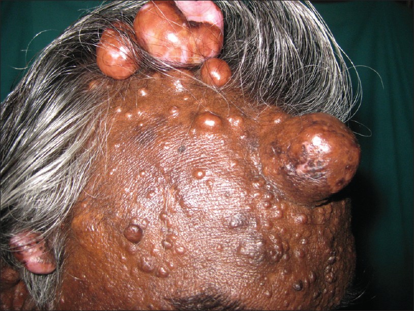 |
| Figure 1: Multiple skin colored nodules and papules seen over the scalp and face |
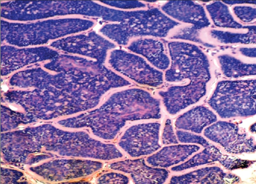 |
| Figure 2: Section shows nests of basaloid tumor cells arranged in a peculiar pattern. Tumor cells secrete thick basement membrane material (H and E, × 100) |
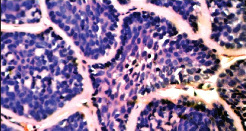 |
| Figure 3: Section shows nests of basaloid tumor cells exhibiting peripheral palisading with center comprised nests of pale cells (H and E, × 400) |
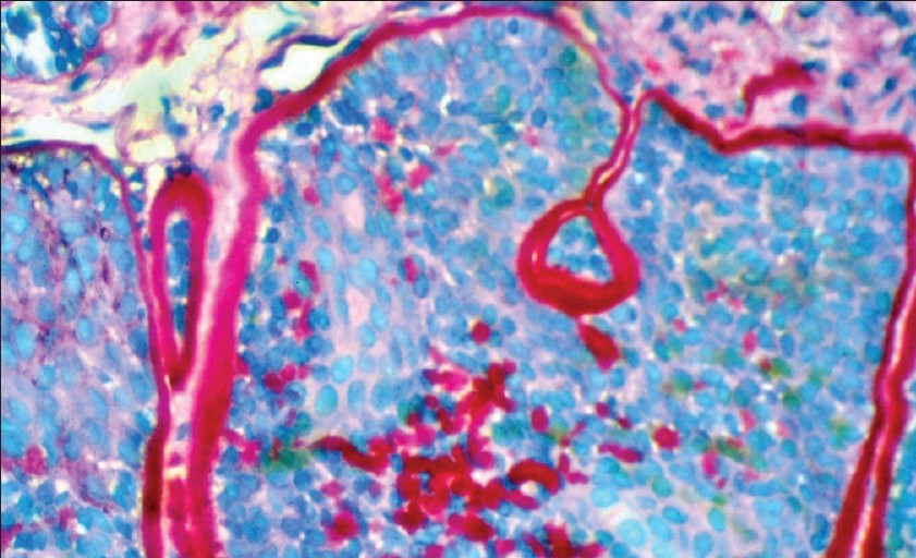 |
| Figure 4: Section shows PAS positivity of basement membrane material secreted by the tumor cells, (PAS stain, × 400) |
What is your diagnosis?
Answer: Cylindroma
Histopathology section showed nests of basaloid tumor cells arranged in jigsaw puzzle pattern. The nests of basaloid tumor cells exhibited peripheral palisading with center of the nests comprised of pale cells. Tumor cells secreted thick basement membrane material, which was PAS positive and diastase resistant. The tumor cells showed nuclear positivity for p63 in the myoepithelial cells of the tumor (IHC DAB stain, × 400) [Figure - 5]. These features were suggestive of cylindroma. Surgical excision of the tumors was done for 2 large nodules after which the patient is on follow-up.
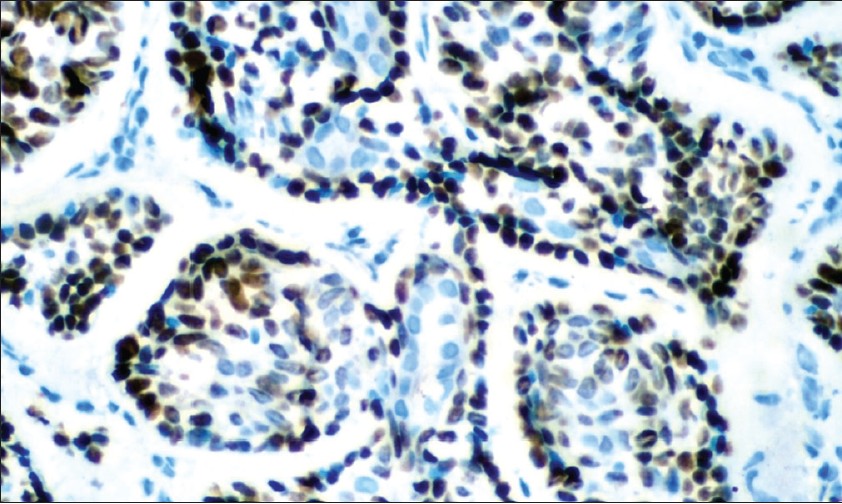 |
| Figure 5: Section shows nuclear positivity for p63 in the myoepithelial cells of the tumor (IHC DAB stain, × 400) |
Discussion
Cylindroma is a rare, benign appendageal tumor arising from eccrine glands. They are also purported to arise from stem cells of hair follicle. [1] They are twice as common in females as compared to males and rarely undergo malignant transformation. Multiple cylindromas can occur as part of familial cylindromatosis (OMIM 313270), Brooke-Spiegler syndrome (OMIM 605041) and multiple familial trichoepitheliomas (OMIM 601606). [1] Brooke-Spiegler syndrome is an autosomal dominant condition caused by mutation in the cylindromatosis CYLD gene, located on 16 q12-q13. [2] The gene product represses the tumor necrosis factor-α (TNF-α) pathway, which increases the expression of nuclear factor-κβ (NF-κβ), which regulates a number of anti-apoptotic genes involved in proliferation of skin appendages. [1],[2] Multiple cylindromas can cover the entire scalp resulting in hair loss, forming "turban tumors". The condition is associated with multiple trichoepitheliomas and spiradenomas in addition to trichoblastomas, basal-cell carcinomas, follicular cysts, organoid nevi etc. [3] Differential diagnoses to be considered eccrine spiradenoma, follicular infundibulum tumor and pilar cyst. Malignant transformation is said to occur more commonly with multiple cylindromas than in solitary cylindroma. 4 Hence, it is important to do a biopsy in patients with multiple cylindromas. Histopathology reveals well-circumscribed dermal lesions with areas of basaloid cell proliferation arranged in jigsaw-puzzle pattern. Rarely, neoplasms with hybrid features are seen including occurrence of all 3 adnexal tumors in a single lesion. [3] Histopathologically, it has to be differentiated from spiradenoma and adenoid cystic carcinoma. Staining with p63 indicates cells of myoepithelial origin, suggesting differentiation towards eccrine secretory cells. In addition to neoplasms of the skin appendages, patients with Brooke-Spiegler syndrome also are at risk for developing tumors of the salivary glands, such as basal-cell adenomas and adenocarcinomas of the parotid glands and minor salivary glands. [1] Reported treatments of cylindromas include excision, dermabrasion, electrodessication, CO 2 laser, cryotherapy and radiotherapy. Wide local excision is also preferred to avoid recurrence. [2] To avoid occurrence of new tumors, topical aspirin derivatives are currently being tried, which reduce the expression of NF-κβ. [1],[2]
| 1. |
Rajan N, Langtry JAA, Ashworth A, Roberts C, Chapman P, Burn J, et al. Tumor mapping in two large multigenerational families with CYLD mutations: Implications for patient management and tumor induction. Arch Dermatol 2009;145:1277-84.
[Google Scholar]
|
| 2. |
Doherty SD, Barrett TL, Joseph AK. Brooke-Spiegler syndrome: Report of a case of multiple cylindromas and trichoepitheliomas. Dermatol Online J 2008;14:8.
[Google Scholar]
|
| 3. |
Kim C, Kovich OI, Dosik J. Brooke-Spiegler syndrome. Dermatol Online J 2007;13:10.
[Google Scholar]
|
Fulltext Views
4,492
PDF downloads
2,246


