Translate this page into:
Proceedings from "Clinical Dermatology Updates - Atopic Dermatitis", 3-4 March, 2012, Mumbai
Correspondence Address:
Rashmi Sarkar
Department of Dermatology, Maulana Azad Medical College, New Delhi
India
| How to cite this article: Sarkar R. Proceedings from "Clinical Dermatology Updates - Atopic Dermatitis", 3-4 March, 2012, Mumbai. Indian J Dermatol Venereol Leprol 2012;78:524-525 |
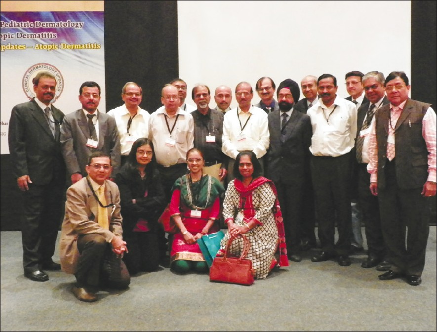
An update on "Atopic Dermatitis" was held in Mumbai on 3 rd and 4 th March, 2012 as a joint collaboration between Indian Society for Pediatric Dermatology and SIG Atopic Dermatitis, which covered the topic of atopic dermatitis (AD) from the basics to counseling. Several experts pooled in their views in this informative academic exercise [Figure - 1]. In his talk on "Diagnostic Criteria and Scoring System" Dr. Sandipan Dhar emphasized that: nummular eczema with 5-6 minor features of AD, can just be AD; severity of AD in children in India is much less than in the West; correlation of disease with IgE is only relevant for prognosis; and need for severity scoring is important mainly for epidemiological study, clinical trials, prognosis, academic and research purposes. Dr. Deepak Parikh delineated several unusual clinical presentations of atopic dermatitis including gluteofemoral or posterior thigh eczema, bilateral symmetrical hypopigmented patches, retroauricular eczema, and cradle cap after 3 months of age, atopic cheilitis, eyelid dermatitis, forefoot eczema and follicular eczema, which have to be observed. Dr. Arun Inamdar brought home the message that filaggrin mutations in AD were important for barrier function of the skin, and is responsible for more severe AD, severe eczema herpeticum, more systemic sensitization, more severe bronchial asthma and more severe association with alopecia areata. [1] Dr. D.M. Thappa, a panelist, also mentioned that aeroallergens as triggers come into the picture in atopics later in life.
 |
| Figure 1: Several experts in "Clinical Dermatology Updates - Atopic Dermatitis" |
There was a discussion whether AD is an epidermal or systemic disease. Dr Parikh discussed that extrinsic AD is seen in 45-80% atopics with raised IgE and is associated with filaggrin gene defects and decreased ceramides in the epidermis unlike intrinsic AD with normal serum IgE, seen in 20-40% atopics. The ceramides bind to the double bilayer of intercellular lamellae and constitute the lipid bilayer. It helps in maintaining the normal pH of the epidermis which when disturbed causes an activation of glucuronidases and can lead to infection. In "Trigger Factors", Dr Sandipan Dhar mentioned that foods commonly implicated in food allergy in infants are milk, egg, tree nut and soy. Most importantly, children usually outgrow these allergies later on in life. Aeroallergens such as animal dander, cockroach and fungus act as a trigger factor in older patients, and these atopics are prone to develop contact dermatitis and contact dermatitis to fragrances, latex, lanolin, and formaldehyde is as frequent as in non-atopics. Psychological triggers such as stress of examination, anxiety, social pressure, indirect stress and anxiety from parents also play an important role. In a panel discussion moderated by Dr. S. Criton, other panelists, Dr. A.J. Kanwar mentioned that atopic march is not seen frequently in Indians and in practice, adult atopic dermatitis is not common. Others, Dr. Rashmi Sarkar and Dr. Abir Saraswat felt that atopic dermatitis is the cutaneous expression of a systemic disease. Dr. Sacchidanand discussed food allergy in atopics mentioning that some of the suggested ways to avoid its development would be to continue breastfeeding, use hydrolyzed formulas, delay exposure to solid foods and introducing easily digestible vegetarian diets. Dr. Krupashankar suggested that very often, one is dealing with atopic dermatitis coexisting with allergic contact dermatitis and patch testing for nickel, chromium, benzocaine, formaldehyde, colophony and parabens may yield results. Regarding, when to investigate a child with atopic dermatitis, Dr. Manish Shah pointed out pertinently that it should be done in a child of AD with failure to thrive. Investigations suggested would be IgG, IgA, IgM, IgE levels; CD3, CD4, CD8, CD19 counts and platelet counts. Out of the investigations done for those with history of food allergy, Skin Prick Test, RAST and Immunocap, Immunocap is much more specific and can be used to determine whether a child has outgrown the allergy. Food specific IgE levels are more important rather than Skin Prick Tests. It is important to address the doubts of the parents and patients and evaluate the tests already done.
While discussing "Topical steroids in AD", Dr. Raghubir Bannerjee emphasized that topical steroids do remain the mainstay of treatment in AD while Dr. Murlidhar Rajgopalan elaborated on topical calcineurin inhibitors in AD, mentioning that data on long-term safety are required. [2] Regarding the place of TCIs in AD, they are not superior to corticosteroids. They are used to prevent relapses, and for maintenance therapy. Topical pimecrolimus is a better agent for application on the face and in those less than 2 years of age; it also is easy to apply and non-sticky. Dr. D.S. Krupashankar talked about the usefulness and safety of narrowband UVB in AD patients. The guidelines for giving PUVA in older children with AD were similar to those for psoriasis. UVA1 therapy is an effective therapy for acute exacerbations of AD. In the future, blue light full body irradiation would be useful for patients of AD. Dr. Sandipan Dhar gave the key messages that systemic therapies of proven efficacy are only few for AD. Systemic cyclosporine is useful for achieving a quick response and the control of AD is better and longer lasting in children than adults. Methotrexate is a cheaper option for AD but the effect is slow. Azathioprine is effective for a small subset of patients but is limited by side effects. Besides these, unproven treatment strategies for AD are delayed introduction of solid foods in infancy, Chinese Herbal Therapy, prolonged breast feeding, reduction of house dust mite, massage therapy and homeopathy. Potential future developments include inhibition of chemokines, Toll Like Receptor Antagonists, antiproteases, anti-IL-31, and blue light therapy. The update ended with the message that counseling and patient empathy and psychological support remained the cornerstone in the management of atopic dermatitis.
| 1. |
Fuiano N, Incorvaia C. Dissecting the causes of atopic dermatitis in children: Less food, more mites. Allergol Int 2012;61:231-43.
[Google Scholar]
|
| 2. |
Katoh N. Future perspectives in the treatment of atopic dermatitis. J Dermatol 2009;36:367-76.
[Google Scholar]
|
Fulltext Views
2,633
PDF downloads
1,813


