Translate this page into:
Primary multicentric cutaneous epithelioid angiosarcoma
Correspondence Address:
Murugan Sundaram
Department of Dermatology and Venereology, Sri Ramachandra University, Chennai
India
| How to cite this article: Sundaram M, Vetrichevvel T P, Subramanyam S, Subramaniam A. Primary multicentric cutaneous epithelioid angiosarcoma. Indian J Dermatol Venereol Leprol 2011;77:111 |
Abstract
Cutaneous epithelioid angiosarcoma is a rare malignant vascular tumor, most commonly affecting elderly men, and is usually located on the extremities. We report a case of an 81-year-old lady who presented with two ulcerated plaques over the right temporal and parietal scalp of 1 year duration. The right submaxillary and submandibular lymph nodes were enlarged and tender. Computed tomography (CT) scan of the head showed soft tissue swelling over parietal and temporal areas and there was no intracranial extension. Ultrasonogram of the abdomen showed hyperechoic areas in liver suggestive of secondaries. Histopathology of the skin lesion showed the dermis and subcutis composed of clusters of atypical epithelioid cells with vesicular nuclei, prominent nucleoli and eosinophilic cytoplasm with increased mitotic figures. Immunohistochemical staining revealed CD-31, 33, 34 and vimentin positivity, while cytokeratin was negative confirming the diagnosis of epitheloid angiosarcoma. This case report highlights the unusual occurrence of multicentric epitheloid angiosarcoma on the scalp with secondaries in the liver.Introduction
Cutaneous epithelioid angiosarcoma is a rare malignant vascular tumor, most commonly affecting elderly men, and is usually located on the extremities. The tumor runs an aggressive course with early metastasis to the viscera. [1] Its unusual multicentric occurrence over the scalp in an elderly lady with secondaries in the liver is reported herewith.
Case Report
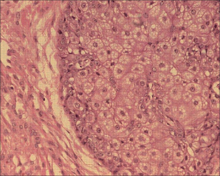
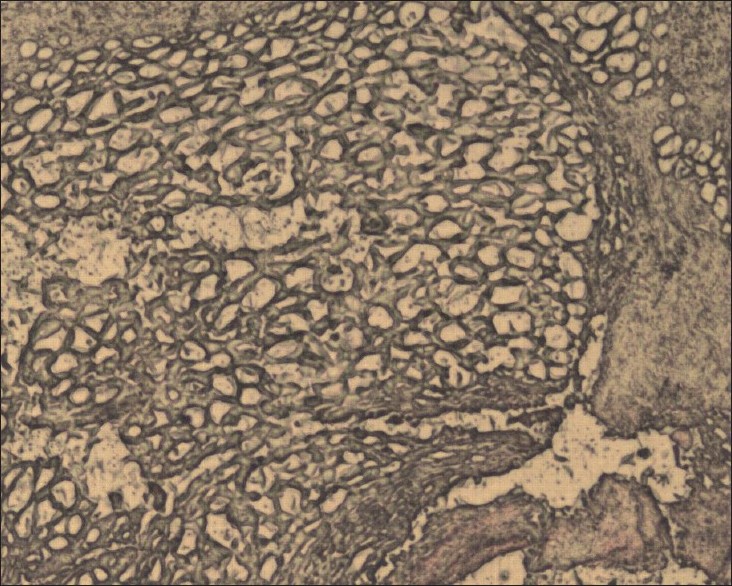
An 80-year-old lady presented with two painful, ulcerated swellings on the scalp of 1 year duration. The first lesion appeared over the right temporal region as pinkish, raised lesions followed by a swelling in the right parieto-occipital region in 3 months and they progressively increased in size and ulcerated. There was no history of weight loss, fever, abdominal pain, early satiety or swellings anywhere else in the body. She was poorly built and nourished; otherwise, the general examination was unremarkable. Cutaneous examination revealed two well-defined ulcerated plaques of size 6.2 × 5.4 cm and 5.3 × 3.2 cm diameter [Figure - 1]. The ulcers were covered with a dark brown eschar, the bases of the ulcers were indurated and the margins were friable. The extension of the lesion to the frontal scalp as purplish indurated plaque was noticed. The right submaxillary and submandibular lymph nodes were enlarged and tender. X-ray of the skull showed soft tissue shadows in the parietal and temporal regions of the scalp and computed tomography (CT) scan of the head showed soft tissue swelling over parietal and temporal areas and there was no intracranial extension. While his chest X-ray was normal, ultrasonogram of the abdomen showed hyperechoic areas in liver, suggestive of secondaries. Histopathology of the lesion showed the dermis and subcutis composed of clusters of atypical epithelioid cells with vesicular nuclei, prominent nucleoli and eosinophilic cytoplasm with increased mitotic figures, suggestive of epithelioid angiosarcoma [Figure - 2]. Immunohistochemical staining revealed CD-31 [Figure - 3], 33, 34, vimentin positivity, while cytokeratin was negative. She was treated with systemic and topical antibiotics as she refused chemotherapy and surgery.
 |
| Figure 1: Ulcerated plaque over right parietal scalp |
 |
| Figure 2: Histopathology showing clusters of atypical epithelioid cells with vesicular nuclei, prominent nucleoli and eosinophilic cytoplasm (H and E, ×400) |
 |
| Figure 3: Immunohistochemical staining showing CD-31 positivity (×400) |
Discussion
Angiosarcomas are rare malignant tumors of the blood vessels and usually arise from the skin and soft tissues but also have been known to arise from bones and internal organs. Cutaneous angiosarcoma of the face and scalp can have varied presentations like bruise, pinkish macules, papules, nodules or plaques with ulceration. Epitheloid angiosarcoma is a rare histological variant of cutaneous angiosarcoma and was first described by Rosai et al. in 1976. It commonly arises from the deep soft tissue [2] and is characterized by oval or polygonal cells with abundant, faintly eosinophilic cytoplasm. [3] Use of thorium dioxide contrast, chronic lymphedema, radiation therapy and sun exposure have been implicated in the causation of cutaneous angiosarcoma. [4] No immunohistochemical staining is specific; however, CD-31, Factor VIII related antigen, Ulex europaeus agglutinin I (UEA I),laminin and CD-34 positivity aids in the diagnosis. CD-31 is reported to be a good specific and sensitive endothelial marker and is helpful in differentiating from amelanotic melanoma and undifferentiated carcinoma, especially if Factor VIII related antigen is negative. [5] Electron microscopy may reveal rod-shaped, microtubulated Weibel-Palade bodies. [4] Prognosis depends on the site, size, stage, cellularity, pleomorphism, and mitotic activity. Other poor prognostic factors include bleeding, pain and lesions of size greater than 5 cm. [4] Epithelioid angiosarcoma usually has a poor prognosis as it grows rapidly with metastases to lung, bone, soft tissue, lymph nodes and brain. [4],[6] Smaller tumors are managed with radical excision with regional lymph node clearance, with or without adjuvant radiotherapy, while multidrug chemotherapy with adriamycin, paclitaxel or vincristine followed by radiotherapy is used for unresectable tumors and metastases. [3],[6]
| 1. |
Chen TJ, Chiou CC, Chen CH, Kuo TT, Hong HS. Metastasis of mediastinal epithelioid angiosarcoma to the finger. Am J Clin Dermatol 2008;9:181-3.
[Google Scholar]
|
| 2. |
Kikuchi A, Satoh T, Yokozeki H. Primary cutaneous epithelioid angiosarcoma. Acta Derm Venereol 2008;88:422-3.
[Google Scholar]
|
| 3. |
Calonje E. Vascular tumors: Tumors and tumor-like conditions of blood vessels and lymphatics. In: Elder DE, Elenitsas R, Johnson BL Jr, Murphy GF, Xu X, editors. Lever's Histopathology of the Skin. 9 th ed. Philadelphia, PA: Lippincott Williams and Wilkins; 2004. p. 1040-3.
th ed. Philadelphia, PA: Lippincott Williams and Wilkins; 2004. p. 1040-3. '>[Google Scholar]
|
| 4. |
Glickstein J, Sebelik ME, Lu Q. Cutaneous angiosarcoma of the head and neck: A case presentation and review of the literature. Ear Nose Throat J 2006;85:672-4.
[Google Scholar]
|
| 5. |
Smith KJ, Lupton GP, Skelton HG. Cutaneous angiosarcomas with a starry-sky pattern. Arch Pathol Lab Med 1997;121:980-4.
[Google Scholar]
|
| 6. |
Kacker A, Antonescu CR, Shaha AR. Multifocal angiosarcoma of the scalp: A case report and review of the literature. Ear Nose Throat J 1999;78:302-5.
[Google Scholar]
|
Fulltext Views
2,865
PDF downloads
2,113


