Translate this page into:
Primary cutaneous endometriosis of the umbilicus
Corresponding author: Dr. Chuan Wan, Department of Dermatology, The First Affiliated Hospital of Nanchang University, 17 Yongwai Zheng Street, Nanchang, China. E-mail: chuanwan@ncu.edu.cn
-
Received: ,
Accepted: ,
How to cite this article: Wan C, Chen L. Primary cutaneous endometriosis of the umbilicus. Indian J Dermatol Venereol Leprol 2021;87:146-146.
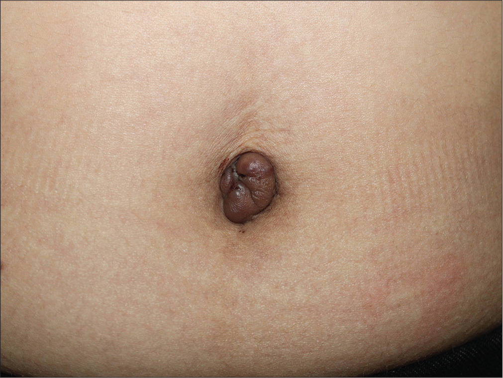
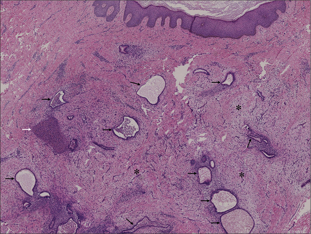
A 29-year-old woman presented with a 1-year history of an umbilical mass, complaining of periodic pain in the umbilical area synchronized with menstruation. She had no history of surgery or abdominal trauma. Examination revealed a firm, 2.2 cm × 1.8 cm, dark-brown mass on her umbilicus [Figure 1a]. Abdominal computed tomography showed an increased density at the umbilicus without connection to any abdominal organs. Histopathological examination showed multiple endometrial glandular structures surrounded by cellular endometrial-type stroma in the dermis [Figure 1b]. Based on these findings, the umbilical lesion was diagnosed as primary cutaneous endometriosis and it was removed by complete surgical excision.

- A well-circumscribed, firm, 2.2 cm × 1.8 cm, dark-brown mass arising from the umbilicus

- Histopathological examination showed, within the dermis, multiple endometrial glandular structures (black arrows) surrounded by cellular endometrial-type stroma (asterisks) in which there were focal hemosiderin deposits (white arrows) (H and E, ×200)
Declaration of patient consent
The authors certify that they have obtained all appropriate patient consent forms. In the form, the patient has given her consent for her images and other clinical information to be reported in the journal. The patient understands that name and initials will not be published and due efforts will be made to conceal identity, but anonymity cannot be guaranteed.
Financial support and sponsorship
Nil.
Conflicts of interest
There are no conflicts of interest.


