Translate this page into:
Localization of varicella around cutaneous leishmaniasis
Correspondence Address:
A H Siadat
Skin Diseases and Leishmaniasis Research Center (SDLRC), Sedigehe Tahereh Research Center, Khoram Street, Isfahan
Iran
| How to cite this article: Siadat A H, Nilforoush Zadeh M A, Moradi S H, Baradaran E H. Localization of varicella around cutaneous leishmaniasis. Indian J Dermatol Venereol Leprol 2007;73:124-126 |
 |
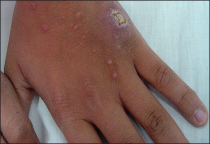
| Vesiculopustular lesions of varicella around leishmaniasis ulcer on the dorsum of the left hand |
 |
| Vesiculopustular lesions of varicella around leishmaniasis ulcer on the dorsum of the left hand |
Sir,
Atypical varicella has been associated with immunosuppression, [1] skin injury, [2] sun exposure [3] and preexisting cutaneous disease. [4] Localization of varicella lesions has also been described after sunburn. [5]
A 13-year-old boy was referred to us for evaluation of an ulcerative erythematous plaque (diameter: 30 x 25 mm) on his hand. His lesion began approximately one month before presentation in our clinic. He lived in an endemic area of leishmaniasis in Isfahan province of Iran. He did not have pruritic or painful lesions. Physical examination revealed an erythematous to violaceous indurated plaque with central ulceration on his left hand.
Based on the characteristics of the lesion and the place of his residence, the diagnosis of cutaneous leishmaniasis was suggested. A direct smear from the skin lesions was positive for Leishman body. Regarding the singularity and diameter of the lesion, it was planned to be treated with intralesional glucantime. The lesion was infiltrated with intralesional glucantime until the lesion and 1 mm surrounding it became blanched. The treatment was repeated every week.
After four sessions of treatments, the size and induration of lesion had decreased by about 60%. At this time, the patient developed fever and after one day, papulo-vesicular and pustular lesions on his face and then his trunk. He was referred to our clinic again and considering his negative past history for varicella, distribution and centrifugal spread of his lesions, a diagnosis of varicella was made for him. The diagnosis was confirmed by Tzanck Smear.
There was aggregation of varicella lesions around the lesion of leishmaniasis. In fact, although his right hand was completely spared during the course of varicella, his left hand (that was involved with cutaneous leishmaniasis) had many varicella lesions that were localized around the leishmaniasis lesion [Figure - 1]. These lesions were positive for multinucleated giant cells in Tzanck smear and did not show any Leishman body on Giemsa stain. There were only a few lesions of varicella on his arms and forearms.
Varicella was treated with oral acyclovir tablet (20 mg/kg/dose) and recovered in seven days. His leishmaniasis lesion healed after several more injections of intralesional glucantime.
In 1987, Shelley and Shelley reported three children to have striking localization of varicella lesions to skin overlying a diphtheria-pertussis-tetanus toxoid (DPT) immunization injection site, a facial abrasion and a sunburn of the trunk, respectively. [6]
Many side effects are attributed to the intralesional administration of the glucantime. Masoudi et al . has reported side-effects such as sporotrichoid nodules, pyoderma, erysipelas, necrosis and urticaria following the use of glucantime for treatment of cutaneous leishmaniasis. [7] Sodium stibogluconate is another pentavalent antimonial compound that is used in the treatment of leishmaniasis. This drug has been reported to have an association with reactivation of varicella zoster virus (VZV). Hartzell et al reported a case of an immunocompetent adult who developed VZV aseptic meningitis and dermatomal herpes zoster during treatment with sodium stibogluconate. [8]
Wortmann et al reviewed 84 patients with cutaneous leishmaniasis treated with sodium stibogluconate and revealed that three had developed herpes zoster during or shortly after receiving therapy. These authors demonstrated that the administration of Pentostam ® for the treatment of cutaneous leishmaniasis results in lymphopenia that may be related to the subsequent occurrence of herpes zoster. [9]
Considering the fact that our patient had negative history for previous varicella infection and also the type of spread of his lesions, his diagnosis was more consistent with primary varicella infection rather than herpes zoster. Localization of varicella lesions around leishmaniasis ulcer has not been reported yet. This localization somewhat simulated satellite lesions of leishmaniasis. Direct smear and also Tzanck smear were used to rule out the satellite leishmaniasis lesions.
Perhaps the increased blood flow in erythematous skin allows greater exposure to viral particles [10] It may be that inflamed skin allows enhanced replication of the virus [11] or that increased capillary permeability allows the virus or viral-infected cells to migrate into the skin in these sites. [12] This possibility is supported by the fact that some reports have described vesicles localized to areas of prior trauma. [6] In addition, this finding may be related to the possible local immunosuppressive effect of the intralesional glucantime. [9]
| 1. |
Nagore E, Sanchez-Motilla JM, Julve N, Lecuona C, Oliver V. Atypical involvement of the palms and soles in a varicella infection. Acta Derm Venereol 1999;79:322.
[Google Scholar]
|
| 2. |
Belhorn TH, Lucky AW. Atypical varicella exanthems associated with skin injury. Pediatr Dermatol 1994;11:129-32.
[Google Scholar]
|
| 3. |
Boyd AS, Neldner KH. Photolocalized varicella in an adult. J Am Med Assoc 1991;266:2204.
[Google Scholar]
|
| 4. |
Egan C, O'Reilly M, Vanderhooft SL, Rallis T. Acute generalized varicella zoster in the setting of pre-existing generalized erythema. Pediatr Dermatol 1999;16:2111-2.
[Google Scholar]
|
| 5. |
Leroy D, Vuillamie M, Verneuil L, Penven K, Letellier P, Dompmartin A. Photodistribution of varicella in an adult. Ann Dermatol Venereol 2004;131:365-7.
[Google Scholar]
|
| 6. |
Shelley WB, Shelley ED. Immunization, abrasion and sunburn as localizing factors in chicken pox. Pediatr Dermatol 1987;4:102-4.
[Google Scholar]
|
| 7. |
Masmoudi A, Maalej N, Boudaya S, Turki H, Zahaf A. Adverse effects of intralesional Glucantime in the treatment of cutaneous leishmaniasis. Med Mal Infect 2006;36:226-8.
[Google Scholar]
|
| 8. |
Hartzell JD, Aronson NE, Nagaraja S, Whitman T, Hawkes CA, Wortmann G. Varicella zoster virus meningitis complicating sodium stibogluconate treatment for cutaneous leishmaniasis. Am J Trop Med Hyg 2006;74:591-2.
[Google Scholar]
|
| 9. |
Wortmann GW, Aronson NE, Byrd JC, Grever MR, Oster CN. Herpes zoster and lymphopenia associated with sodium stibogluconate therapy for cutaneous leishmaniasis. Clin Infect Dis 1998;27:509-12.
[Google Scholar]
|
| 10. |
Castrow FF 2 nd , Wolf JE Jr. Photolocalized varicella. Arch Dermatol 1973;107:628.
[Google Scholar]
|
| 11. |
Boyd AS, Neldner KH. Photolocalized varicella in an adult. JAMA 1991;266:2204.
[Google Scholar]
|
| 12. |
Feder HM Jr, Luchetti ME. Varicella mimicking a vesiculobullous sun eruption. J Infect Dis 1988;158:243.
[Google Scholar]
|
Fulltext Views
2,091
PDF downloads
1,812


