Translate this page into:
Squamous cell carcinoma of the thumb nail bed
Correspondence Address:
Manohar Arumugam
Department of Orthopaedic Surgery, Faculty of Medicine and Health Science, University Putra Malaysia, Clinical Campus, Jalan Masjid 50586, Kuala Lumpur
Malaysia
| How to cite this article: Arumugam M. Squamous cell carcinoma of the thumb nail bed. Indian J Dermatol Venereol Leprol 2007;73:445 |
Abstract
Squamous cell carcinoma arising from the nail bed is not common. This condition can be easily misdiagnosed, especially if there is preceding trauma. We present here a case of squamous cell carcinoma of the right thumb in a 70 year-old man. The distal phalanx and part of the proximal phalanx were also involved. We performed a disarticulation of the metacarpophalangeal joint of the right thumb. The wound healed well. If an early diagnosis is made, then Moh's micrographic surgery or wide local excision with the use of a local flap could be advocated. In late stages, amputation or disarticulation is the treatment of choice. |
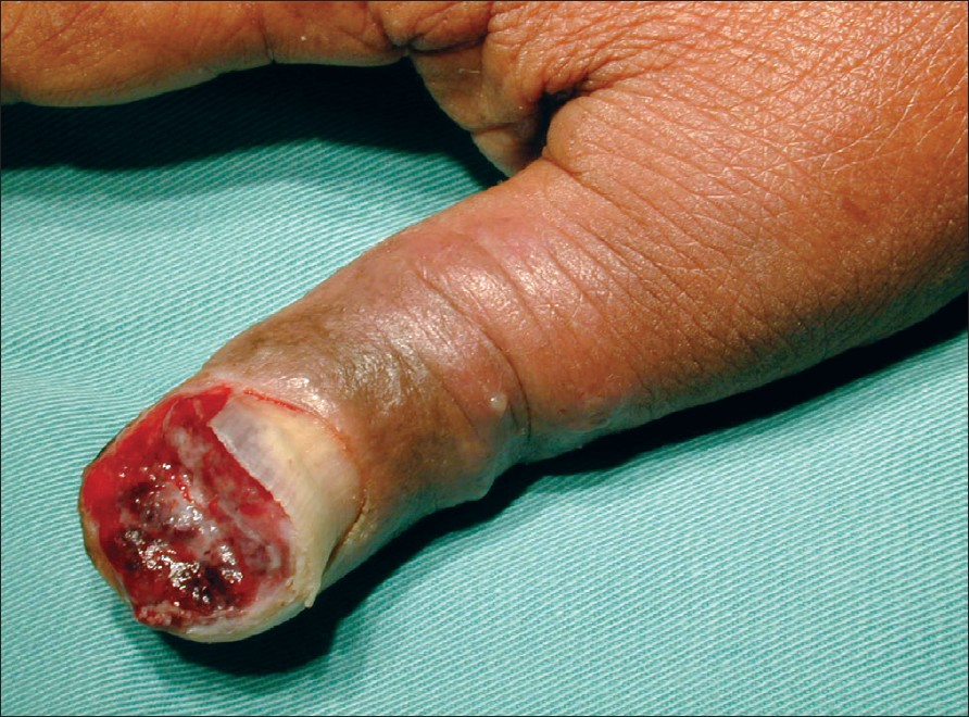
| Figure 1: Squamous cell carcinoma lifting off the nail plate from the nail bed |
 |
| Figure 1: Squamous cell carcinoma lifting off the nail plate from the nail bed |
Squamous cell carcinoma of the nail bed is uncommon. The clinical presentation can mimic several benign conditions such as paronychia, pyogenic granuloma or verruca vulgaris. A high degree of suspicion is needed to diagnose this condition. Any chronic or recurring lesion that fails to respond to initial treatment should be biopsied. The treatment depends on the extent of the lesion at the time of presentation and can vary from Moh′s microsurgery to digital amputation. We report here a case of subungual squamous cell carcinoma of the thumb with bone involvement treated with disarticulation of the metacarpophalangeal joint.
Case Report
A 70 year-old man presented with pain and swelling of the right thumb after accidentally hitting it with a hammer. He was initially treated by a general practitioner. The subungual hematoma was drained and analgesics were prescribed for pain.
Six months after the initial injury, he presented with a nonhealing wound with a nonhealing ulcerated growth measuring 2.5 x 2.1 cm in size at the tip of the right thumb with a raised nail plate [Figure - 1]. Our provisional diagnosis was pyogenic granuloma. Histopathology of this mass showed an ulcerated, moderately differentiated squamous cell carcinoma arranged in sheets and large clumps. The cells showed nuclear pleomorphism, increased abnormal mitosis and areas of keratin formation. No vascular or lymphatic permeation was seen. The margins of the tumor were clear. A diagnosis of moderately differentiated squamous cell carcinoma was made. Radiographs revealed the involvement of the distal phalanx and part of the proximal phalanx. There was no evidence of distant metastasis. We performed a disarticulation of the metacarpophalangeal joint of the thumb, which healed uneventfully. The patient did not want any further reconstructive surgery for the thumb.
Discussion
Squamous cell carcinoma of the nail bed was first described in 1850 by Velpeau. [1],[2] The actual rate of incidence is not known and the common age of incidence appears to be in the 5 th decade. [3] The youngest patient reported to have squamous cell carcinoma of the nail bed was 25 years old. [4] The actual cause is not known. A few possible causes have been suggested. Chronic infections, radiation exposure, human papillomavirus (HPV), burn scars, chronic exposure to sun and chronic dermatitis have been implicated. [7],[8],[9],[10] Squamous cell carcinoma arising from a psoriatic nail bed has been reported. [11] There is an association between squamous cell carcinoma and HPV. Genital-digital transmission has been suggested as a mechanism of transmission. [12] This is further supported by the fact that no HPV DNA was detected using polymerase chain reaction (PCR) in patients with subungual squamous cell carcinoma of the toe. [13] PCR was not performed on the sample obtained from our patient. In our patient, there was no past history of viral warts, although he did have preceding trauma. A few authors have reported crush injury, fishbone penetration and paper staple puncture as the cause of trauma preceding subungual squamous cell carcinoma. [4],[14] Our patient accidentally hit his thumb with a hammer. The exact relation between trauma and squamous cell carcinoma is not known, however this observation could be explained by coincidence, pain causing increased attention to an area of the digit and bleeding of the tumor after trauma. Squamous cell carcinoma of the nail bed can have different clinical presentations, it can resemble chronic paronychia, verruca vulgaris, pyogenic granuloma, ulcerative lesion or present as a small swelling. Unlike subungual melanoma, the involvement of axillary lymph nodes and distant metastasis are rare since the tumor is slow-growing and low-grade in nature. Bone tends to be involved in late cases. Diagnosis is often delayed since this is not a common condition. In our patient, preceding trauma drew attention to the tumor. Most of the reported cases involve the thumb. However, involvement of the other digits like the index finger and middle finger have also been reported. [6],[9]
Differential diagnoses include delayed healing of traumatic wound, chronic nail biting, paronychia and fungal infections. Others include pyogenic granuloma, verrucae vulgaris, verrucous carcinoma, subungual metastasis, acral amelanotic melanoma, subungual keratoacanthoma. [4] Pyogenic granuloma is relatively quite common. It is usually a small, red, oozing and bleeding growth that looks like raw meat. There is often preceding trauma and it grows rapidly over a period of a few weeks to an average size of a half an inch. Verrucae vulgaris can appear on the palmar or dorsal aspect of the fingers, presenting as multiple, raised, hyperkeratotic lesions. Verrucous carcinoma is rare-it usually occurs in the 6 th decade of life and it presents as a slow-growing, fungating, recalcitrant and exophytic mass. [15] The clinical presentation of subungual metastasis is variable-it may present as an erythematous swelling of the digit or it may appear as a violaceous nodule which may cause distortion of either the nail plate or the surrounding soft tissue. Since it is frequently painful in nature, it may be mistaken for an acute infection. [16] Acral amelanotic melanoma presents as a small bluish lesion in the subungual area. The skin around the nail is edematous and is usually slightly tender. [17] Subungual keratoacanthoma is a rapidly growing tumor, which presents as a rapidly growing painful mass. X rays show a lytic, cup-shaped erosion of the distal phalanx. Healing occurs rapidly. [18] A plain X ray therefore is useful to determine if there is bone involvement. A biopsy often clinches the diagnosis.
The treatment of squamous cell carcinoma of the nail bed depends on the extent of the tumor. Moh′s micrographic surgery has been used in the early stage of the disease to minimize tissue loss. [10],[19] For lesions with no bone involvement, wide local excision with no less than 4 mm of normal tissue from the margins of the tumor should be done along with reconstruction of the remaining digit. [20] Reconstruction can be done with full-thickness skin graft or local flaps. Among the local flaps that have been described are a dorsal V-Y flap, a Brunneli flap and a lateral pulp flap. [21],[22],[23] For lesions with bone involvement, amputation is the treatment of choice. The level of amputation depends on the extent of bone involvement. Radiation therapy has been recommended for the salvage of unresectable subungual squamous cell carcinoma. [24]
Squamous cell carcinoma of the nail bed is an uncommon condition. Awareness about the disease and a high index of suspicion is necessary to make an early diagnosis. Nail lesions not responding after initial treatment should be biopsied.
| 1. |
Sigel H . Squamous cell carcinoma epithelioma of the left fourth finger. Am J Cancer 1937;30:108.
[Google Scholar]
|
| 2. |
Eibel P. Squamous cell carcinoma of the nail bed. A report of two cazes and a discussion of the literature. Clin Orthop Relat Res 1971;74:155-60.
[Google Scholar]
|
| 3. |
Yip KM, Lam SL, Shee BW, Shun CT, Yang RS. Subungual squamous cell carcinoma: Report of 2 cases. J Formos Med Assoc 2000;99:646-9.
[Google Scholar]
|
| 4. |
Figus A, Kanitkar S, Elliot D. Squamous cell carcinoma of the lateral nail fold. J Hand Surg Br 2006;31:216-20.
[Google Scholar]
|
| 5. |
Ashinoff R, Li JJ, Jacobson M, Friedman-Kien AE, Geronemeus RG. Detection of human papilloma virus DNA in squamous cell carcinoma of the nail bed and finger determined by polymerase chain reaction. Arch Dermatol 1991;127:1813-8.
[Google Scholar]
|
| 6. |
Zabawski EJ Jr, Washak RV, Cohen JB, Cockerell CJ, Brown SM. Squamous cell carcinoma of the nail bed: Is finger predominance another clue to etiology? A report of 5 cases. Cutis 2001;67:59-64.
[Google Scholar]
|
| 7. |
Phatak SV, Kolwadkar PK. Images: Squamous cell carcinoma of the hand and wrist developing in burn scar. Indian J Radiol Imaging 2005;15:467-8.
[Google Scholar]
|
| 8. |
Kumar N, Saxena YK. Two cases of rare presentation of basal cell and squamous cell carcinoma on the hand. Indian J Dermatol Venereol Leprol 2002; 68:349-51.
[Google Scholar]
|
| 9. |
Carroll RE. Squamous cell carcinoma of the nail bed. J Hand Surg Am 1976;1:92-7.
[Google Scholar]
|
| 10. |
Mikhail GR. Bowen disease and the squamous cell carcinoma of the nail bed . Arch Dermatol 1974;110:267-70.
[Google Scholar]
|
| 11. |
Dobson CM, Azurdia RM, King CM. Squamous cell carcinoma arising in a psoriatic nail bed: Case report with discussion of diagnostic difficulties and therapeutic options. Br J Dermatol 2002;147:144-9
[Google Scholar]
|
| 12. |
Dalle S, Depape L, Phan A, Balme B, Ronger-Savle S, Thomas L. Squamous cell carcinoma of the nail apparatus: Clinicopathological study of 35 cases. Br J Dermatol 2007;156:871-4.
[Google Scholar]
|
| 13. |
Nasca MR, Innocenzi D, Micali G. Subungual squamous cell carcinoma of the toe: Report on three cases. Dermatol Surg 2004;30:345-8.
[Google Scholar]
|
| 14. |
Wong TC, IP FK, Wu WC. Squamous cell carcinoma of the nail bed: Three case reports. J Orthop Surg 2004;12:248-52.
[Google Scholar]
|
| 15. |
Wright PK, Vidyadharan R, Jose RM, Rao GS. Plantar verrucous carcinoma continues to be mistaken for verruca vulgaris. Plast Reconstruct Surg 2004;113:1101-3.
[Google Scholar]
|
| 16. |
Cohen PR. Metastatic tumors to the nail unit: Subungual metastases. Dermatol Surg 2001;27:280-93.
[Google Scholar]
|
| 17. |
Grunwald MH, Yerushalmi J, Glesinger R, Lapid O, Zirkin HJ. Subungual amelanotic melanoma. Cutis 2000;65:303-4.
[Google Scholar]
|
| 18. |
Levy DW, Bonakdarpour A, Putong PB, Mesgarzadeh M, Betz RR. Subungual Keratoacanthoma. Skeletal Radiol 1985;13:287-90.
[Google Scholar]
|
| 19. |
Goldminz D, Bennettr RG. Mohs micrographic surgery of the nail unit. J Dermatol Surg Oncol 1992;18:721-6.
[Google Scholar]
|
| 20. |
Thomas DJ, King AR, Peat BG. Excision margins for non melanotic skin cancer. Plast Reconst Surg 2003;112:57-63.
[Google Scholar]
|
| 21. |
Yii NW, Elliot D. Dorsal V-Y advancement flaps in digital reconstruction. J Hand Surg Br 1994;19:91-7.
[Google Scholar]
|
| 22. |
Brunneli F, Vigasio A, Valenti P, Brunneli GR. Arterial anatomy and clinical application of dorsoulnar flap of the thumb. J Hand Surg Am 1999;24:803-11.
[Google Scholar]
|
| 23. |
Elliot D, Jigjinni VS. The lateral pulp flap. J Hand Surg Br 1993;18:423-6.
[Google Scholar]
|
| 24. |
Yaparpalvi R, Mahadeia PS, Gorla GR, Beitler JJ. Radiation therapy for the salvage of unresectable subungual squamous cell carcinoma. Dermatol Surg 2003;29:294-6.
[Google Scholar]
|
Fulltext Views
4,727
PDF downloads
1,925


