Translate this page into:
A burning orange-brown plaque in a country man
Corresponding author: Dr. Felipe Tavares Rodrigues, 51 Visconde de Abaeté, 111, Rio de Janeiro, Rio de Janeiro, 20551-080, Brazil. medftr@yahoo.com.br
-
Received: ,
Accepted: ,
How to cite this article: Rodrigues FT, Andrade LG, D’Acri AM. An orange-brownish burning plaque in a country field man. Indian J Dermatol Venereol Leprol 2022;88:428-9.
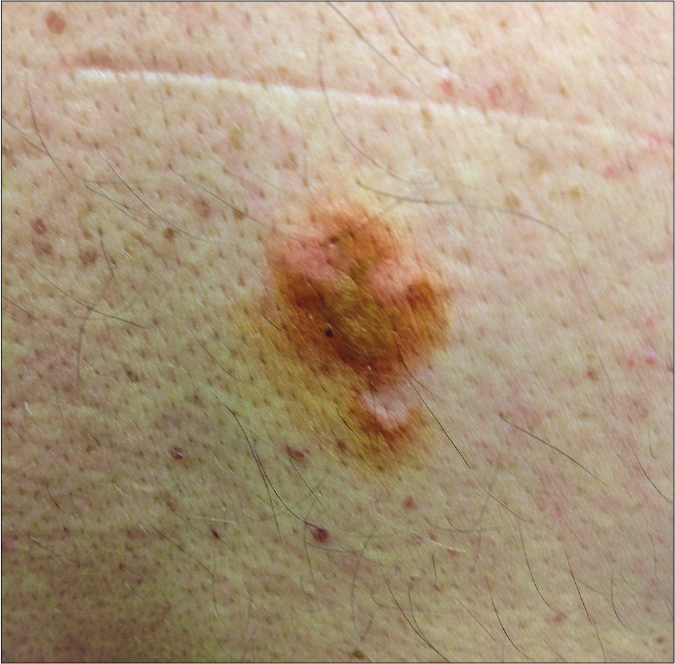
A 45-year-old man complained of slightly erythematous and pruriginous orange-brownish asymmetrical plaques after lying on a riverside lawn for fishing near to his house. The plaques were sudden in onset, had well-defined borders and measured approximately 4 cm in diameter [Figure 1]. He lived in the countryside – a peripheral city in the metropolitan region of Rio de Janeiro, Brazil. He had no known medical problems. At the time of the event, the patient complained of a mild burning sensation which persisted after self-medication of an antihistamine drug.

- An orange-brown plaque affecting a country man’s back
Question
What is the diagnosis?
Answer
Millipede dermatitis
Discussion
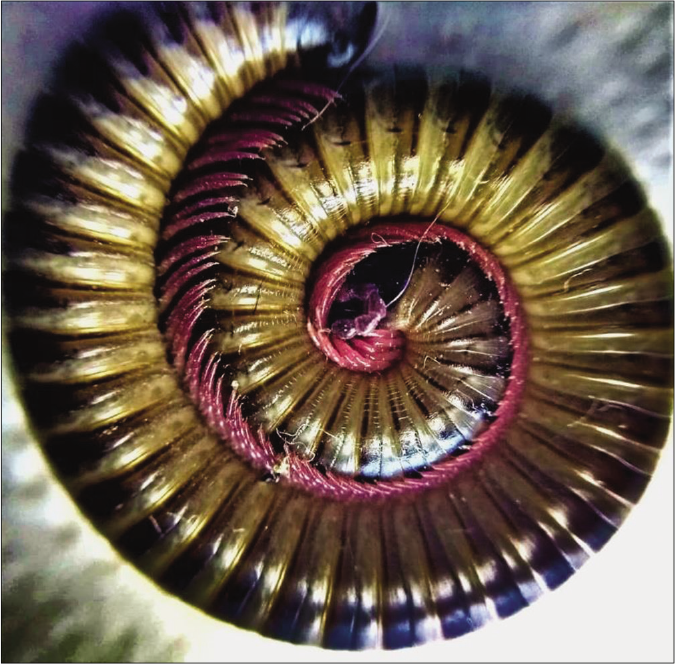
The patient reported direct skin contact with a millipede while he was fishing. After two days, he consulted a dermatologist at Gaffrée e Guinle’s University Hospital and provided a sample of the arthropod to the dermatology attendant [Figure 2].

-
Rhinocricus padbergi sample brought by the patient at presentation
With regard to treatment, cold compresses and a topical cream (0.5 mg/g desonide, twice daily for seven days) were applied. Consequently, the lesions subsided and there was symptomatic improvement. Post-inflammatory pigmentation lasted for four months in our patient which, according to literature, can persist for months together.
The differential diagnoses for millipede dermatitis could be irritant contact dermatitis and exogenous pigmentary conditions such as phytophotodermatitis. Contact dermatitis can also occur by exposure to other arthropods, such as the Paederus sp. beetle which is predominantly seen in India and Asia. This insect causes dermatitis linearis which differs from millipede dermatitis because it is more intense, more painful and can elicit vesicle formation more easily; its lesions have a typical linear patch pattern, not exhibiting an orange-brown discoloration.1-3
In more exuberant cases, millipede dermatitis can present with cyanosis and can mimic an ischemic tissue condition, clinical peripheral vascular disease, or even cutaneous burns.
Millipedes belong to the phylum Arthropoda and are members of the Diplopoda class. They are commonly found in South America. In Brazil, the species with the highest prevalence is Rhinocricus padbergi (family Rhinocricidae). Accidental brushing against or crushing the animal over the skin provokes the release of benzoquinone which is an irritating substance that triggers contact dermatitis and residual dyspigmentation. During the rainy summer season in the Southeast of Brazil, this species invades the urban areas, resulting in numerous cases of dermatitis, mainly affecting the feet because millipedes commonly hide in footwear.4,5
Case reports of millipede dermatitis are usually published from tropical countries, considering the fact it is a self-limiting disease and most cases are less severe. Its diagnosis is predominantly clinical. In rare cases where biopsies are required, the histopathologic features resemble irritant contact dermatitis which shows a partial-thickness epidermal necrosis with adjacent inflammatory infiltrate.6
Arthropod-related dermatitis is treated by washing the area, applying cold wet compresses and topical steroid. Rarely, oral antibiotics may be prescribed in case of secondary infection.5
Declaration of patient consent
The authors certify that they have obtained all appropriate patient consent.
Financial support and sponsorship
Nil.
Conflicts of interest
There are no conflicts of interest.
References
- Millipede accident with unusual dermatological lesion. An Bras Dermatol. 2019;94:765-7.
- [CrossRef] [PubMed] [Google Scholar]
- Paederus dermatitis: An outbreak on a medical mission boat in the Amazon. J Clin Aesthet Dermatol. 2011;4:44-6.
- [Google Scholar]
- Paederus dermatitis. Indian J Dermatol Venereol Leprol. 2007;73:13-5.
- [CrossRef] [PubMed] [Google Scholar]
- Composition of the defensive secretion of the Neotropical millipede Rhinocricus padbergi. Verhoeff 1938 (Diplopoda: Spirobolida: Rhinocricidae) Entomotropica. 2003;18:79-82.
- [Google Scholar]
- Millipede burn masquerading as trash foot in a paediatric patient. ANZ J Surg. 2013;84:388-90.
- [CrossRef] [PubMed] [Google Scholar]


