Translate this page into:
Eruptive collagenoma: A rare connective tissue hamartoma presenting with an unusual morphology
Corresponding author: Dr Jaspriya Sandhu, Associate Professor, Department of Dermatology, Venereology & Leprology, Dayanand Medical College & Hospital, Tagore Nagar, Civil Lines, Ludhiana, India. sandhu.jaspriya@gmail.com
-
Received: ,
Accepted: ,
How to cite this article: Sandhu J, Gupta SK, Kaur H, Kaur H. Eruptive collagenoma: A rare connective tissue hamartoma presenting with an unusual morphology. Indian J Dermatol Venereol Leprol. 2024;90:215–7. doi: 10.25259/IJDVL_210_2022
Dear Editor,
Collagenoma is a connective tissue nevus predominantly composed of collagen.1 Collagenomas represent benign hamartomatous lesions of the dermis of unknown aetiology. They are classified as inherited or acquired; inherited include familial cutaneous collagenomas and shagreen patches of tuberous sclerosis and the acquired include eruptive collagenomas and isolated collagenomas.2 We report here an unusual case of eruptive collagenoma in a middle-aged man.
A 51-year-old man presented to us with gradually progressive, asymptomatic symmetric eruption of multiple, skin-coloured, raised lesions present on chest, abdomen, upper back and bilateral arms for seven months. There was no past history of trauma or skin eruptions at the site of lesions. Systemic examination showed no abnormality. On dermatological examination, multiple partly circumscribed, discrete to coalescent, firm, waxy flesh-coloured to dusky red nodules and plaques of varying sizes, 0.5 × 0.5 cm2 to 2.0 × 2.0 cm2 were present symmetrically on chest, upper back (scapular region) and proximal upper limbs [Figures 1a and 1b]. The overlying skin showed no surface changes; mild tenderness was present over larger coalescent lesions. The mucosae, scalp and nails were normal.

- Multiple, well-defined nodules coalescing to form plaques present symmetrically on upper chest, proximal limbs and abdomen

- Multiple well- to ill-defined, discrete to coalescent, firm, waxy flesh-coloured to dusky red nodulo-plaques varying from 0.5 × 0.5 cm2 to 2.0 × 2.0 cm2 in size present symmetrically on upper back (scapular region) and proximal upper limbs
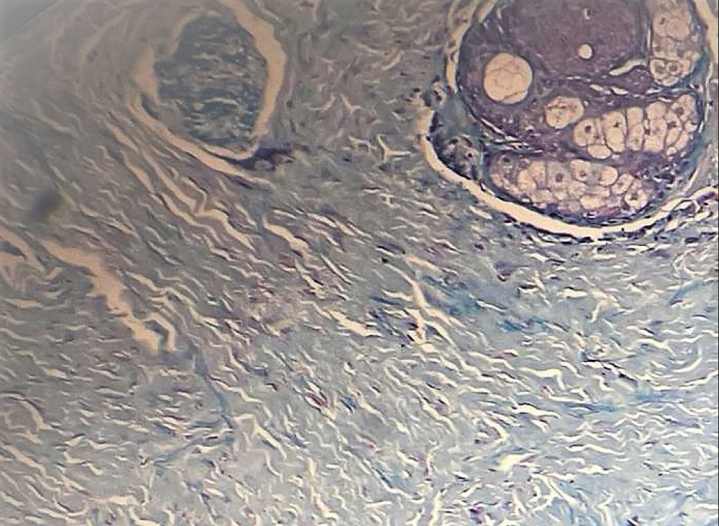
Differential diagnoses of mycosis fungoides, primary cutaneous B-cell lymphoma, amyloidosis, leiomyomas, multiple keloids, papular mucinosis/lichen myxedematosus and histoid leprosy were considered. A skin biopsy was done and on histopathological examination mild acanthosis of epidermis, dense collagenization in the reticular dermis with a mild perivascular infiltrate of lymphocytes and plasma cells were observed [Figure 2a]. Zeihl neelsen and Alcian Blue stain were negative for acid fast bacilli and mucin deposition. Masson’s trichrome and Verhoeff-van-Gieson stains showed markedly increased collagen arranged in a haphazard pattern in the dermis with a reduction in elastin fibres [Figure 2b]. With clinical and the histopathology findings, two differential diagnoses were considered, familial cutaneous collagenoma and eruptive collagenoma.

- (Low power, 10× lens total magnification 100×) Mild acanthosis, reticular dermis shows dense collagenization, scantly infiltrate

- (Masson’s Trichrome, 400×) Dense collagen fibres throughout dermis
Eruptive collagenoma was first described by Colomb in 1955.3 They usually present in the first two decades of life and lesions tend to be asymptomatic, cutaneous nodules which are distributed symmetrically on head and neck, upper and lower limbs.4 Histologically, it is characterised by dense coarse collagen fibres arranged haphazardly in dermis with a decrease in elastic fibres.5 Eruptive collagenoma needs to be differentiated from familial cutaneous collagenoma which is inherited in an autosomal dominant pattern with a positive family history.6 Familial cutaneous collagenoma (FCC) may be associated with cardiac abnormalities such as progressive cardiomyopathy and conduction defects attributed to fibrotic process affecting various structures of the heart.7 Lesions of FCC usually affect the trunk (upper back) and tend to be more numerous than the lesions of eruptive collagenoma which mainly affect the periphery including head, neck, upper and lower limbs.4 In our case, there was no family history and there was no evidence of any cardiovascular disease. Based on these findings, we diagnosed this as a case of eruptive collagenoma. A review of literature of similar cases has been presented in Table 1. We report this case of eruptive collagenoma due to its unusual distribution and morphology where histopathology helped us clinch the diagnosis.
| Author | Age (years)/Sex | Sites | Clinical morphology | Histopathology |
|---|---|---|---|---|
| Mukhi et al.8 | 42/M | Face, back, upper limb, abdomen | Skin coloured, firm, non-tender nodules and plaques | Lobules of densely collagenized acellular connective tissue in dermis; elastic fibres |
| Sharma et al.4 | 14/F | Lower back, chest and upper thighs | Skin-coloured to slightly hypopigmented, firm, non-tender papules | Randomly arranged coarse collagen bundles; elastic fibres |
| Barad et al.5 | 4/F | Bilateral axilla, upper back and face | Symmetrical brownish papules and plaques with leathery surface | Dermis with haphazardly arranged thickened collagen bundles; elastic fibres |
| Brar et al.9 | 18/F | Upper 2/3 of back & mons pubis | Skin coloured, firm, non-tender nodules | Focal acanthosis with significantly increased density of collagen bundles; elastic fibres |
| Sonkusale et al.10 | 5/F | Face and back | Skin coloured discrete-to-confluent papules and plaques | Haphazardly arranged thick collagen bundle in the dermis; elastic fibres |
| Present case | 51/M | Chest, abdomen, upper back and bilateral arms | Flesh coloured to dusky red, discrete to coalescent, firm, waxy nodulo-plaques | Mild acanthosis with dense collagenization in the reticular dermis and mild perivascular infiltrate; elastin fibres |
Declaration of patient consent
The authors certify that they have obtained all appropriate patient consent.
Financial support and sponsorship
Nil.
Conflicts of interest
There are no conflicts of interest.
References
- Isolated pedunculated collagenoma (collagen nevi) of the scalp. Indian J Dermatol. 2013;58:411.
- [CrossRef] [PubMed] [PubMed Central] [Google Scholar]
- Isolated collagenoma on the face: a rare occurrence. Acta Dermatovenerol Alp Pannonica Adriat. 2019;28:41-43. Erratum for: Acta Dermatovenerol Alp Pannonica Adriat. 2019;28:95
- [PubMed] [Google Scholar]
- Female with eruptive collagenoma clustered in the left lateral aspect of the abdomen. J Dermatol. 2010;37:843-5.
- [CrossRef] [PubMed] [Google Scholar]
- Eruptive collagenoma. Indian J Dermatol Venereol Leprol. 2013;79:256-8.
- [CrossRef] [PubMed] [Google Scholar]
- Eruptive collagenoma: a rarely reported entity in Indian literature. Indian J Dermatol. 2015;60:104.
- [CrossRef] [PubMed] [PubMed Central] [Google Scholar]
- Familial cutaneous collagenoma: genetic studies on a family. Br J Dermatol. 1979;101:185-95.
- [CrossRef] [PubMed] [Google Scholar]
- Eruptive Ccollagenoma in a Mongol Girlmongol girl: A Rare Association.rare association. International Journal of Case Reports and Images (IJCRI). 2015;6:427-30.
- [Google Scholar]
- Eruptive collagenoma: A rare entity in pediatric age. Indian Journal of Paediatric Dermatology. 2019;20:240.
- [Google Scholar]


