Translate this page into:
Onychomatricoma: Clinical, dermoscopy and ultrasound findings
Correspondence Address:
Denise Gam�
Department of Dermatology, Hospital Germans Trias I Pujol, Carretera De Canyet S/N 08916, Badalona
Spain
| How to cite this article: Gam� D, Jaka A, Ferr�ndiz C. Onychomatricoma: Clinical, dermoscopy and ultrasound findings. Indian J Dermatol Venereol Leprol 2019;85:190-191 |
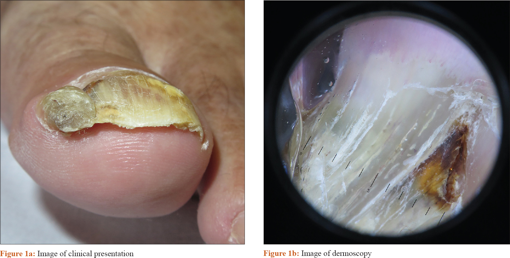
A 58-year-old man presented with a 5-year history of an asymptomatic thickening of the nail at his right great toe. Physical examination and dermoscopy revealed a well-demarcated tubular thickening of the nail plate, along with yellow and white longitudinal bands with splinter hemorrhages [Figure - 1]a and [Figure - 1]b. Ultrasonography showed a nonvascularized hyper and hypoechoic tumor located in the nail matrix and projecting towards the nail bed and nail plate. Hyperechoic linear dots within the hypoechoic areas were also seen. No erosive changes on the underlying bone were identified [Figure - 2]. Histopathologic examination showed “gloved finger” papillary projections covered by matrix type epithelium consistent with onychomatricoma. A complete surgical excision was performed.
 |
| Figure 1: |
 |
| Figure 2: Ultrasound findings: Showing a cross section of the distal part of nail |
Declaration of patient consent
The authors certify that they have obtained all appropriate patient consent forms. In the form, the patient has given his consent for his images and other clinical information to be reported in the journal. The patient understand that name and initials will not be published and due efforts will be made to conceal identity, but anonymity cannot be guaranteed.
Financial support and sponsorship
Nil.
Conflicts of interest
There are no conflicts of interest.
Fulltext Views
4,722
PDF downloads
3,065


