Generic selectors
Exact matches only
Search in title
Search in content
Post Type Selectors
Search in posts
Search in pages
Filter by Categories
15th National Conference of the IAOMFP, Chennai, 2006
Abstract
Abstracts from current literature
Acne in India: Guidelines for management - IAA Consensus Document
Addendum
Announcement
Art & Psychiatry
Article
Articles
Association Activities
Association Notes
Award Article
Book Review
Brief Report
Case Analysis
Case Letter
Case Letters
Case Notes
Case Report
Case Reports
Clinical and Laboratory Investigations
Clinical Article
Clinical Studies
Clinical Study
Commentary
Conference Oration
Conference Summary
Continuing Medical Education
Correspondence
Corrigendum
Cosmetic Dermatology
Cosmetology
Current Best Evidence
Current Issue
Current View
Derma Quest
Dermato Surgery
Dermatopathology
Dermatosurgery Specials
Dispensing Pearl
Do you know?
Drug Dialogues
e-IJDVL
Editor Speaks
Editorial
Editorial Remarks
Editorial Report
Editorial Report - 2007
Editorial report for 2004-2005
Errata
Erratum
Focus
Fourth All India Conference Programme
From Our Book Shelf
From the Desk of Chief Editor
General
Get Set for Net
Get set for the net
Guest Article
Guest Editorial
History
How I Manage?
IADVL Announcement
IADVL Announcements
IJDVL Awards
IJDVL AWARDS 2015
IJDVL Awards 2018
IJDVL Awards 2019
IJDVL Awards 2020
IJDVL International Awards 2018
Images in Clinical Practice
Images in Dermatology
In Memorium
Inaugural Address
Index
Knowledge From World Contemporaries
Leprosy Section
Letter in Response to Previous Publication
Letter to Editor
Letter to the Editor
Letter to the Editor - Case Letter
Letter to the Editor - Letter in Response to Published Article
LETTER TO THE EDITOR - LETTERS IN RESPONSE TO PUBLISHED ARTICLES
Letter to the Editor - Observation Letter
Letter to the Editor - Study Letter
Letter to the Editor - Therapy Letter
Letter to the Editor: Articles in Response to Previously Published Articles
Letters in Response to Previous Publication
Letters to the Editor
Letters to the Editor - Letter in Response to Previously Published Articles
Letters to the Editor: Case Letters
Letters to the Editor: Letters in Response to Previously Published Articles
Media and news
Medicolegal Window
Messages
Miscellaneous Letter
Musings
Net Case
Net case report
Net Image
Net Images
Net Letter
Net Quiz
Net Study
New Preparations
News
News & Views
Obituary
Observation Letter
Observation Letters
Oration
Original Article
ORIGINAL CONTRIBUTION
Original Contributions
Pattern of Skin Diseases
Pearls
Pediatric Dermatology
Pediatric Rounds
Perspective
Presedential Address
Presidential Address
Presidents Remarks
Quiz
Recommendations
Regret
Report
Report of chief editor
Report of Hon : Treasurer IADVL
Report of Hon. General Secretary IADVL
Research Methdology
Research Methodology
Resident page
Resident's Page
Resident’s Page
Residents' Corner
Residents' Corner
Residents' Page
Retraction
Review
Review Article
Review Articles
Reviewers 2022
Revision Corner
Self Assessment Programme
SEMINAR
Seminar: Chronic Arsenicosis in India
Seminar: HIV Infection
Short Communication
Short Communications
Short Report
Snippets
Special Article
Specialty Interface
Studies
Study Letter
Study Letters
Supplement-Photoprotection
Supplement-Psoriasis
Symposium - Contact Dermatitis
Symposium - Lasers
Symposium - Pediatric Dermatoses
Symposium - Psoriasis
Symposium - Vesicobullous Disorders
SYMPOSIUM - VITILIGO
Symposium Aesthetic Surgery
Symposium Dermatopathology
Symposium-Hair Disorders
Symposium-Nails Part I
Symposium-Nails-Part II
Systematic Review and Meta-Analysis
Systematic Reviews and Meta-analyses
Systematic Reviews and Meta-analysis
Tables
Technology
Therapeutic Guideline-IADVL
Therapeutic Guidelines
Therapeutic Guidelines - IADVL
Therapeutics
Therapy
Therapy Letter
Therapy Letters
View Point
Viewpoint
What’s new in Dermatology
View/Download PDF

Translate this page into:

Letter to the Editor - Case Letter
2019:85:5;506-508
doi: 10.4103/ijdvl.IJDVL_16_19
PMID: 31368452

Chromoblastomycosis with a sporotrichoid distribution

Arpita Nibedita Rout, Kananbala Sahu, Chandra Sekhar Sirka
 Department of Dermatology, All India Institute of Medical Sciences, Bhubaneswar, Odisha, India

Correspondence Address:
Arpita Nibedita Rout
Department of Dermatology, All India Institute of Medical Sciences, Bhubaneswar - 751 019, Odisha
India
How to cite this article:
Rout AN, Sahu K, Sirka CS. Chromoblastomycosis with a sporotrichoid distribution. Indian J Dermatol Venereol Leprol 2019;85:506-508
Copyright: (C)2019 Indian Journal of Dermatology, Venereology, and Leprology

Sir,

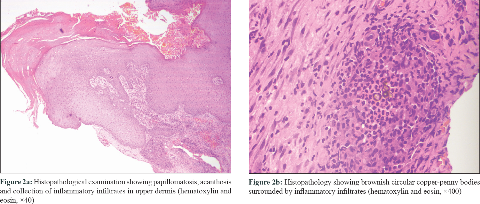
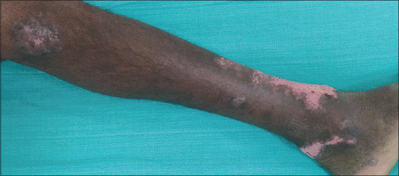
An 82-year-old farmer presented with multiple asymptomatic verrucous growths on the left lower limb of 23 years duration. He could not recall any definite history of trauma, before the appearance of the lesions. On examination, there were three discrete verrucous plaques on the left leg [Figure - 1]a. Multiple black spots were seen on the verrucous plaques extending from medial malleolus to lateral malleolus covering the entire anterior aspect of left ankle [Figure - 1]b. Possibility of cutaneous chromoblastomycosis, tuberculosis verrucosa cutis and lichen planus hypertrophicus was considered. There was no regional or generalized lymphadenopathy. Complete blood count, liver function test and renal function test were within normal range. X-ray of the limb did not reveal any abnormality. Examination of the black dots in 10% potassium hydroxide (KOH) preparation did not show any spores. Histopathological examination of the lesion showed hyperkeratosis and papillomatosis with brownish thick-walled copper-penny bodies within mixed inflammatory infiltrates composed of neutrophils and lympho-histiocytes in the superficial dermis [Figure - 2]a and [Figure - 2]b. Culture of the tissue biopsy specimen did not grow any organism. A diagnosis of chromoblastomycosis in sporotrichoid pattern was made based on morphology of the lesions and histopathology findings. The patient was treated with itraconazole 200mg twice daily. There was a marked response after 2 months of treatment [Figure - 3]. He is on regular follow-up and continuing the treatment.
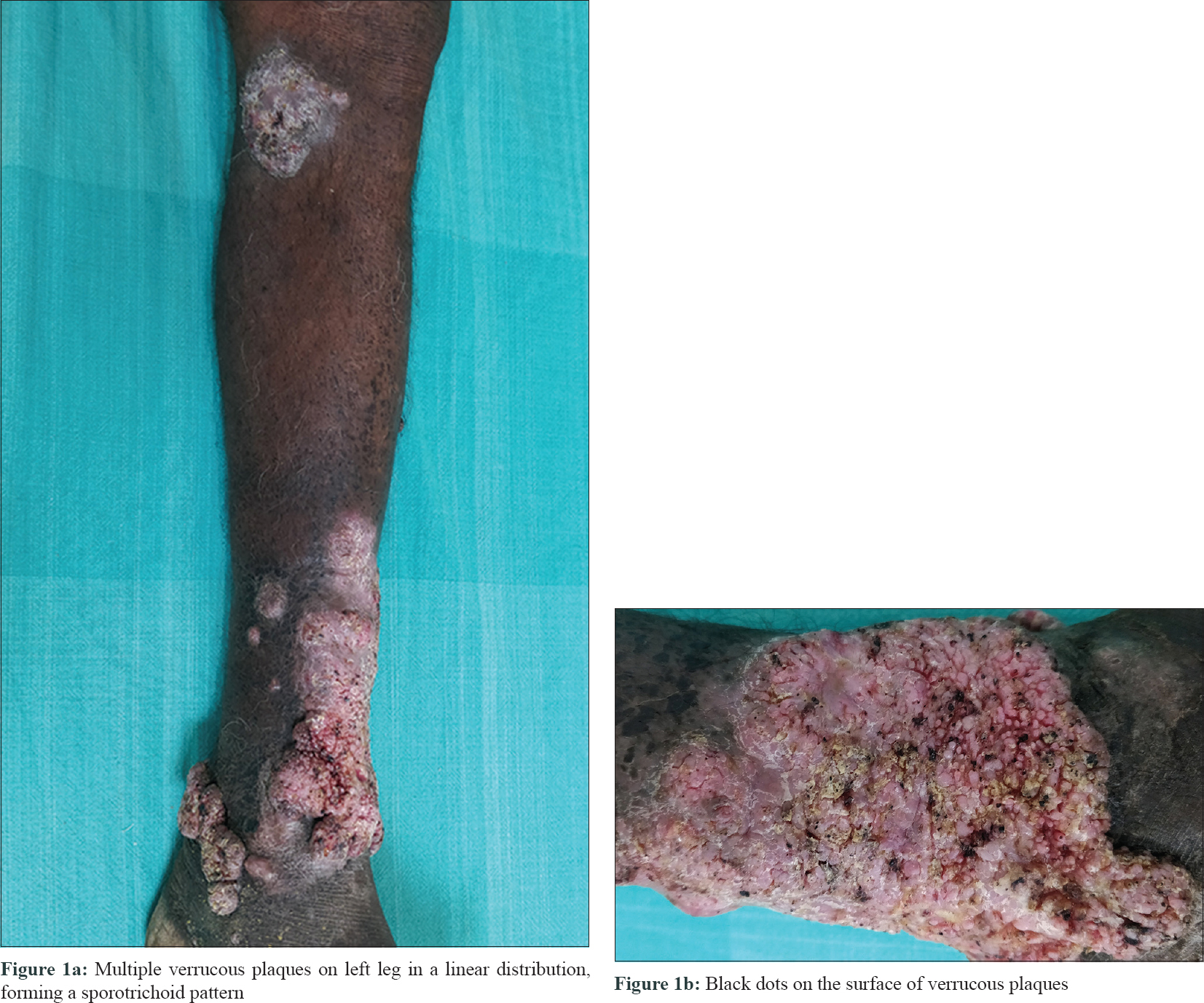
Figure 1
Figure 2
Figure 3: Marked response after 2 months of oral itraconazole 200 mg twice daily

Chromoblastomycosis is a chronic cutaneous and subcutaneous fungal infection caused by dematiaceous fungi, following traumatic implantation of the organism.[1] It can spread by autoinoculation and rarely through lymphatics.[2],[3]

Cutaneous lesions begin as erythematous papules, cauliflower-like nodules or warty growths. They gradually enlarge to form large plaques with brown or black dots on the surface. The most common sites of involvement are the distal limbs. Rarely, it occurs on buttocks, trunk and face.[1] In advanced disease, it can spread to adjoining skin as satellite lesions. Such spread occurs from autoinoculation or transmission via the lymphatic system.[4] In a review of literature, we found four cases of chromoblastomycosis with sporotrichoid distribution.[1],[4],[5],[6]

The sporotrichoid distribution of lesions is seen in sporotrichosis, cutaneous tuberculosis and atypical mycobacterial infection. Chromoblastomycosis can be differentiated from these conditions by identification of the copper-penny bodies, histopathology and culture.

Usually, histopathology reveals acanthosis and may demonstrate pseudo-carcinomatous hyperplasia. Neutrophils and giant cells may be seen infiltrating the epidermis with occasional formation of micro-abscesses. The dermis reveals a granulomatous tissue reaction with a mixed focal or diffuse inflammatory infiltrate consisting of lymphocytes, neutrophils, monocytes, plasma cells, eosinophils and giant cells of the foreign body and Langhans types. The copper-penny bodies may be seen both intracellularly and extracellularly.

The diagnosis of chromoblastomycosis is from microscopic identification of Medlar bodies/fumagoid bodies/muriform bodies/copper-penny bodies/sclerotic bodies on scrapings from the lesion in 20% KOH and/or histological examination of a biopsy specimen and by culture of scrapings or biopsy material.[1] Our patient had multiple nodules in a linear fashion distant from the primary plaque and the diagnosis was confirmed by demonstration of copper-penny bodies in tissue section.

Treatment options include itraconazole, terbinafine and potassium iodide. Our patient was treated with oral itraconazole 200mg twice daily and after 2 months there was good response without any side effects of therapy.

In chromoblastomycosis with satellite lesions, presentation in a sporotrichoid fashion is rare.[1] In our case, based on the morphology, autoinoculation following scratching or minor trauma might have been the cause rather than lymphatic spread. We could not isolate the organism in culture, but the histopathology and response to treatment proved the diagnosis retrospectively. Though isolation of organism in culture is regarded as the gold standard for diagnosis, in resource-poor centers, classical morphology and histopathology can be the pointers to diagnosis. We have compiled and compared the four cases with our case in regards to clinical morphology, site of involvement, investigations and treatment outcomes [Table - 1].[1],[4],[5],[6] We report this case because of its rarity and response to monotherapy with itraconazole.
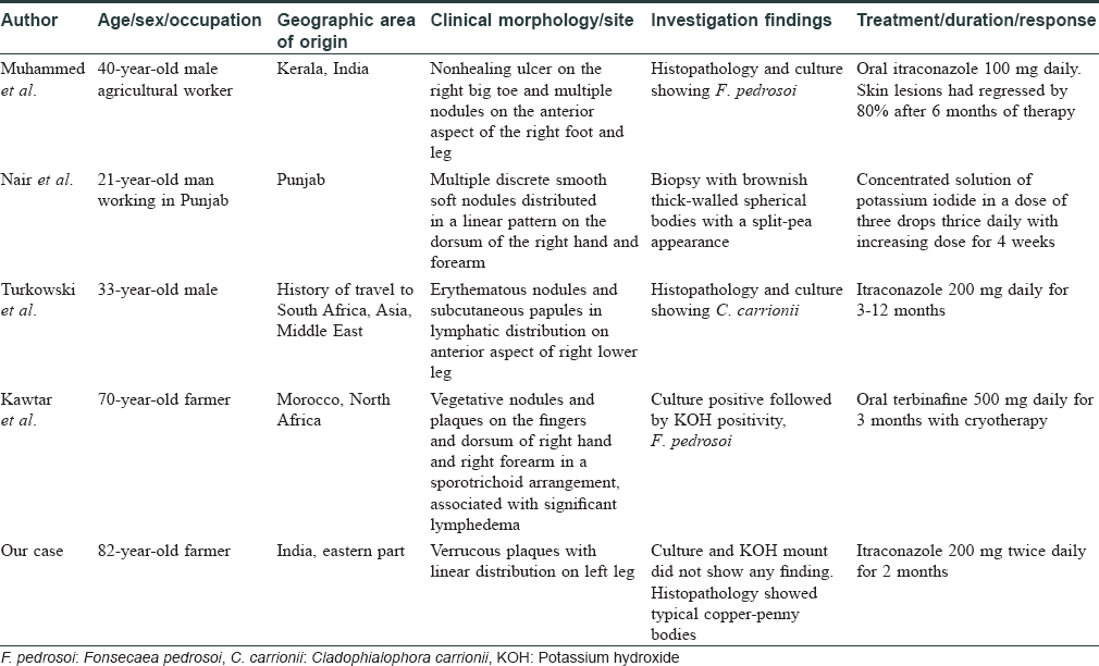
Table 1: Comparison of cases of chromoblastomycosis with sporotrichoid pattern

Declaration of patient consent

The authors certify that they have obtained all appropriate patient consent forms. In the form, the patient has given his consent for his images and other clinical information to be reported in the journal. The patient understands that name and initials will not be published and due efforts will be made to conceal identity, but anonymity cannot be guaranteed.

Financial support and sponsorship

Nil.

Conflicts of interest

There are no conflicts of interest.

References
1.
Muhammed K, Nandakumar G, Asokan KK, Vimi P. Lymphangitic chromoblastomycosis. Indian J Dermatol Venereol Leprol 2006;72:443-5.
[Google Scholar]
2.
Queiroz-Telles F, Esterre P, Perez-Blanco M, Vitale RG, Salgado CG, Bonifaz A. Chromoblastomycosis: An overview of clinical manifestations, diagnosis and treatment. Med Mycol 2009;47:3-15.
[Google Scholar]
3.
Kondo M, Hiruma M, Nishioka Y, Mayuzumi N, Mochida K, Ikeda S. A case of chromomycosis caused by Fonsecaea pedrosoi and a review of reported cases of dematiaceous fungal infection in Japan. Mycoses 2005;48:221-5.
[Google Scholar]
4.
Nair PS, Sarojini PA. Chromoblastomycosis resembling sporotrichosis. Indian J Dermatol Venereol Leprol 1993;59:125-6.
[Google Scholar]
5.
Turkowski Y, Aleissa S, Plotnikova N, Tse J, Rosmarin D. Sporotrichoid chromoblastomycosis on right lower leg. J Am Acad Dermatol 2018;79:AB268.
[Google Scholar]
6.
Kawtar I, Salim G, Mariame M, Fatimazahra M, Imane T, Salma B, et al. Sporotrichoid chromomycosis. Dermatol Online J 2013;19:20394.
[Google Scholar]

Fulltext Views
3,486

PDF downloads
1,724
Show Sections