
Translate this page into:
Atypical clinical and dermoscopic features of hobnail hemangioma
Corresponding author: Dr. Laxmisha Chandrashekar, Department of Dermatology, Venereology and Leprology, JIPMER, Puducherry - 605 006, India. laxmishac@gmail.com
-
Received: ,
Accepted: ,
How to cite this article: Behera B, Chandrashekar L, Thappa DM, Gochhait D, Ayyanar P. Atypical clinical and dermoscopic features of hobnail hemangioma. Indian J Dermatol Venereol Leprol 2021;87:93-7.
Sir,
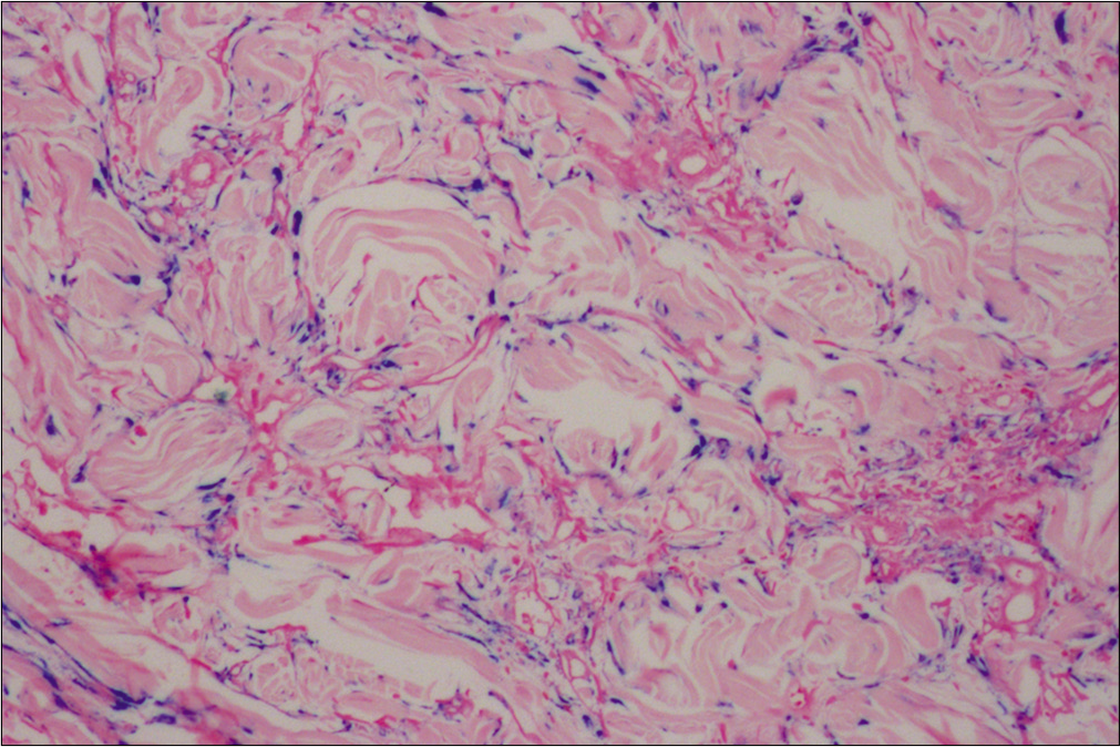
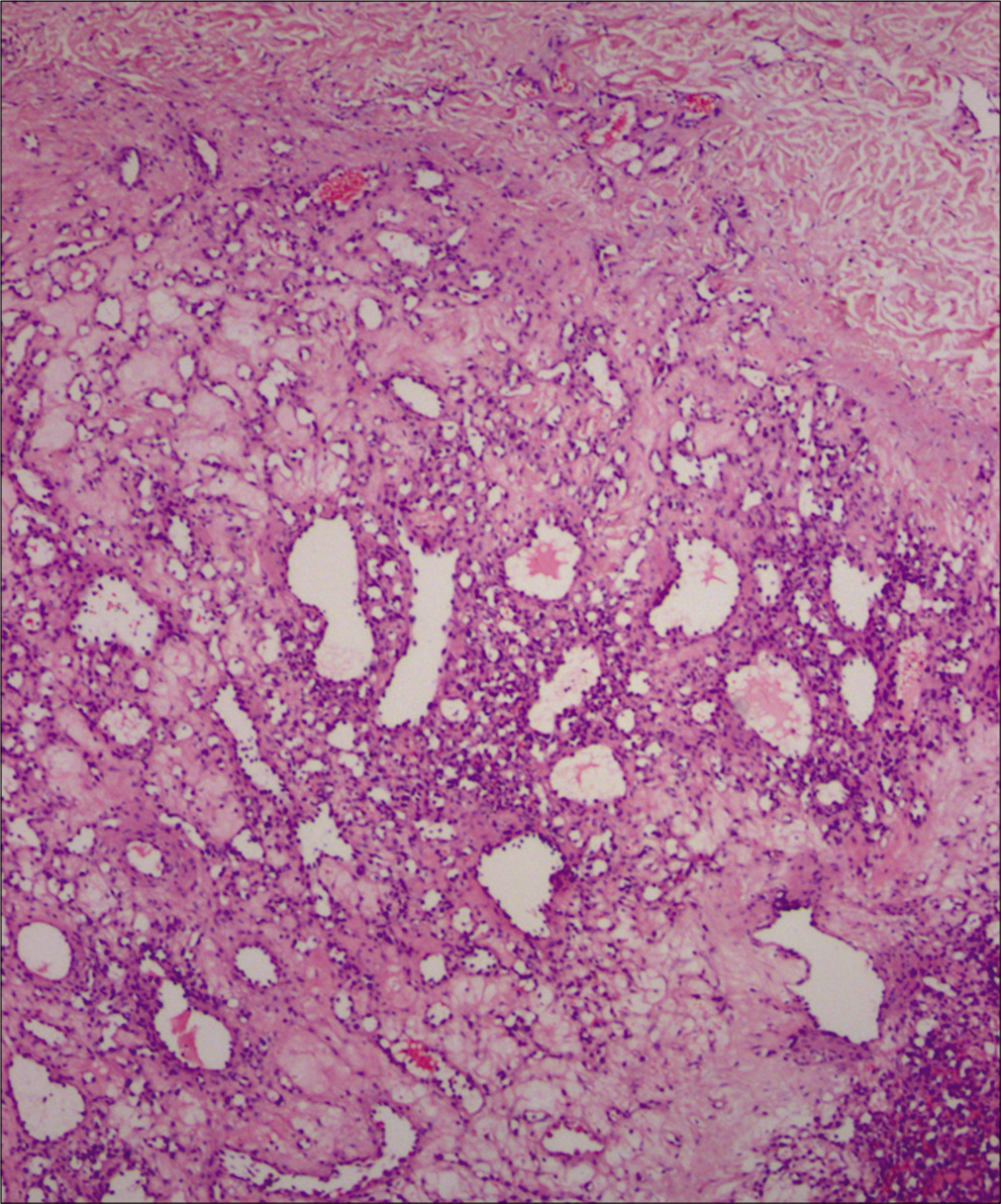
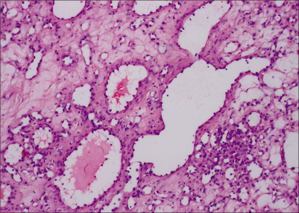
A 36-year-old woman (Patient 1) with skin phototype-V, presented with a 10-month history of an asymptomatic pigmented lesion on the abdomen. Cutaneous examination revealed a solitary 1.5 cm × 1 cm, firm, non-tender, pigmented plaque on the abdomen [Figure 1a]. Differential diagnosis of dermatofibroma, dermatomyofibroma and melanocytic nevus was considered. Dermoscopic examination under nonpolarized contact mode (HEINE DELTA20® Dermatoscope, Germany, 10 × magnification) showed a cerebriform pattern. The dermoscopic features observed were a gray background, comedo-like openings, linear irregular crypts and keratotic plugs [Figure 1b]. The excisional biopsy of the lesion showed multiple lymphangioma-like dilated spaces with intraluminal projections lined by hobnail endothelial cells in the upper dermis, and slit-like vascular spaces dissecting the collagen bundles along with perivascular lymphocytic infiltration in the lower dermis. Perl’s Prussian blue stain demonstrated dermal hemosiderin [Figure 2].
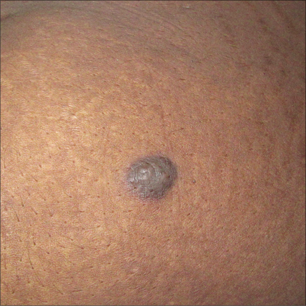
- Solitary firm pigmented plaque on the abdomen
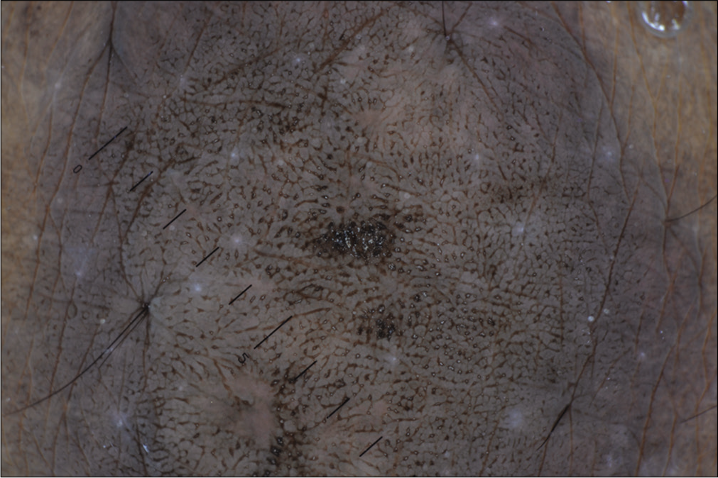
- Dermoscopic examination (under nonpolarized contact mode, HEINE DELTA20® Dermatoscope, Germany, ×10) demonstrating a cerebriform pattern, a gray background, comedo-like openings, linear irregular crypts and keratotic plugs
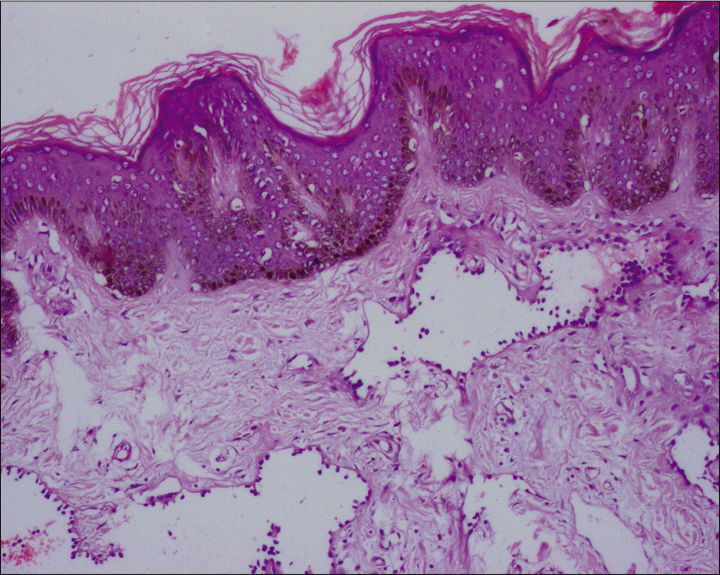
- Multiple lymphangioma-like dilated spaces in the upper dermis lined by hobnail endothelial cells with an overlying hypertrophic and hyperpigmented epidermis (H and E, ×100)
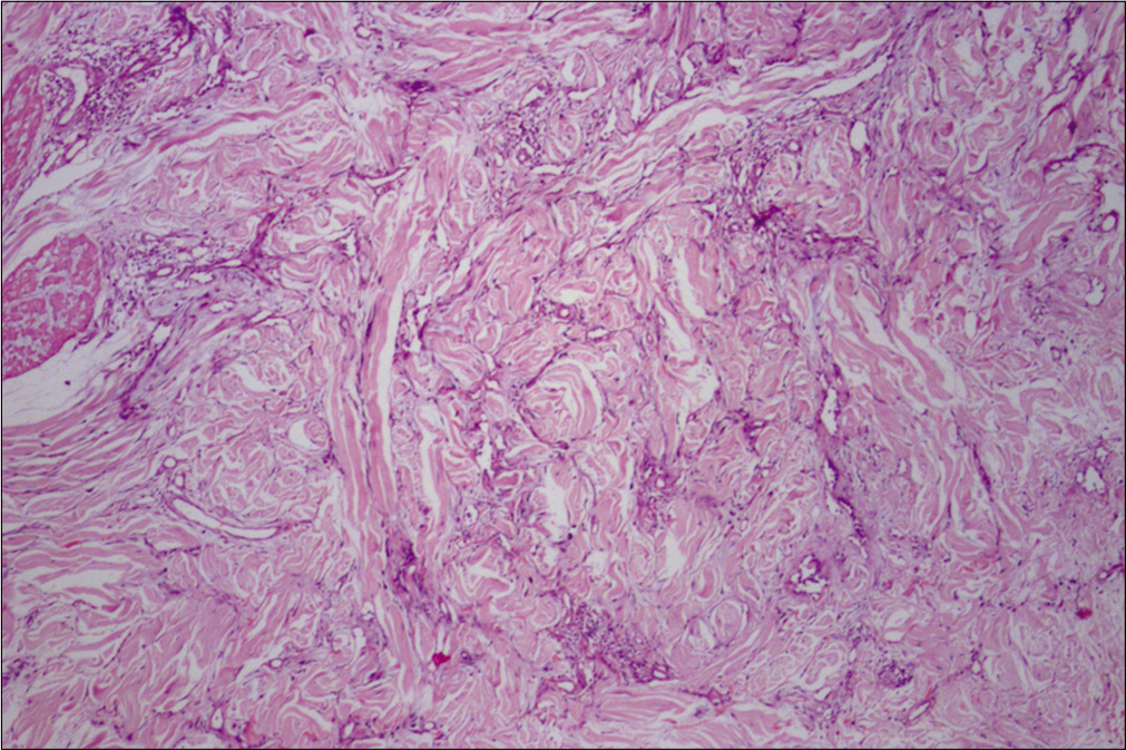
- Slit-like vascular spaces dissecting the collagen bundles in the lower dermis (H and E, ×100)
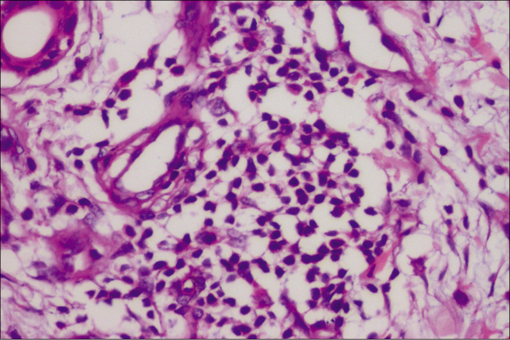
- Perivascular lymphocytic infiltration (H and E, ×400)
- Special stain highlighting hemosiderin (Perl’s Prussian blue stain, ×100)
A 44-year-old man (Patient 2) with skin phototype-V had a slow-growing lesion on the abdomen for the last 6 months. Cutaneous examination revealed a solitary 5 mm × 5 mm firm non-tender shiny dome-shaped skin-colored nodule on the abdomen [Figure 3a]. Differential diagnosis of solitary neurofibroma, solitary circumscribed neuroma and fibroepithelial polyp was considered. Dermoscopy of the nodule demonstrated a gray homogenous area, along with brown peppering [Figure 3b]. Shave biopsy of the lesion showed, proliferating endothelial cells admixed with multiple dilated vascular spaces lined by hobnail endothelial cells. Perl’s Prussian blue showed occasional hemosiderin deposition [Figure 4].
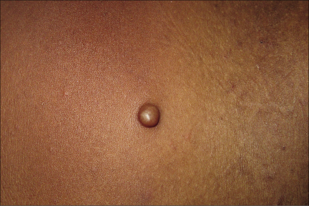
- Solitary firm non-tender dome-shaped nodule on the abdomen
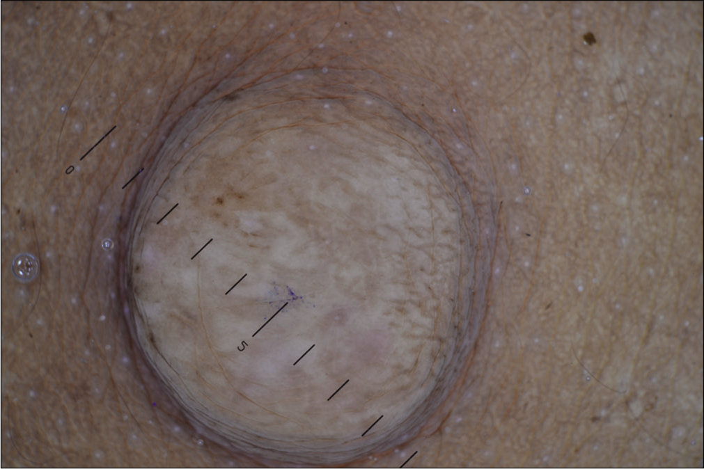
- Dermoscopic examination (under nonpolarized contact mode, HEINE DELTA20® Dermatoscope, Germany, ×10x) demonstrating a gray homogenous area and brown peppering
- Proliferating endothelial cells admixed with multiple dilated vascular spaces in the dermis (H and E, ×50)
- Dilated vascular spaces lined by hobnail endothelial cells (H and E, ×100)
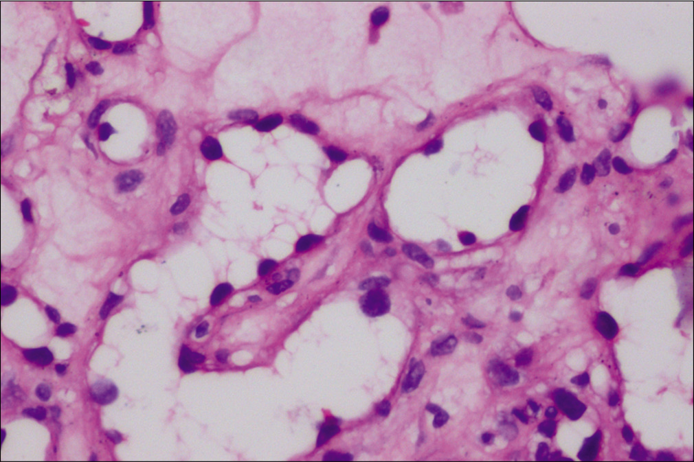
- Hobnail endothelial cells (H and E, ×400)
In both cases, a final diagnosis of hobnail hemangioma was made based upon the histopathological findings.
Hobnail hemangioma, also known as targetoid hemosiderotic hemangioma is a benign cutaneous vascular lesion. It commonly occurs in young to middle-aged persons and has a slight female preponderance. A solitary asymptomatic papule on the extremities and trunk, in a young to middle-aged adult, is the most common presentation of hobnail hemangioma. The size ranges from 0.6 cm to 2 cm. As the name suggests, a targetoid appearance with a central violaceous to red macule or papule surrounded by a pale rim, and an outermost ecchymotic ring, usually provides a clue to the diagnosis. The targetoid appearance is not universal; cases with nontargetoid appearance have been described.1,2 In the case of a nontargetoid or atypical presentation, it can be mistaken for other cutaneous vascular (hemangioma, angiokeratoma, Kaposi’s sarcoma), melanocytic (melanocytic nevus, dysplastic nevus, melanoma) and fibrohistiocytic (dermatofibroma, fibroma) tumors. Both of the described cases neither had a targetoid pattern nor any clinical clue like reddish-violaceous color, to suggest the vascular nature of the lesions. In the first case, a pigmented firm plaque, we considered differentials of dermatofibroma, dermatomyofibroma and melanocytic nevus. In the second case, a firm skin-colored dome-shaped nodule, we considered differentials of solitary neurofibroma, solitary circumscribed neuroma and fibroepithelial polyp.
The dermoscopic examination is a rapid, noninvasive procedure that provides subtle clues to the diagnosis of a cutaneous lesion. For a vascular malformation/ tumor, the presence of lagoons/clods of varying colors during dermoscopic examination is a constant feature. Although not specific to any particular vascular tumor/ malformation, their presence guides the clinicians to rule out nonvascular etiology. A recent review of dermoscopic features of hobnail hemangioma described red lacunae, dark lacunae, whitish structures, a central homogeneous area, intermediate yellowish areas, peripheral red-violaceous ring, vascular structures and pigment network.2 Zaballos et al. analyzed 35 cases of hobnail hemangioma and reported dermoscopic features such as reddish-violaceous, and brown homogenous areas (85.7%), red and dark lacunae (74.3%), chrysalis (42.8%), vascular structure (40%) and delicate pigment network (14.3%).3 The various patterns observed by them were; a central lacuna and peripheral homogenous area with or without intermediate skin-colored, yellow or white circular homogeneous area (71.4%), diffuse reddish-violaceous homogenous area and delicate pigment network and red lacuna alone (single report). The other dermoscopic features reported for hobnail hemangioma are enlisted in Table 1.4-10 From the various reports, it is evident that a lagoon of variable color, with or without a targetoid pattern, is a consistent feature of hobnail hemangioma. In the two of our described cases, dermoscopy neither demonstrated a targetoid pattern nor had any dermoscopic clue to the diagnosis of a vascular lesion, thus mislead the diagnosis. The first case dermoscopically mimicked a dermatofibroma or seborrheic keratosis due to the presence of cerebriform pattern, and gray background. Another rare clinical mimic, aneurysmal fibrous histiocytoma, commonly demonstrates a blue homogenous area that may accompany other shades of color like pink, reddish-brown or white. The blue homogenous area corresponds to the dermal hemosiderin deposition. Other features described are pigment network, branched streaks, white crystalline structure, a rainbow pattern and vessels ranging from dotted, polymorphous and linear irregular vessels. The second case dermoscopically mimicked a neurofibroma due to a featureless pattern. The dermoscopic difference between hobnail hemangioma, neurofibroma and dermatofibroma is mentioned in Table 2.2,3,11-14 In case-1, the hyperplastic and hyperpigmented epidermis masked the dermoscopic vascular structures. In case-2, along with mild hyperplastic and hyperpigmented epidermis, the relatively lower dermal location of the vascular lesion may be responsible for the absence of the dermoscopic vascular feature. The final diagnosis of both cases was revealed by histopathological examination.
| Report | Dermoscopic features |
|---|---|
| Sahin et al.1 | Red lagoons sharply demarcated black macule and violaceous maculae ring A homogenous red area with indistinct violaceous ring Red to reddish-blue lagoon, black macule surrounded by erythema |
| Morales- Callaghan et al.4 |
A central round bright red round structures, smaller round pale-pink structures admixed with a diffuse pink-white pigmentation and peripheral pigment network |
| Lacarrubba et al.5 | A central round red lagoon-like area with a peripheral thin pigment network |
| Ghibaudo et al.6 | A central closely packed red-violaceous ovoid structures, whitish-veil and peripheral ecchymotic ring |
| Ertam et al.7 | Red lagoon-like areas, red homogenous area, linear white structure and a peripheral pigment network |
| Avci et al.8 | Sharply demarcated blue-red lagoons |
| Ibrahim and Shwayder9 | Well-defined red circular blebs |
| Piccolo et al.10 | A central reddish structureless area with chrysalis- like structures and a peripheral pigment network |
| Tumour | Dermoscopic features |
|---|---|
| Hobnail hemangioma | A lagoon of varying color (red/reddish-blue/dark) A targetoid pattern: Acentral lacuna and peripheral homogenous area with or without intermediate skin-colored/yellow/white circular homogeneous area Other features: Diffuse reddish-violaceous homogenous area, chrysalis, pigment network |
| Dermatofibroma | Typical appearance: Acentral scar-like area with peripheral delicate pigment network Different dermoscopic patterns: As a result of various combinations of the following features; white scar-like area, pigment network, homogenous pigmentation, white network, brown dots and globules, cerebriform pattern, linear irregular crypts It can vary depending upon histopathological subtypes |
| Neurofibroma | A yellow, yellow-brown, brown, skin-colored to gray background/homogenous area with exaggerated skin markings Other features: Pigment network-like area, blanchable erythema, brown globules, fingerprint-like structures |
In conclusion, we describe two cases of hobnail hemangioma with atypical clinical and dermoscopic features. The vascular nature of the hobnail hemangioma was concealed due to the absence of clinical and dermoscopic vascular features like reddish-violaceous color and vascular lagoons, respectively. At times, the dermoscopic examination can be misleading, especially in cases of atypical clinical presentation, in which case a histopathological examination is essential for the diagnosis.
Declaration of patient consent
The authors certify that they have obtained all appropriate patient consent forms. In the form, the patients have given their consent for their images and other clinical information to be reported in the journal. The patients understand that their names and initials will not be published and due efforts will be made to conceal their identity but anonymity cannot be guaranteed.
Financial support and sponsorship
Nil.
Conflicts of interest
There are no conflicts of interest.
References
- Targetoid haemosiderotic haemangioma: Dermoscopic monitoring of three cases and review of the literature. Clin Exp Dermatol. 2005;30:672-6.
- [CrossRef] [PubMed] [Google Scholar]
- A pigmented papule acting like a playful ghost: dermoscopy of three targetoid hemosiderotic hemangiomas. G Ital Dermatol Venereol. 2018;153:685-91.
- [CrossRef] [PubMed] [Google Scholar]
- Dermoscopy of targetoid hemosiderotic hemangioma: A morphological study of 35 cases. Dermatology. 2015;231:339-44.
- [CrossRef] [PubMed] [Google Scholar]
- Targetoid hemosiderotic hemangioma: clinical and dermoscopical findings. J Eur Acad Dermatol Venereol. 2007;21:267-9.
- [CrossRef] [Google Scholar]
- A red-violaceous papular lesion in a young girl, Targetoid haemosiderotic haemangioma. Acta Derm Venereol. 2015;95:121-3.
- [CrossRef] [PubMed] [Google Scholar]
- Fully regressive targetoid haemosiderotic haemangioma. J Eur Acad Dermatol Venereol. 2009;23:722-3.
- [CrossRef] [PubMed] [Google Scholar]
- Hobnail haemangioma: dermatoscopic findings facilitating the differential diagnosis. Clin Exp Dermatol. 2009;34:e231-3.
- [CrossRef] [PubMed] [Google Scholar]
- Targetoid hemosiderotic hemangioma. J Eur Acad Dermatol Venereol. 1998;11:186-7.
- [CrossRef] [PubMed] [Google Scholar]
- Hobnail hemangioma in a nine-year-old boy: A rare case presented with dermoscopy. Dermatol Online J. 2010;16:7.
- [Google Scholar]
- Dermoscopic misdiagnosis of melanoma in a patient with targetoid hemosiderotic hemangioma. J Am Acad Dermatol. 2014;71:e179-81.
- [CrossRef] [PubMed] [Google Scholar]
- Dermoscopy of dermatofibromas: a prospective morphological study of 412 cases. Arch Dermatol. 2008;144:75-83.
- [CrossRef] [Google Scholar]
- Different dermoscopic faces of dermatofibromas. J Am Acad Dermatol. 2007;57:401-6.
- [CrossRef] [PubMed] [Google Scholar]
- Multicoloured rainbow pattern in a case of aneurysmal dermatofibroma [published online ahead of print 2020 Apr 28] Australas J Dermatol 2020
- [CrossRef] [PubMed] [Google Scholar]
- Dermoscopic features of solitary neurofibroma: A retrospective analysis of 32 cases [published online ahead of print 2020 May 29] Australas J Dermatol 2020
- [CrossRef] [PubMed] [Google Scholar]


