Generic selectors
Exact matches only
Search in title
Search in content
Post Type Selectors
Search in posts
Search in pages
Filter by Categories
15th National Conference of the IAOMFP, Chennai, 2006
Abstract
Abstracts from current literature
Acne in India: Guidelines for management - IAA Consensus Document
Addendum
Announcement
Art & Psychiatry
Article
Articles
Association Activities
Association Notes
Award Article
Book Review
Brief Report
Case Analysis
Case Letter
Case Letters
Case Notes
Case Report
Case Reports
Clinical and Laboratory Investigations
Clinical Article
Clinical Studies
Clinical Study
Commentary
Conference Oration
Conference Summary
Continuing Medical Education
Correspondence
Corrigendum
Cosmetic Dermatology
Cosmetology
Current Best Evidence
Current Issue
Current View
Derma Quest
Dermato Surgery
Dermatopathology
Dermatosurgery Specials
Dispensing Pearl
Do you know?
Drug Dialogues
e-IJDVL
Editor Speaks
Editorial
Editorial Remarks
Editorial Report
Editorial Report - 2007
Editorial report for 2004-2005
Errata
Erratum
Focus
Fourth All India Conference Programme
From Our Book Shelf
From the Desk of Chief Editor
General
Get Set for Net
Get set for the net
Guest Article
Guest Editorial
History
How I Manage?
IADVL Announcement
IADVL Announcements
IJDVL Awards
IJDVL AWARDS 2015
IJDVL Awards 2018
IJDVL Awards 2019
IJDVL Awards 2020
IJDVL International Awards 2018
Images in Clinical Practice
Images in Dermatology
In Memorium
Inaugural Address
Index
Knowledge From World Contemporaries
Leprosy Section
Letter in Response to Previous Publication
Letter to Editor
Letter to the Editor
Letter to the Editor - Case Letter
Letter to the Editor - Letter in Response to Published Article
LETTER TO THE EDITOR - LETTERS IN RESPONSE TO PUBLISHED ARTICLES
Letter to the Editor - Observation Letter
Letter to the Editor - Study Letter
Letter to the Editor - Therapy Letter
Letter to the Editor: Articles in Response to Previously Published Articles
Letters in Response to Previous Publication
Letters to the Editor
Letters to the Editor - Letter in Response to Previously Published Articles
Letters to the Editor: Case Letters
Letters to the Editor: Letters in Response to Previously Published Articles
Media and news
Medicolegal Window
Messages
Miscellaneous Letter
Musings
Net Case
Net case report
Net Image
Net Images
Net Letter
Net Quiz
Net Study
New Preparations
News
News & Views
Obituary
Observation Letter
Observation Letters
Oration
Original Article
ORIGINAL CONTRIBUTION
Original Contributions
Pattern of Skin Diseases
Pearls
Pediatric Dermatology
Pediatric Rounds
Perspective
Presedential Address
Presidential Address
Presidents Remarks
Quiz
Recommendations
Regret
Report
Report of chief editor
Report of Hon : Treasurer IADVL
Report of Hon. General Secretary IADVL
Research Methdology
Research Methodology
Resident page
Resident's Page
Resident’s Page
Residents' Corner
Residents' Corner
Residents' Page
Retraction
Review
Review Article
Review Articles
Reviewers 2022
Revision Corner
Self Assessment Programme
SEMINAR
Seminar: Chronic Arsenicosis in India
Seminar: HIV Infection
Short Communication
Short Communications
Short Report
Snippets
Special Article
Specialty Interface
Studies
Study Letter
Study Letters
Supplement-Photoprotection
Supplement-Psoriasis
Symposium - Contact Dermatitis
Symposium - Lasers
Symposium - Pediatric Dermatoses
Symposium - Psoriasis
Symposium - Vesicobullous Disorders
SYMPOSIUM - VITILIGO
Symposium Aesthetic Surgery
Symposium Dermatopathology
Symposium-Hair Disorders
Symposium-Nails Part I
Symposium-Nails-Part II
Systematic Review and Meta-Analysis
Systematic Reviews and Meta-analyses
Systematic Reviews and Meta-analysis
Tables
Technology
Therapeutic Guideline-IADVL
Therapeutic Guidelines
Therapeutic Guidelines - IADVL
Therapeutics
Therapy
Therapy Letter
Therapy Letters
View Point
Viewpoint
What’s new in Dermatology
View/Download PDF

Translate this page into:

Case Letter
90 (
5
); 646-649
doi:
10.25259/IJDVL_935_2022
pmid:
37609733

Dermoscopic and in vivo reflectance confocal microscopic features of endogenous ochronosis

Mardin Educational and Research Hospital Dermatology Clinic, Mardin, Turkey
Department of Dermatology, Medical Faculty, Ege University, Izmir, Turkey
Department of Pathology, Medical Faculty, Ege University, Izmir, Turkey

Corresponding author: Dr. Fatmagul Dirican Ulusoy, Department of Dermatology, Mardin Educational and Research Hospital Dermatology Clinic, Mardin, Turkey. gdirican@hotmail.com

Licence
This is an open-access article distributed under the terms of the Creative Commons Attribution-Non Commercial-Share Alike 4.0 License, which allows others to remix, transform, and build upon the work non-commercially, as long as the author is credited and the new creations are licensed under the identical terms.

How to cite this article: Ulusoy FD, Yaman B, Ceylan C, Karaarslan I. Dermoscopic and in vivo reflectance confocal microscopic features of endogenous ochronosis. Indian J Dermatol Venereol Leprol. 2024;90:646-9. doi: 10.25259/IJDVL_935_2022

Dear Editor,

Endogenous ochronosis (EO) is a rare hereditary metabolic disease caused by the decreased activity of the enzyme, homogentisate 1,2 dioxygenase. Bluish-grey discoloration of the skin and mucosa is typically seen in this disorder. 1 The features of ochronosis by in vivo reflectance confocal microscopy (RCM) have scarcely been reported and that too, in only the exogenous type. The present case demonstrates the clinical features, dermoscopic findings, RCM findings and the response to the treatment in a case of EO. We were unable to find any previous reports of RCM features of EO.

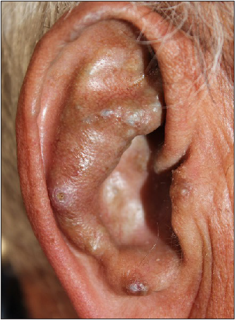
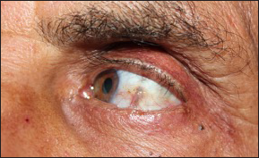
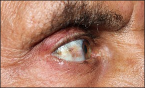
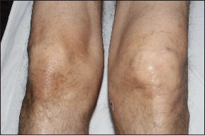
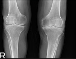
A 49-year-old male presented with pigmented skin lesions on his ears and pain and limitation of movement in both knees since five years. There was no history of intake of systemic drugs or topical medications. There were bluish-grey nodules seen on both the auricles [Figures 1a and 1b] and yellowish-brown discoloration of the conjunctivae [Figures 1c and 1d]. The left knee was swollen and had a limited range of motion [Figure 1e]. The magnetic resonance imaging and X-ray of the knee revealed osteophytic degenerative changes [Figure 1f]. Liver and kidney function tests, complete blood count, and rheumatoid factor were within normal limits. ANA was positive at a titer of 1/160. The patient’s urine darkened after two hours of exposure to the air. The level of homogentisic acid detected in urine or blood could not be evaluated for technical reasons. The homozygous mutation was detected in the HGD gene (exon 10, c.731_749del p.Thr244ArgfsTer30). Renal or cardiovascular involvement was not detected.

Bluish-grey nodular lesions on the left auricle.
Figure 1a:
Bluish-grey nodular lesions on the left auricle.
Bluish-grey nodular lesions on the right auricle.
Figure 1b:
Bluish-grey nodular lesions on the right auricle.
Yellow-brown discoloration in left conjunctiva.
Figure 1c:
Yellow-brown discoloration in left conjunctiva.
Yellow-brown discoloration in right conjunctiva.
Figure 1d:
Yellow-brown discoloration in right conjunctiva.
Swelling of the left knee joint.
Figure 1e:
Swelling of the left knee joint.
Osteophytic degenerative changes on direct radiography.
Figure 1f:
Osteophytic degenerative changes on direct radiography.

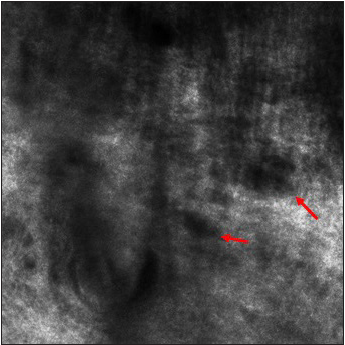
Dermoscopic examination of the ear lesions revealed a homogeneous brown to grey-blue pigmentation and linear vessels on a pinkish background [Figure 2a and 2b]. On reflectance confocal microscopy (Vivascope 3000; CALIBER ID, Rochester, NY, USA), the epidermis exhibited a normal honeycombed pattern with inflammatory and dendritic cells. The dermo-epidermal junction had a normal ridged pattern with normal-edged papillae. The dermis had hypo-refractile irregular, disorganised dark areas of different shapes and sizes [Figures 3a and 3b]. There were thin irregular collagen fibres with increased inflammatory cells and vessels between these dark areas. There were no atypical cells.
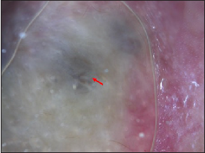
Homogeneous brown to grey-blue pigmentation (red arrow) (Dermlite DL4, polarised light)
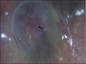
Figure 2a:
Homogeneous brown to grey-blue pigmentation (red arrow) (Dermlite DL4, polarised light)
Linear vessels on the pinkish background (black arrow) (Dermlite DL4, polarised light).
Figure 2b:
Linear vessels on the pinkish background (black arrow) (Dermlite DL4, polarised light).

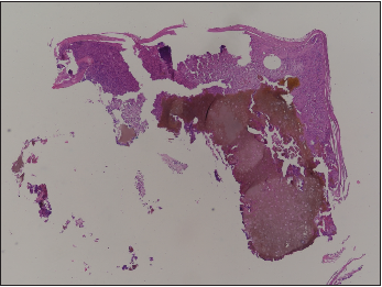
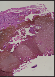
Histopathological examination revealed both small and large, numerous, yellowish brown, oval to round amorphous acellular substances (ochre pigment) in the reticular dermis along with increased vascularization and inflammatory cell infiltrates. Small amorphous deposits were seen very close to the flattened epidermis in some areas [Figures 3c and 3d]. A-low protein diet and nitisinone treatment (30 mg/day) were given. No progression was seen during the ensuing six-month period.
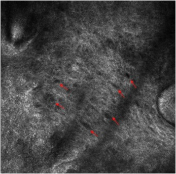
Oval dark spaces between irregular collagen fibers at lower magnification (red arrow).
Figure 3a:
Oval dark spaces between irregular collagen fibers at lower magnification (red arrow).
Oval dark spaces between irregular collagen fibers at higher magnification (red arrow).
Figure 3b:
Oval dark spaces between irregular collagen fibers at higher magnification (red arrow).
Yellowish brown amorphous material in the dermis; increased vascularity and inflammatory cell infiltrates (H&E, 40x).
Figure 3c:
Yellowish brown amorphous material in the dermis; increased vascularity and inflammatory cell infiltrates (H&E, 40x).
Yellowish brown amorphous material in the dermis; increased vascularity and inflammatory cell infiltrates (H&E, 100x).
Figure 3d:
Yellowish brown amorphous material in the dermis; increased vascularity and inflammatory cell infiltrates (H&E, 100x).

Dermoscopic findings of EO have been reported in only two cases, which included pseudo-network and partial occlusion of the follicular openings on the face and bluish-grey and white globular areas on the acral region. 1,2 In the present case, homogeneous brown to blue-grey pigmentation and linear vessels on a pinkish background were the dermoscopic features on the ears.

Evaluating only the superficial layers of the skin including the superficial dermis may be a major limitation of RCM in cases of EO with deep deposits. However, it may be possible to demonstrate deposits close to the surface. This possibility has been shown clearly in the present case; ochre pigment was observed as hypo-refractile, irregular, disorganized dark areas with different shapes and sizes at the dermal level on RCM. In the literature, available data on RCM findings of ochronosis are based on observations in some cases of exogenous ochronosis. In exogenous ochronosis, brown-grey reticulated macules with grey-black coarse spots are seen on the skin, most commonly on the malar regions of the patients treated for melasma with topical hydroquinone. 3 In these cases, the accumulation is expected to be small and superficial. 47 Dark, well-defined, round-to-oval and banana-shaped spaces in the papillary and middle dermis were the reported features on RCM. 46

Dermoscopy and RCM are particularly important in considering the clinical differential diagnoses of bluish-grey nodular lesions. In this case, the multiple symmetrical lesions on the ears suggest a possible diagnosis of EO. The differential diagnosis of a single bluish-grey nodule may include, besides EO, nodular melanoma, melanoma metastasis, blue nevus, pigmented basal cell carcinoma, and skin appendageal tumors. With the typical dermoscopic findings, it may be possible to differentiate between these entities. However, in a solitary lesion of EO, the diagnosis of ochronosis could be more convincing by demonstrating the deposits in the dermis and excluding the differentials demonstrating atypical cells.

In conclusion, RCM may be helpful in the diagnosis of EO with very superficial deposits, as seen in the present case. However, it must be noted that RCM findings should be taken in tandem with the suggestive clinical features and the diagnosis still needs to be confirmed by histopathology.

Declaration of patient consent

The authors certify that they have obtained all appropriate patient consent.

Financial support and sponsorship

Nil.

Conflicts of interest

There are no conflicts of interest.

References

  1. , , . Endogenous ochronosis: A dermoscopic view. Int J Dermatol. 2019;58:e175-e178.
    [CrossRef] [PubMed] [Google Scholar]
  2. , , , , , . Endogenous ochronosis: When clinical suspicion prevails over histopathology. Dermatol Online J. 2019;25:13030/qt5q2763pj.
    [PubMed] [Google Scholar]
  3. , , , , , . Exogenous ochronosis. Indian J Dermatol. 2015;60:537-43.
    [CrossRef] [PubMed] [PubMed Central] [Google Scholar]
  4. , , , , , , et al. Dermoscopic and reflectance confocal microscopic features of exogenous ochronosis. Arch Dermatol. 2010;146:1021-5.
    [CrossRef] [PubMed] [Google Scholar]
  5. , , , , , . Apport de la dermoscopie et de la microscopie confocale par réflectance dans le diagnostic d’ochronose exogène [Role of dermoscopy and reflectance confocal microscopy as an aid in the diagnosis of exogenous ochronosis] Ann Dermatol Venereol. 2016;143:318-20.
    [CrossRef] [PubMed] [Google Scholar]
  6. , , , , , . Dermoscopic and laser confocal features of an exogenous ochronosis case. G Ital Dermatol Venereol. 2020;155:367-8.
    [CrossRef] [PubMed] [Google Scholar]
  7. , , . Exogenous ochronosis in melasma: A study from South India. Pigment International. 2014;1:17-22.
    [Google Scholar]

Fulltext Views
1,579

PDF downloads
1,423
View/Download PDF
Download Citations
BibTeX
RIS
Show Sections