Translate this page into:
A case of cutis verticis gyrata related to pregnancy
Corresponding author: Prof. Yongqiong Deng, No. 25, Taiping Street, Luzhou 646000, Sichuan Province, China. dengyongqiong1@126.com
-
Received: ,
Accepted: ,
How to cite this article: Li X, Chen L, Xu J, Xiong X, Deng Y. A case of cutis verticis gyrata related to pregnancy. Indian J Dermatol Venereol Leprol 2021;87:533-4.
Sir,
Cutis verticis gyrata is a benign disorder of the scalp characterized by convoluted folds and deep furrows which resemble cerebral sulci and gyri.1 Throughout the literature review, we were unable to find any reports of cutis verticis gyrata that transiently occurred during pregnancy and spontaneously regressed after delivery. We report a case of pregnancy-related cutis verticis gyrata-like features on the forehead of a young woman.
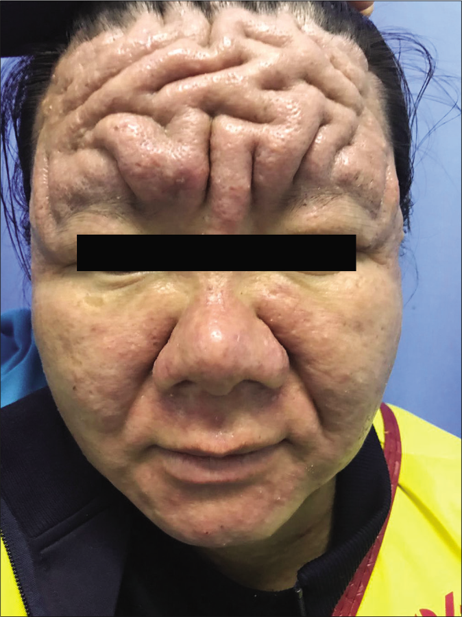
A 32-year-old woman at her third month of pregnancy presented with thickening of skin on her forehead with cerebriform convoluted folds and furrows [Figure 1a]. There were erythematous papules along with greasy scales on the central face. The trunk, limbs and joints were normal. There was no hirsutism, deepening of voice, acromegaly, headache, impaired vision, or hypertension. She had similar lesions during pregnancy three years ago which spontaneously subsided within a few months after delivery. Family history was noncontributary. Laboratory investigations revealed high levels of total serum testosterone [128.54 ng/dl: normal levels: (9.81-82.10ng/dl)]. Other parameters like blood pressure, serum insulin levels, blood sugars, thyroid function tests and other sex hormones were normal. Ultrasound examination of pelvis and fetus were normal. She refused further investigations like skin biopsy and imaging studies citing concerns about the health of her child.

- Thickening of skin on forehead with cerebriform convoluted folds and furrows at the 3rd month of pregnancy
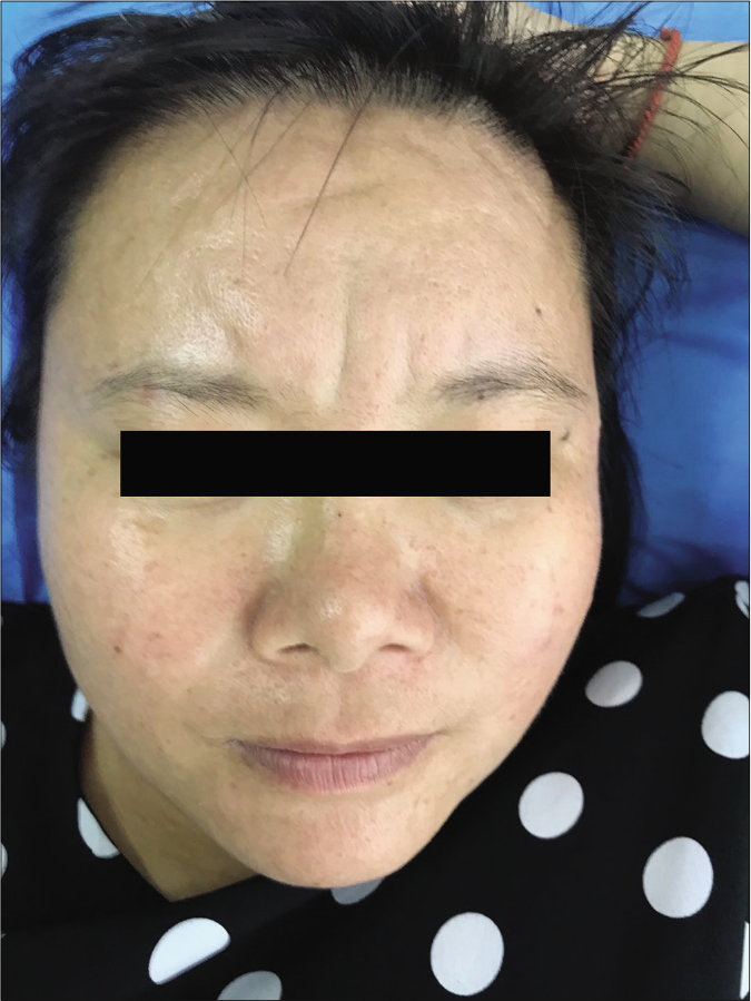
The patient was reviewed at two months after full-term normal delivery. The thickening of the forehead along with folds and furrows had visibly reduced without complete resolution [Figure 1b]. The X-ray of the limbs, ultrasound examination of abdomen, and the serum testosterone retest were normal. The magnetic resonance imaging of the skull revealed a partially empty sella.

- Mild thickening of skin on forehead seen after delivery
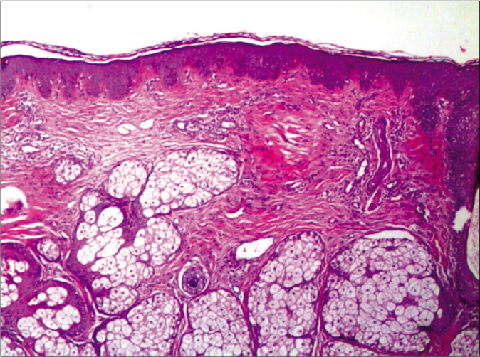
Skin biopsy from the forehead showed hyperplasia of sebaceous glands. [Figure 2]. Immunohistochemical studies showed mild intensity of staining for androgen receptors and negative staining for estrogen and progesterone receptors. Alcian blue staining was negative for mucin. Both gynaecologist and endocrinologist were of the opinion that no treatment is required as no underlying tumor was detected and regular follow-up during pregnancy was suggested.

- Sebaceous gland hyperplasia (H and E ×100)
The histopathological findings in the secondary cutis verticis gyrata show a significant variation due to the different underlying disorders.2 Sebaceous gland hyperplasia has been observed in the lesions of patients diagnosed with cutis verticis gyrata which is the target tissue of testosterone.3,4 The present case showed elevated serum testosterone and sebaceous gland hyperplasia. Association of serum testosterone and cutis verticis gyrata has long been discussed based on its preponderance in males, the occurrence after puberty, and the disappearance after castration, suggesting that secondary, hormone-induced cutis verticis gyrata could subside after the normalization of hormone secretion.4 In this case, the serum testosterone returned to normal after delivery, accompanied by the regression of the skin lesions. However, the causal relationship between androgen and the disease needs further experimental verification.
The proposed mechanism that causes empty sella involves the initial enlargement of the pituitary gland due to the elevation of sex hormones during pregnancy, followed by the subsequent regression in pituitary volume due to the normalized hormones after delivery which creates an empty space, in which cerebrospinal fluid accumulates.5 The cause of empty sella and the transient cutis verticis gyrata in the present case could be explained by the change in hormones during pregnancy.
In summary, this is a case report of cutis verticis gyrata, that developed and spontaneously subsided, probably due to hormonal changes during pregnancy. The present case may reveal the existence of a novel pregnancy-related syndrome which may manifest as a transient cutis verticis gyrata, androgen elevation and empty sella.
Declaration of patient consent
The authors certify that they have obtained all appropriate patient consent.
Financial support and sponsorship
Nil.
Conflicts of interest
There are no conflicts of interest.
References
- Is cutis verticis gyrata-intellectual disability syndrome an underdiagnosed condition? A case report and review of 62 cases. Am J Med Genet A. 2017;173:638-46.
- [CrossRef] [PubMed] [Google Scholar]
- Cutis verticis gyrata in association with vemurafenib and whole-brain radiotherapy. J Clin Oncol. 2014;32:e54-6.
- [CrossRef] [PubMed] [Google Scholar]
- Pathological characterization of pachydermia in pachydermoperiostosis. J Dermatol. 2015;42:710-4.
- [CrossRef] [PubMed] [Google Scholar]
- An endocrinological study of patients with primary cutis verticis gyrata. Acta Derm Venereol. 1993;73:348-9.
- [Google Scholar]
- Primary empty sella (PES): A review of 175 cases. Pituitary. 2013;16:270-4.
- [CrossRef] [PubMed] [Google Scholar]


