Translate this page into:
A case of disseminated cutaneous rhinosporidiosis presenting with multiple subcutaneous nodules and a warty growth
Correspondence Address:
Biju Vasudevan
Department of Dermatology, Command Hospital, Pune Wanowrie, Pune- 411040, Maharasthra
India
| How to cite this article: Verma R, Vasudevan B, Pragasam V, Deb P, Langer V, Rajagopalan S. A case of disseminated cutaneous rhinosporidiosis presenting with multiple subcutaneous nodules and a warty growth. Indian J Dermatol Venereol Leprol 2012;78:520 |
Sir,
Rhinosporidiosis is a rare chronic granulomatous condition, acquired by contact with stagnant water in places like swimming pools. Mucosal involvement of the nose, nasopharynx and also soft palate is common. Cutaneous involvement is rare and usually presents as friable warty growths. We herein, report a patient of rhinosporidiosiswho had multiple subcutaneous nodules and a warty growth in addition to nasal involvement. Such a morphological presentation is not very commonly reported in literature.
A 33-year-old male, resident of Kerala, India had a previous history of nasal blockage on and off since the last 10 years. Since the last 3 months, he noticed multiple lumps over the body which gradually increased in size. The lumps on the right foot were grouped. After a period of 1 month, these lesions on the right foot developed multiple bleeding points, and he had pain and difficulty in walking.
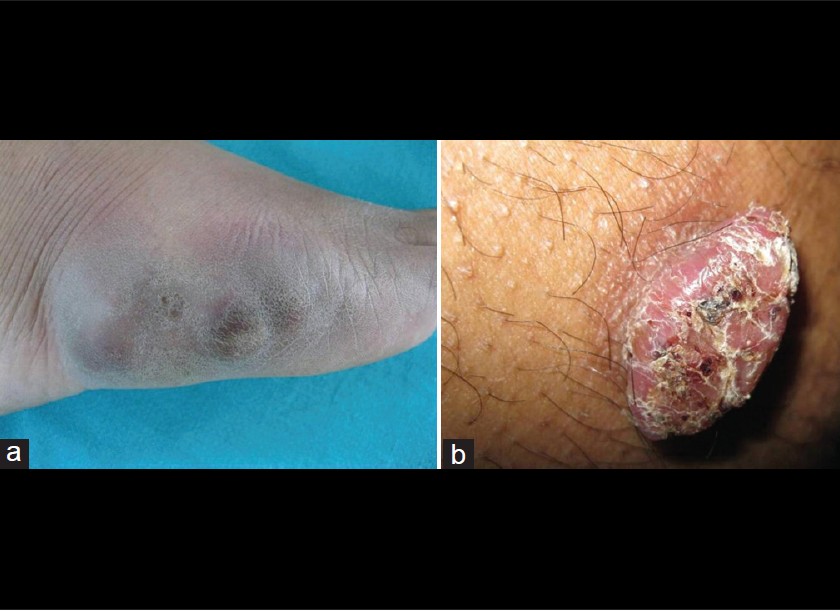
General physical and systemic examination was normal. Dermatological examination revealed multiple well-defined, soft, skin-colored, discrete, non-tender subcutaneous nodules over both forearms, thighs, legs and chest. Similar, but grouped nodules were present on dorsum of right foot [Figure - 1]a. There was a solitary, non-tender, warty growth measuring 3 × 1.5 cm situated over upper part of right thigh [Figure - 1]b. An examination of the nasal cavity revealed multiple pedunculated polypoidal masses, studded with small white dots on the surface. Diagnosis of disseminated cutaneous along with nasal rhinosporidiosis was considered.
 |
| Figure 1: Skin lesions of rhinosporidiosis: (a) Multiple grouped nodules on right foot (b) Solitary warty growth on right thigh |
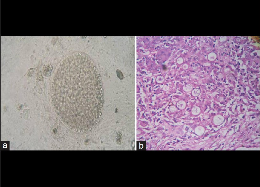
Investigations including complete blood counts, erythrocyte sedimentation rate, blood sugar, liver with renal function tests, chest X-ray and abdominal ultrasonography were within normal limits. ELISA for HIV was non-reactive. Smear from the skin lesion mounted in 10% KOH revealed spherules and large numbers of endospores on microscopy [Figure - 2]a. Cutaneous and nasopharyngeal lesions microscopically revealed numerous thick-walled sporangia in a vascular connective tissue along with a granulomatous inflammation [Figure - 2]b. This confirmed the diagnosis of cutaneous and nasopharyngeal rhinosporidiosis. He was started on Dapsone therapy 100 mg/day and was advised regular follow-up to assess response to the therapy. Surgical excision of foot lesions was done followed by split skin grafting. An excision of the warty growth was done, and endoscopic removal with cauterization of base was done for the nasopharyngeal masses.
 |
| Figure 2: Investigations revealed (a) Endospores on 10% KOH mount (40X) (b) Multiple sporangia on Histopathology (H and E, ×10) |
Rhinosporidiosis was initially thought to be caused by Rhinosporidium seeberi, a fungus. However, a cyanobacterium called Microcystic aeruginosa and an aquatic protistan parasite belonging to Mesomycetozoea class are the present contenders for the role of etiological agent. [1] Rhinosporidiosis has previously been reported in several countries, but most commonly from India and Sri Lanka. [2] It is more common in the southern states in India. Nasal, ocular, cutaneous and disseminated forms are the 4 common varieties mentioned. [3] Cutaneous lesions are very rare, and if present, are generally associated with mucosal lesions.
3 main types of cutaneous rhinosporidiosis have been reported, namely satellite skin lesions associated with nasal involvement, disseminated skin lesions with or without the nasal involvement and primary cutaneous type. [4] Skin lesions resembling warts, lipomas, verrucous tuberculosis, pyogenic granulomas, donovanosis and also subcutaneous nodules have been described in literature. Other rarer forms include those resembling soft tissue tumors, furuncles, ecthyma and giant cutaneous lesions. [5]
Disseminated cutaneous rhinosporidiosis has been described in both immunocompetent and HIV patients. [6] Our patient had disseminated cutaneous rhinosporidiosis in association with recurrent nasopharyngeal rhinosporidiosis in an immunocompetent setting. The other interesting fact was that he had polymorphic lesions- a warty growth and subcutaneous nodules, both grouped and solitary appearing. Only one case of polymorphic lesions in a single patient has been described earlier. [7] This presentation with its morphology and setting is an uncommon one and is, therefore, being reported.
| 1. |
Fredricks DN, Jolley JA, Lepp PW, Kosek JC, Relman DA. Rhinosporidium seeberi: A human pathogen from a novel group of aquatic protistan parasites. Emerg Infect Dis 2000;6:273-82.
[Google Scholar]
|
| 2. |
Thappa DM, Venkatesan S, Sirka CS, Jaisankar TJ, Gopalkrishnan, Ratnakar C. Disseminated cutaneous rhinosporidiosis. J Dermatol 1998;25:527-32.
[Google Scholar]
|
| 3. |
Kumari R, Nath AK, Rajalakshmi R, Adityan B, Thappa DM. Disseminated cutaneous rhinosporidiosis: Varied morphological appearances on the skin. Indian J Dermatol Venerol Leprol 2009;75:68-71.
[Google Scholar]
|
| 4. |
Acharya PV, Gupta RL, Darbari BS. Cutaneous rhinosporidiosis. Indian J Dermatol Venereol Leprol 1973;39:22-5.
[Google Scholar]
|
| 5. |
Anjunwala P, Teissera DA, Dissanaike AS. Rhinosporidiosis presenting with two soft tissue tumors followed by dissemination. Pathology 1999;31:57-8.
[Google Scholar]
|
| 6. |
Nayak S, Acharjya B, Devi B, Sahoo A, Singh N. Disseminated cutaneous rhinosporidiosis. Indian J Dermatol Venereol Leprol 2007;73:185-7.
[Google Scholar]
|
| 7. |
Ghorpade A. Polymorphic cutaneous rhinosporidiosis. Eur J Dermatol 2006;16:190-2.
[Google Scholar]
|
Fulltext Views
1,958
PDF downloads
1,450


