Translate this page into:
A case of subungual exostosis
2 Department of Orthopaedics, Duzce University Medical Faculty, Duzce, Turkey
3 Department of Pathology, Duzce University Medical Faculty, Duzce, Turkey
Correspondence Address:
Hakan Turan
Department of Dermatology, Duzce University Medical Faculty, Konuralp 81620, Duzce
Turkey
| How to cite this article: Turan H, Uslu M, Erdem H. A case of subungual exostosis. Indian J Dermatol Venereol Leprol 2012;78:186 |
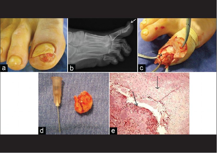
A sixteen-year-old male patient applied to our outpatient clinic with a painful mass on his right big toe. His complaint had been present for four months. There was no history of preceding trauma or bleeding. Cryotherapy had been applied to the lesion under the assumption that it was a pyogenic granuloma. Personal and family histories were unremarkable. Routine physical examination findings were normal. Dermatological examination revealed a hard, fixed, painful, 1 × 1 cm nodule with superficial erosion on the medial aspect of the tip of his right big toe [Figure - 1]a. A radiographic examination revealed a calcified lesion on the dorsomedial part of the distal phalanx continuing along the underlying bone [Figure - 1]b. The mass was resected completely from the underlying bone [Figure - 1]c and d. Histopathological examination showed evidence of mature trabecular bone [[Figure - 1]e, bottom arrow] covered by a fibrocartilaginous cap [[Figure - 1]e, top arrow]. A diagnosis of subungual exostosis was made in the light of these findings. Clinical and radiological recurrence was not detected after six months of follow-up.
 |
| Figure 1: (a) Hard, fixed, 1 × 1 cm nodule with superficial erosion on the medial aspect of the tip of right big toe of the patient under study. (b) Calcified lesion on the dorsomedial part of the distal phalanx of the patient under study. (c) Appearance of subungual exostosis during surgical excision. (d) Clinical appearance of excised mass after surgical removal. (e) Mature trabecular bone (bottom arrow) covered by a fibrocartilaginous cap (top arrow) (H and E, ×40) |
Subungual exostosis is a benign osteocartilaginous tumor described by Dupuytren in 1817. It is located on the distal phalanx of a digit, usually the big toe, commonly beneath or adjacent to the nail plate, and occurs predominantly in children and young adults. While the etiopathogenesis of subungual exostosis has not yet been clearly established, trauma and chronic infections are considered to be the causative factors of fibrocartilaginous metaplasia. Subungual exostosis is best diagnosed by radiographic examination. Histopathologically, it consists of mature trabecular bone structure covered by a fibrocartilaginous cap. The characteristic triad is subungual tumor leading nail plate deformity, digital pain and typical radiologic findings. The differential diagnosis mainly includes verruca, pyogenic granuloma, fibroma, glomus tumor, osteochondroma, myositis ossificans, and keratoacanthoma. Treatment is in the form of complete surgical resection of the lesion. Misdiagnosis is common for subungual exostosis, as in our case. Therefore, radiological examination must be carried out on these types of lesions, especially those located on the distal phalanx, adjacent to the nail.
Fulltext Views
3,548
PDF downloads
3,476


