Translate this page into:
A historical note on the evolution of “ringworm”
Correspondence Address:
Amiya Kumar Mukhopadhyay
“Pranab,” Ismile (Near Dharma Raj Mandir), Burdwan, Asansol - 713 301, West Bengal
India
| How to cite this article: Mukhopadhyay AK. A historical note on the evolution of “ringworm”. Indian J Dermatol Venereol Leprol 2019;85:125-128 |
Introduction
RINGWORM. The vulgar designation of the Herpes circinatus of Bateman. It appears in small circular patches, in which the vesicles arise only round the circumference.
A Dictionary of the Medical Terms, 1845.[1]
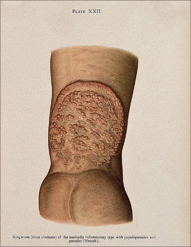
It is not even 200 years since the lines quoted above attributed to the disease that has become an enigma to the academicians and nightmare to the practicing dermatologists today. The incidence of ringworm infection has suddenly surged to an alarming level in the last 4–5 years.[2] The “specter of dermatophytosis” is certainly haunting the Indian dermatology.[3] Indeed, this common skin disease has been described since antiquity in the religious and medical treatises, illustrated under many headings, confused with many unrelated diseases, bare a number of names, but the systematic study started only less than two centuries back. This article is a brief sketch of the historical aspect of the evolution of the knowledge about disease: Ringworm [Figure - 1]. It should be admitted here that the immunological and therapeutic aspects have not been touched upon in this article as they merit separate treatment.
 |
| Figure 1: “Ringworm; lesions on inside right wrist, 1905” by Franz Mracek (credit: Wellcome Collection. CC BY 4.0 http://creativecommons.org/licenses/by/4.0/) |
“Ringworm:” Disease or Diseases? Nosological Confusions…
In the past, a number of diseases used to be lumped under a common expression. The terms like lichen, lepra, lupus, herpes, psora are few examples. Ringworm was no exception. Various diseases, particularly of annular configuration, were put together under some common headings. Guy de Chauliac (AD 1300–1368), a French physician, used the word Tinea as a generic term for many such ailments. Willan replaced it by Porrigo scutulata. Alibert classified tinea and his Tigne granulée was again a doubtful entity.[4] Other authors designated it in various forms like Herpes squamosus (Cazenev), Tinea tondante (Mahon), Phyto-alopecia (Melmsten), Tinea circinata (Anderson), Trichinosis furfuracea (Devergie) to name a few.[5] The early discussions were centered around the scalp ringworms [Figure - 2] and the most used designation was Favus. Other terms used were Porrigo scutulata, Trichophytica capitis, Tinea ficosa, Achores even Scabies capitis simplex (Plenck).[6] Even David Gruby, the pioneer of dermatophytological research, named it Porrigo decalvans – a term already used by Bateman for alopecia areata – an altogether different disease.
 |
| Figure 2: “A boy with a skin disease of the scalp. By W. Bagg, 1847.” (Credit: Wellcome Collection. CC BY 4.0 http://creativecommons.org/licenses/by/4.0/) |
In 1835, Rayer in his famous ‘A theoretical and practical treatise on the diseases of the skin’ described ringworm under the banner Impetigo annulata.[7] Schamberg in 1896 also lumped ringworm with some other diseases under the title Impetigo contagiosa annulata et serpiginosa.[8] The confusion also hovered around the etiology. It was believed that a common organism was responsible for different varieties of ringworms. This trend went on till 1894, when Sabouraud published his notable findings in Les Trichophyties Humaines, avec Atlas establishing the plurality of the etiological agent of ringworm.[9] In 1910, Sabouraud published his magnum opus Les Tiegnes and a new era begun.
Ringworms in the Antiquity
The fossil evidence of ringworm infection goes back to 125 million years back when “curious stumpy hairs on the back” of the fossil remains of Spinolestes xenarthrosus, one of the oldest mammals, were discovered at Las Hoyas, East-Central Spain in July, 2011.[10] As far as the ringworm in humans is concerned, the early evidence can be noted in the Charaka Samhita (c. second century BC) which mentioned a term Dadru in the seventh chapter of the Chikitsa Sthanam, whose description resembled ringworm.[11] Other ancient Indian authorities like Susruta (c. BC 600) and Vagbhata (c. sixth century AD) also mentioned Dadru under the broad caption of Mahakustha.[12] The Bower manuscript of third or fourth century AD mentioned about various remedies of ringworm.[13]
In the first century AD, Celsus described the cerion ulcer (kerion) – a ringworm of the scalp in the Chapter XXVII of Book V of his renowned treatise De Medicina.[14] Pliny in his Historia Naturalis (first century AD) termed it Lichen.[15] In AD 60, Dioscordes wrote about scalp ringworm in children. Galen of Pergamum (AD 130–210) also mentioned about it. Ringworm attracted the attention of the physicians of the Arabic medical world. Rabban Tabri (AD 810–895) talked about ringworm as Qooba and prescribed its management in his Firdaus ul hikmat. The legendry physician Zakariya Razi (AD 810–923) divided Qooba into two groups in his Al Hawi fit Tibb.[16] But practically speaking, nothing much happened until the beginning of the 19th century and till then ringworm infection was variously lumped with other diseases under the terms porrigo, lichen, lepra, psora, favus, etc.
Beginning of the Story of Modern Day Mycology and Ringworm
Ringworm had attracted attention of the physicians of all ages. Skin diseases had started gaining a separate shape of a subject since the time of Mercurialis (1530–1606). The founding text of British dermatology (and first such in English language) the De Morbis Cutaneis: A treatise of diseases incident to the diseases of the skin by Daniel Turner (1667–1741) mentioned about ringworm under the term Lichen (or Tettar) in 1714.[17] The study of fungus received a major thrust when Robert Hooke detected some filamentous organism on the leaves of damask rose with his newly constructed magnifying glass in 1677. In 1729, Michelli mentioned about Aspergillus amongst other fungus. A good deal of development happened in the human fungal disorder in the early 19th century. Robert Willan described the affection of the scalp by ringworms in 1817 and subsequently Plumbey told about it in 1821. During this period, it was confounded with other scalp disorders such as alopecia, seborrhea, etc.[18] In 1835, Agostino Bassi (1773–1856), an Italian entomologist, rendered an idea with experimental proof that fungus, a living organism, is responsible for the muscardine disease of the silkworm.[19] The story of ringworm took a turn in 1835 with the observation of Robert Remak, when he first observed the peculiar structures resembling rods and buds in the favus lesion. Remak never claimed that he recognized these structures to be fungus nor did he publish his observations. Remak credited his mentor Johann Lukas Schӧenlein (1793–1864) instead. Remak also allowed Xavier Hube to use his observation in a doctoral dissertation in 1837. He went ahead to culture it on apple slices and described the agent as Achorion schoenleinii in reverence to his guide Schӧenlein.[20] Schӧenlein in 1839 described about the favus fungus.[21] Melmsten described the fungus of the ringworm in Stockholm in 1845 and named Trichophyton tonsurans.[5] Tillbury Fox described Tinea pedis in 1870.[22] The ball was thus rolling and few more years passed.
“Gruby's Disease” and Thereafter…
It was 1841. David Gruby (1810–1898), a Hungarian physician, was working on the microscopic anatomy in France [Figure - 3]. He was unaware about the works of Remak or Schӧenlien. He isolated the fungus from the favus. The monumental discovery of agar as culture medium was yet to come but Gruby cultured the matter on the potato slices and inoculated the growth on the healthy tissue to reproduce the lesion of favus on it. The fungal element was later named Achorion schoenleinii in the honor of Schӧenlein. On the following year, he described Trichophyton ectothrix which caused sycosis barbae. In the year 1843, Gruby described another fungus named Microsporum audouinii and endothrix invasion as Porrigo decalvans[23] As previously mentioned, this term created confusion as the same term was already used by Bateman for alopecia areata.[18] This confusion was cleared later by Sabouraud.
 |
| Figure 3: David Gruby (1810–1898) by Mathieu Deroch (c. 1880). (Credit: David_Gruby_Portrait.jpg: Mathieu Deroche derivative work: Itzuvit. CC BY 3.0, http://creativecommons.org/licenses/by/3.0) |
Unearthing of the “Worms”
It was 1910 that a new era had begun. Raymond Sabouraud (1864–1938), a French physician, a painter, and sculptor of repute published Les Tiegnes – the first ever treatise on mycology, not only designed a culture medium with dextrose and agar but also was the first to propound a taxonomy and classification of the fungi responsible for “ringworm.” This cleared the air of confusion that was hovering around the correct nomenclature of the dermatophytes since antiquity. He classified the dermatophytes into four genera depending on the clinical, cultural, and microscopic characteristics: Achrion, Epidermophyton, Microsporum, and Trichophyton. In 1934, Emmons modernized the classification on the basis of morphology of the spores and accessory organs and dropped the genus Achorion from the list.[24] Further work of George and others about the identification based on the routine nutritional tests made the task much simplified.[25]
Further Researches and Newer Revealations…
Sabouaud made further work on dermatophytes much easier for the future workers. Now, one can grow ringworms in the laboratory and gain entry into their world. Bloch's experiment with guinea pig provided much knowledge on the pathology and immunology of the ringworm disease. Quincke and his student Bodin recognized that the organisms for mouse and human favus are different.[26] Dawson and Gentles described the telemorph in 1959. Studies on sexual reproduction and pleomorphism led to the better understanding of the taxonomy and behavior of the different varieties of ringworms.[27]
The Final Call
The colossal works of Sabouraud during 1894–1895 supported the view held by Gruby. With these works, the important but neglected subject of medical mycology generated new waves of interest. This phenomena attended pinnacle when a full session was held in the afternoon of August 1896 in London at the Third International Congress of Dermatology. The session was devoted to the topic Ringworms and the Trichophytons where Sabouraud presented his epoch making study of 300 cultures. The luminaries like Malcolm Morris, Colcot Fox, Unna, Rosenbach contributed and the official transactions led to a volume exceeding hundred pages! Ainsworth has rightly commented: “…(with this session) the main lines for study of these fungi during the next fifty years laid down.”[28] And it really happened.
Epilogue
As already mentioned, studies on ringworm started since long and Gruby's discovery during 1841–1843 laid the foundation stone, but the important contributions came from the works of the European giants like Schӧenlein, Sabouraud, Malmsten, and others. If we consider the work of Willan (1817) as the beginning of the modern study of dermatophytosis, then the Les Tignes of Raymond Sabouraud in 1910 marks the peak. More than a century has elapsed since then and we still are facing the riddle as to the peculiar behavior of the ringworms every moment at present. Much work is yet to be done. There is a long way to go before we sleep!
| 1. |
Hoblyn RD. A Dictionary of Terms Used in Medicine and the Collateral Sciences. Philadelphia: Lea and Blanchard; 1845. p. 311.
[Google Scholar]
|
| 2. |
Verma S, Madhu R. The great Indian epidemic of superficial dermatophytosis: An appraisal. Indian J Dermatol 2017;62:227-36.
[Google Scholar]
|
| 3. |
Panda S, Verma S. The menace of dermatophytosis in India: The evidence that we need. Indian J Dermatol Venereol Leprol 2017;83:281-4.
[Google Scholar]
|
| 4. |
Morris M. Ringworm in the Light of Recent Research. London: Cassell and Co., Ltd.; 1898. p. 1-4.
[Google Scholar]
|
| 5. |
Aldersmith H. Ringworm and Alopecia Areata. London: H. K. Lewis; 1897. p. 2-4.
[Google Scholar]
|
| 6. |
Bateman T. A Practical Synopsis of Cutaneous Diseases. London: Longman, Rees, Orme, Brown and Green; 1829. p. 226.
[Google Scholar]
|
| 7. |
Rayer P. A Theoretical and Practical Treatise on the Diseases of the Skin. Philadelphia: Carey and Hart; 1845. p. XIV.
[Google Scholar]
|
| 8. |
Schamberg JF. Impetigo contagiosa annulata et sarpiginosa. J Cutanoeus Genitourin Dis 1896;14:168-73.
[Google Scholar]
|
| 9. |
Sabouraud R. Les Trichophyties Humaines. Paris: Rueff and Co., Editeurs; 1894.
[Google Scholar]
|
| 10. |
Martin T, Marugán-Lobón J, Vullo R, Martín-Abad H, Luo ZX, Buscalioni AD, et al. Acretaceous eutriconodont and integument evolution in early mammals. Nature 2015;526:380-4.
[Google Scholar]
|
| 11. |
Mukhoadhyay AK. Skin Diseases ('Dermatology') in India: History and Evolution. Calcutta: Allied Book Agency; 2011. p. 51.
[Google Scholar]
|
| 12. |
Melashankar SM, Sharada. Clinical evaluation of the efficacy of Laghu Manjisthadi Kwatha and Chakramardadi Lepa in Dadru (Tinea). J Auyrveda Intger Med Sci 2016;1:24-8.
[Google Scholar]
|
| 13. |
Hoernle AF. The Bower Manuscript. Calcutta, India: Office of the Superintendent of Government Printing; 1897.
[Google Scholar]
|
| 14. |
Grieve J. A Cornelius Celsus of Medicine (In eight Books). London: D. Wilson and T. Durham; 1756. p. 316.
[Google Scholar]
|
| 15. |
Pliny. Natural History. English Translation by Jones WH. Vol. 6. Libri XX-XVI. Cambridge: Harvard University Press; 1951. p. viii.
[Google Scholar]
|
| 16. |
Firdaus S, Ali F, Sultana N. Dermatophytosis (Qooba) a misnomer infection and its management in modern and Unani perspective – A comparative view. J Med Plants Stud 2016;4:109-14.
[Google Scholar]
|
| 17. |
Turner D. De Morbis Cutaneis: A Treatise of Diseases Incident to the Diseases of the Skin. 3rd ed. London: R and J Bonwicke and Others; 1726. p. 73.
[Google Scholar]
|
| 18. |
Turner JP. Ringworm and its Successful Treatment. Philadelphia: FA Davis Company, Publishers; 1921. p. 15-8.
[Google Scholar]
|
| 19. |
Ainsworth GC. Medical Mycology: An Introduction to its Problems. London: Sir Isaac Pitman and Sons Ltd.; 1952. p. 1.
[Google Scholar]
|
| 20. |
Weitzman I, Summerbell RC. The dermatophytes. Clin Microbiol Rev 1995;8:240-59.
[Google Scholar]
|
| 21. |
Lewis GM, Hopper ME. An Introduction to Medical Mycology. 3rd ed. Chicago: The Year Book Publishers; 1948. p. 3-4.
[Google Scholar]
|
| 22. |
Swartz JH. Elements of Medical Mycology. 2nd ed. London: William Heinemann Medical Books Ltd.; 1949. p. xiii-xiv.
[Google Scholar]
|
| 23. |
Gruby D. Available from: http://www.whonamedit.com/doctor.cfm/3066.html. [Last accessed on 2018 Jan 15].
[Google Scholar]
|
| 24. |
Emmons CW. Dermatophytes: Natural groupings based on the form of the spores and accessory organs. Arch Dermatol Syphylol 1934;30:337-62.
[Google Scholar]
|
| 25. |
Georg LK, Camp LB. Routine nutritional tests for the identification of dermatophytes. J Bacteriol 1957;74:113-21.
[Google Scholar]
|
| 26. |
Kligman AM. Pathophysiology of ringworm infections in animals with skin cycles. J Invest Dermatol 1956;27:171-85.
[Google Scholar]
|
| 27. |
Weitzman I. Variation in Microsporum gypseum. I. A genetic study of pleomorphism. Sabouraudia 1964;3:195-204.
[Google Scholar]
|
| 28. |
Ainsworth GC. Introduction to the History of Mycology. Cambridge: Cambridge University Press; 1976. p. 140-83.
[Google Scholar]
|
Fulltext Views
28,892
PDF downloads
4,104


