Translate this page into:
A novel missense mutation in ADAR1 gene causing dyschromatosis symmetrica hereditaria in a Chinese patient
Correspondence Address:
Cheng-Rang Li
Jiangwangmiao Street 12, Nanjing, Jiangsu 210042
People's Republic of China
| How to cite this article: Li ZL, Zhang GY, Hui Y, Yu RX, Li Q, Xu HX, Li CR. A novel missense mutation in ADAR1 gene causing dyschromatosis symmetrica hereditaria in a Chinese patient. Indian J Dermatol Venereol Leprol 2015;81:327 |
Dyschromatosis symmetrica hereditaria, also called reticulate acropigmentation of Dohi, is an autosomal dominant, cutaneous disease characterized by asymptomatic, hyperpigmented and hypopigmented macules on the dorsal aspects of the extremities. Heterozygous mutations in the adenosine deaminase acting on RNA 1 (ADAR1) gene were identified as the molecular basis of dyschromatosis symmetrica hereditaria. [1]
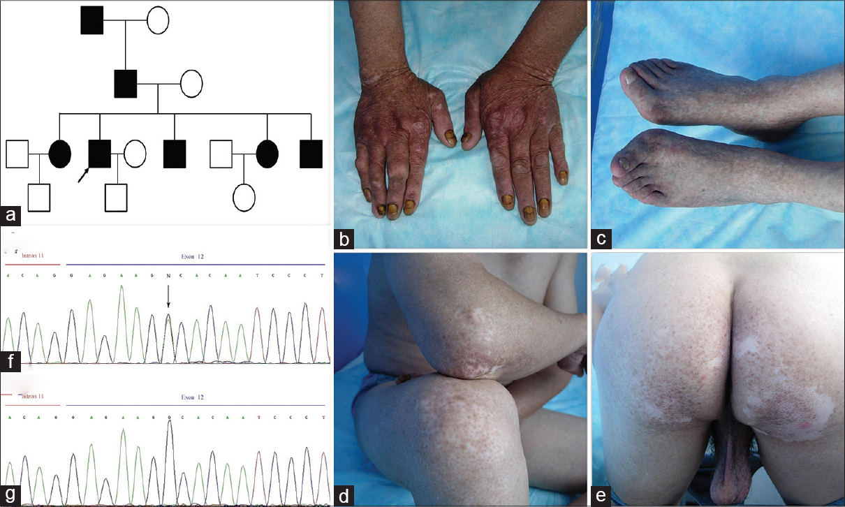
In this study, we investigated the members of a four-generation family from Jiangsu province of China with typical dyschromatosis symmetrica hereditaria [Figure - 1]a. The proband of this family is a 60-year-old male with a history, from 6 to 7 months age, of hyperpigmented and hypopigmented macules on the distal extremities [Figure - 1]b and c. The typical lesions were also observed on the knees, elbows [Figure - 1]d, and buttocks [Figure - 1]e. After informed consent, we collected peripheral blood from the patient and sequenced all exons of the ADAR1 gene. We detected a heterozygous missense mutation (c. 3026G > A) in the ADAR1 gene in the patient [Figure - 1]f. The ADAR1 gene mutation was, however, not detected in 100 unrelated controls [Figure - 1]g.
 |
| Figure 1: Identifying lesions and characterizing a novel mutation in the ADAR1 gene in a proband of a family affected with dyschromatosis symmetrica hereditaria. (a) Pedigree of the family. Affected family members are represented by black symbols. The proband is indicated by an arrow. (b) Hypopigmented and hyperpigmented macules on the back of the hand. (c) Hypopigmented and hyperpigmented macules on the lower limbs. (d) Hypopigmented and hyperpigmented macules on the elbows. (e) Hypopigmented and hyperpigmented macules on the buttocks. (f) Novel c. 3026G > A mutation of the ADAR1 gene observed in the patient. (g) Normal ADAR1 gene sequence |
A previous study demonstrated that a heterozygous mutation of ADAR1 causes dyschromatosis symmetrica hereditaria, [1] and so far, more than 120 mutations in the ADAR1 gene have been reported. [2] The deaminase domain, located between positions 886 to 1221 of the ADAR1 gene, covers more than 50% of the reported mutations. [2] This domain is critical for enzyme function and is thought to play an important role in the pathogenesis of dyschromatosis symmetrica hereditaria. The missense mutation c. 3026G > A, which was identified in the patient, is located in the deaminase domain of the ADAR1 protein in exon 12. Mutations in exon 12 have also been reported in other unrelated families, [2] and this result further supported the speculation that the deaminase domain might be a hot spot for mutations. The new missense mutation c. 3026G > A noted in our patient changes the neutral glycine residue to an acidic aspartate residue at position 1009 of the protein sequence (p.G1009D). This amino acid residue at position 1009 may play a vital role in maintaining the conformation of the catalytic site of ADAR1 and a mutation at this locus may influence the normal function of the ADAR1 enzyme.
The role of ADAR1 mutation in the development of dyschromatosis symmetrica hereditaria is still unknown. One may speculate that the dysfunction of A-to-I editing induced by ADAR1 mutation might affect differentiation of melanoblasts to melanocytes when melanoblasts migrate from the neural crest to the skin during development. Previous in vitro studies have indicated that ADAR1 is related to viral inactivation, [3] so there is also a viewpoint that ADAR1 mutation may increase susceptibility to viral infection and trigger apoptosis of melanocytes; this leads to the formation of hypopigmented lesions. Then the remnant melanocytes in the bulge area of hair follicles migrate toward the epidermis to form hyperpigmented macules. [2]
In our patient, typical lesions were observed on the elbows, knees, and buttocks other than dorsal extremities. The fact that typical lesions extended to the proximal extremity was also reported by Bilen et al.[4] and Kondo et al.[5] The variety of clinical phenotypes caused by mutation of the same gene suggests that other factors may influence the extent of the clinically apparent skin lesions.
Acknowledgment
The authors are indebted to Foundation of PUMC innovation team, Teaching and Research Fund Project of PUMC (Grant No. 2011zlgc0114, 303-05-8050).
| 1. |
Miyamura Y, Suzuki T, Kono M, Inagaki K, Ito S, Suzuki N, et al. Mutations of the RNA-specific adenosine deaminase gene (DSRAD) are involved in dyschromatosis symmetrica hereditaria. Am J Hum Genet 2003;73:693-9.
[Google Scholar]
|
| 2. |
Hayashi M, Suzuki T. Dyschromatosis symmetrica hereditaria. J Dermatol 2013;40:336-43.
[Google Scholar]
|
| 3. |
Toth AM, Li Z, Cattaneo R, Samuel CE. RNA-specific adenosine deaminase ADAR1 suppresses measles virus-induced apoptosis and activation of protein kinase PKR. J Biol Chem 2009;284:29350-6.
[Google Scholar]
|
| 4. |
Bilen N, Akturk AS, Kawaguchi M, Salman S, Ercin C, Hozumi Y, et al. Dyschromatosis symmetrica hereditaria: A case report from Turkey, a new association and a novel gene mutation. J Dermatol 2012;39:857-8.
[Google Scholar]
|
| 5. |
Kondo T, Suzuki T, Mitsuhashi Y, Ito S, Kono M, Komine M, el al. Six novel mutations of the ADAR1 gene in patients with dyschromatosis symmetrica hereditaria: Histological observation and comparison of genotypes and clinical phenotypes. J Dermatol 2008;35:395-406.
[Google Scholar]
|
Fulltext Views
2,140
PDF downloads
2,109


