Translate this page into:
A novel multipurpose dermoscope adapter
Corresponding author: Dr. Sandip Agrawal, Department of Dermatology, LTMMC and LTMGH, Sion, Mumbai, Maharashtra, India. sandipagrawaliggmc@gmail.com
-
Received: ,
Accepted: ,
How to cite this article: Cheng CY. Intratherapeutic dermoscopy assists nevus removal by laser therapy. Indian J Dermatol Venereol Leprol 2022;88:128-30.
Problem
Dermoscopy, also known as surface microscopy, has evolved as a noninvasive technique allowing rapid and magnified in vivo observation of the skin. The increase in popularity has expanded the scope of utility of the dermoscope by leaps and bounds in recent times. However, the need for close contact with the lesional skin and restricted field of vision, has limited the use of the dermoscope.
Solution
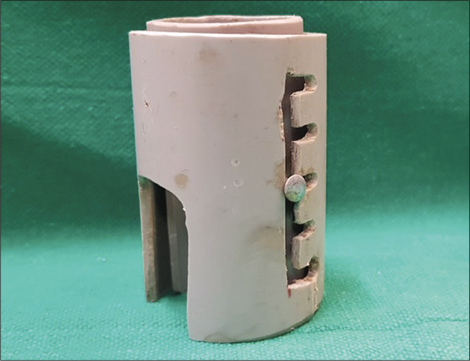
We suggest a simple, reproducible adapter that expands the use of the dermoscope for various diagnostic and therapeutic modalities. Two plastic pipes of length 6.1 cm are fitted into each other. The inner pipe has a screw attached and the outer pipe has sockets created for the same, to enable change in the effective height of the adapter when required. Two windows of dimension 3 × 2 cm are created at the base for easy passage of any instrument through it [Figures 1 and 2].

- The dermoscope adapter with two plastic pipes fitted into each other

- Two pipes unassembled and all the parts shown separately

- Two pipes unassembled and all the parts shown separately
Principle
When a video dermoscope is placed over the adapter, the field of vision increases due to subsequent decrease in the magnification from × 500 to × 200 and increase in focal length to the created height of the adapter, i.e., 6.1 cm [Figure 3]. Further change in field of vision can be done by simply changing the length of the adapter.

- Field of vision using a video dermoscope at (×200)

- Increase in the field of vision using the adapter
Various other proposed uses of the adapter include:1,2
Specific sample collection can be achieved by inserting forceps through the created windows under direct vision of the dermoscope, e.g., collection of corkscrew hair for KOH examination in tinea capitis.
A specific site for biopsy can be marked, e.g., a white dot or vellus hair.
The biopsy sample can be cut through a precise level with help of blade and forceps which can be passed through the windows in the probe. e.g., cutting a scalp biopsy at the level of isthmus [Figure 4].
By introducing the probe inside the mouth, mucoscopy can be performed more easily. It is patient friendly because it becomes easier for the patient to hold their mouth open for a longer period. Also, the clenched tube indirectly provides stability to the dermatoscope aiding in better and easier dermoscopic visualization of the oral mucosa. With the use of this plastic tube holder, good quality dermoscopic images can be obtained.3
Dermoscopic guided biopsy can be done with the help of a motorized punch mounted on an angled hand piece.

- Cutting a scalp biopsy at the level of bulb of terminal hair
Although there are many adapters described till now, none include the scope for intervention and reusability as described with this multipurpose dermoscope adapter. Through the use of this adapter we aim to ease and expand the potential of the humble dermoscope which has become an essential tool in everyday practice.
Financial support and sponsorship
Nil.
Conflicts of interest
There are no conflicts of interest.
References
- Possible applications of video dermatoscopy beyond pigmented lesions. Int J Dermatol. 2003;42:430-3.
- [CrossRef] [PubMed] [Google Scholar]
- Dermatoscopy in Clinical Practice: Beyond Pigmented Lesions Vol 20. London: Informa Healthcare Ltd.;
- [CrossRef] [Google Scholar]
- Innovative modification of the USB dermatoscope for mucoscopy. J Am Acad Dermatol. 2018;78:e3-4.
- [CrossRef] [PubMed] [Google Scholar]


